
Enterobius (Colobenterobius) serratus Hasegawa, Matsuo et Onuma, 2003
|
publication ID |
https://doi.org/ 10.11646/zootaxa.4722.3.6 |
|
publication LSID |
lsid:zoobank.org:pub:FB95CB62-8499-4080-9F16-E0082BB021FB |
|
DOI |
https://doi.org/10.5281/zenodo.5621003 |
|
persistent identifier |
https://treatment.plazi.org/id/656C87EC-6A21-FFDE-08A4-FD4EFC5EB3E7 |
|
treatment provided by |
Plazi |
|
scientific name |
Enterobius (Colobenterobius) serratus Hasegawa, Matsuo et Onuma, 2003 |
| status |
|
Enterobius (Colobenterobius) serratus Hasegawa, Matsuo et Onuma, 2003
(ZooBank registration: urn:lsid:zoobank.org:pub:69E6C6D0-A122-4AB0-AE90-747E941EF26A )
( Nematoda : Oxyuridae : Enterobiinae)
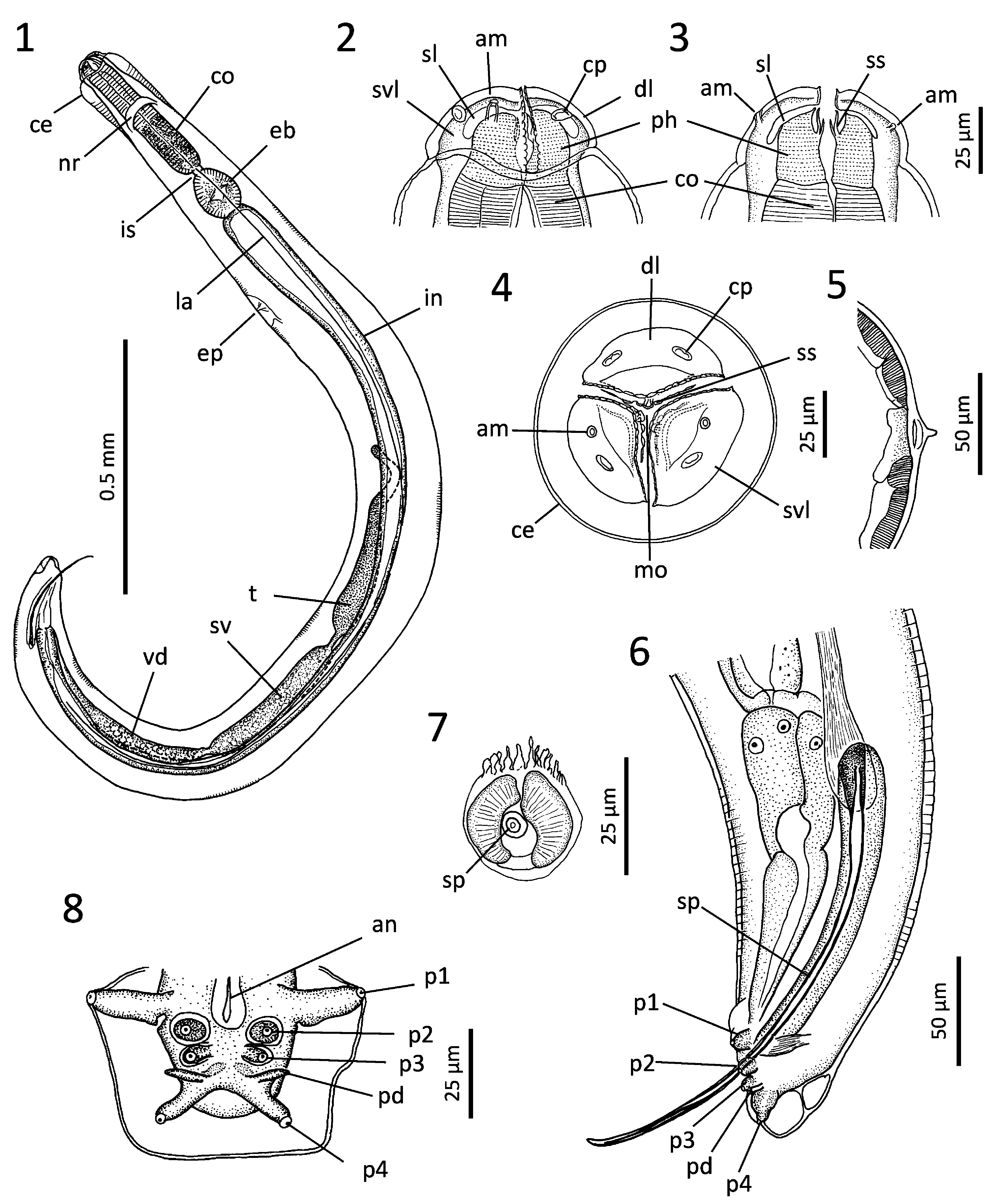
Male (based on 6 males): Body minute, 1.96–2.91 (x̄ =2.54) mm long and 163–178 (172) µm wide in midbody. Posterior end bent ventrally ( Fig. 1 View FIGURES 1–8 ). Cuticle transversely striated ( Figs. 1, 6 View FIGURES 1–8 ). Lateral alae single-crested, commencing at level of esophageal bulb and terminating ca. 200 µm anterior to posterior end ( Figs. 1, 5 View FIGURES 1–8 ). Cephalic expansion 150–200 (175) µm long by 100–128 (116) µm wide, with inner septa in posterior half. Cephalic end with three lips of almost equal size, forming round head with diameter of 63–70 (68) µm. Lips well set off from body, each with serrated inner margin ( Figs. 2, 4 View FIGURES 1–8 ). Dorsal lip with two cephalic papillae; subventral lips each with one cephalic papilla and amphidial pore; distance between amphidial pores 44–49 µm (n=2) ( Fig. 3, 4 View FIGURES 1–8 ). Slots present between lips and pharynx ( Fig. 3 View FIGURES 1–8 ). Pharynx with specific teeth composed of one large median and three pairs of side projections just beneath each lip ( Figs. 2–4 View FIGURES 1–8 ). Pharynx 23–35 (30) µm long; esophageal corpus 255–285 (271) µm long by 51–65 (58) µm wide with dark granules in posterior half; esophageal isthmus short, 5–10 (7) µm long by 21–25 (23) µm wide; esophageal bulb valved, 95–113 (102) µm long by 83–103 (90) µm wide ( Fig. 1 View FIGURES 1–8 ). Distance from cephalic apex to nerve ring 158–170 (165) µm and excretory pore 534–750 (662) µm. Testis extending to middle of body ( Fig. 1 View FIGURES 1–8 ). Spicule thin, slender, with ellipsoid light-refractile mass basally; distal portion pointed, slightly bent ventrally, 227–248 (241) µm long (n=4) ( Fig. 6 View FIGURES 1–8 ). Spicule portion inside body housed in spicular pouch with paired muscular bands ( Figs. 6, 7 View FIGURES 1–8 ). Caudal papillae comprised of four pairs: 1 st pair large, pedunculated, projecting laterally at level of anus; 2 nd and 3 rd pairs slightly posterior to 1 st pair, mostly flat, directing ventrally, surrounded by transparent cuticular thickenings; 4 th pair pedunculated, smaller than 1 st pair, directing posterolaterally ( Figs. 6, 8 View FIGURES 1–8 ). Phasmids arising from anterior base of 4 th papillae, directing laterally ( Figs. 6, 8 View FIGURES 1–8 ).
No known copyright restrictions apply. See Agosti, D., Egloff, W., 2009. Taxonomic information exchange and copyright: the Plazi approach. BMC Research Notes 2009, 2:53 for further explanation.