
Gebiacantha sagamiensis, Komai, Tomoyuki, 2017
|
publication ID |
https://doi.org/ 10.11646/zootaxa.4263.3.9 |
|
publication LSID |
lsid:zoobank.org:pub:83B58CB6-249C-4257-8EFA-F9AFE01A9E68 |
|
DOI |
https://doi.org/10.5281/zenodo.6028413 |
|
persistent identifier |
https://treatment.plazi.org/id/246C2E14-6197-45DD-9C7C-B1B5C088DEF7 |
|
taxon LSID |
lsid:zoobank.org:act:246C2E14-6197-45DD-9C7C-B1B5C088DEF7 |
|
treatment provided by |
Plazi |
|
scientific name |
Gebiacantha sagamiensis |
| status |
sp. nov. |
Gebiacantha sagamiensis View in CoL n. sp.
[New Japanese name: Sagami-toge-ana-jyako] Figs 1–4 View FIGURE 1 View FIGURE 2 View FIGURE 3 View FIGURE 4
Material examined. Holotype: male (cl 3.2 mm), RV “ Rinkai-maru ”, west of Misaki , Miura, Sagami Bay, 35°08.43’N, 139°33.01’E, 106– 101 m, 25 June 2015, dredge, coll. T. Komai, CBM-ZC 13874. GoogleMaps
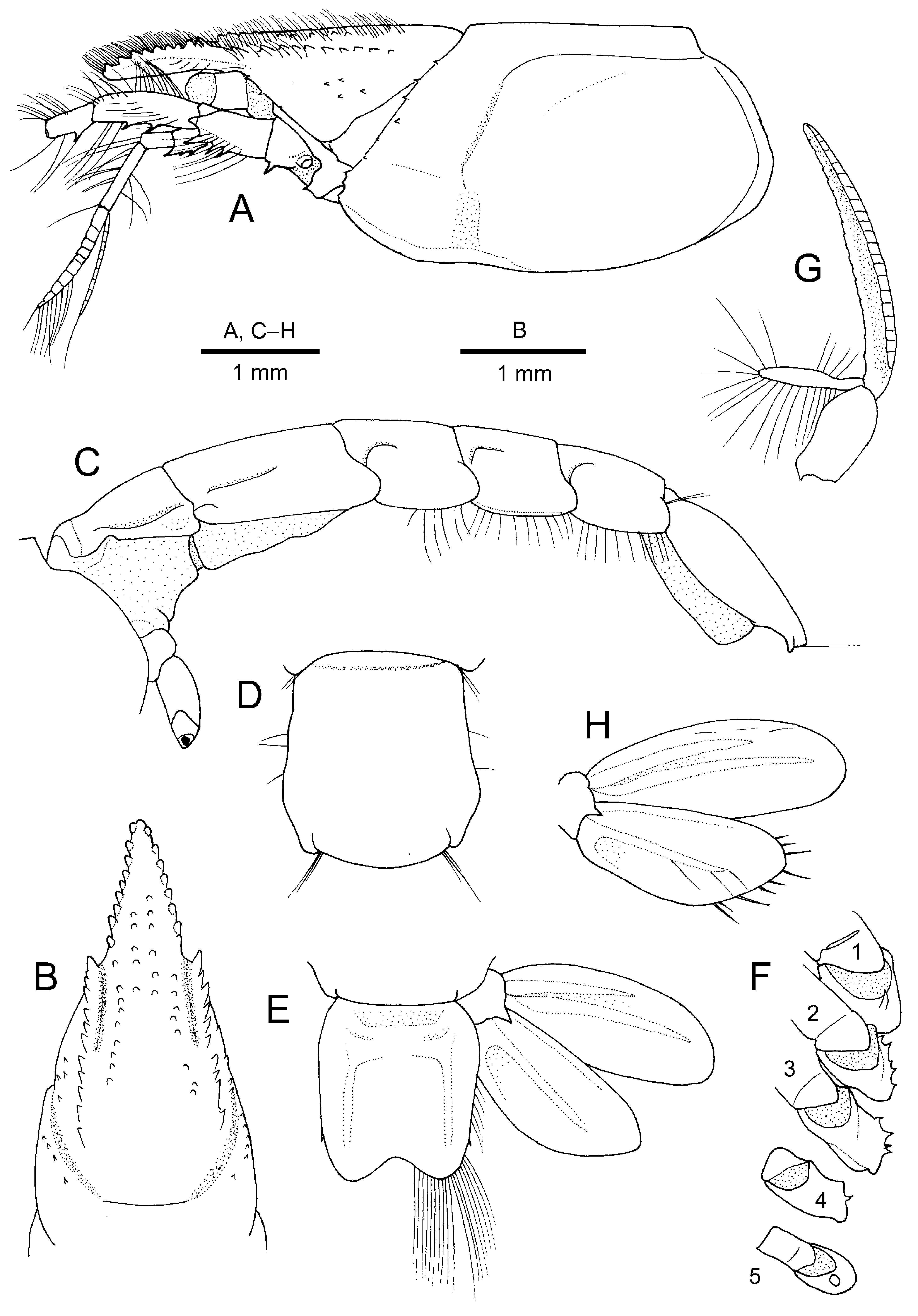
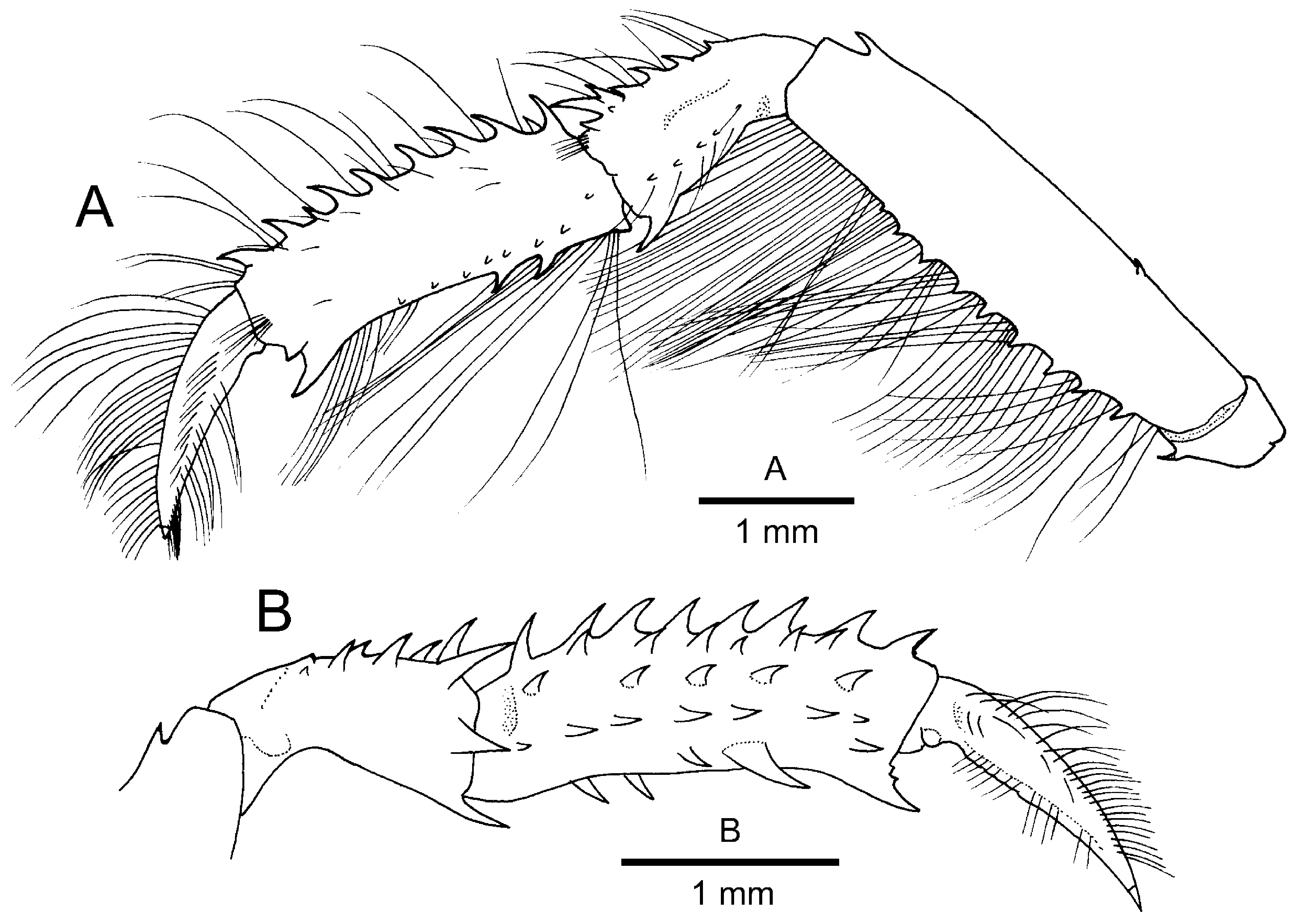
Description. Holotype male. Rostrum ( Fig. 1 View FIGURE 1 A, B) long, reaching nearly to distal end of article 4 of antennal peduncle, tapering distally in blunt apex, straight and directed anteriorly in lateral view, flattened dorsoventrally, tip far exceeding distal corneal margins; dorsolateral margins with 9 (left) and 7 (right) narrowly spaced, small tubercles; dorsal surface with numerous short setae and longitudinal row of 5 or 6 small tubercles on either side of midline in proximal half; ventral surface gently convex transversely, unarmed. Anterior carapace ( Fig. 1 View FIGURE 1 A, B) with lateral gastric ridges very slightly diverging posteriorly and bearing row of 10 tiny spines (including terminal one) decreasing in size and sharpness toward posterior; anterior end of gastric lateral ridge slightly produced, separated from carapace by small U-shaped notch; median part of anterior carapace flanked by distinct grooves running along gastric lateral ridges and having longitudinal row of tiny blunt tubercles continuing from dorsolateral margins of rostrum accompanied by tufts of short setae on either side of midline, midline neither carinate or sulcate; anterolateral margin with 3 (left) or 5 (right) spinules, including postocular spine; postorbital region with some scattered spinules; pterygostomial margin spinulose. Posterior carapace ( Fig. 1 View FIGURE 1 A) with shoulder along cervical groove bearing row of spinules; hepatic region with short longitudinal groove and obliquely transverse groove extending dorsally subparallel to cervical groove and curved posteriorly and connected with linea thalassinica. Linea thalassinica widely interrupted, anterior part slightly sinuous, extending from anterolateral margin somewhat superior to anterolateral notch to cervical groove, posterior part extending from posterior to cervical groove to posterodorsal margin of carapace.
Pleon ( Fig. 1 View FIGURE 1 C) fairly flattened dorsoventrally. Pleuron 1 narrow, slightly elevated, demarcated from tergum by distinct longitudinal groove; pleuron 2 also narrow, anterior part demarcated from tergum by distinct longitudinal groove reaching to midlength of somite, ventral margin slightly upturned. Pleomeres 3–5 each with short curved groove on anterior part; pleural margins with row of seta directed laterally, each posterolateral margin rounded. Pleomere 6 ( Fig. 1 View FIGURE 1 D) longest, 1.1 times as long as greatest width at 0.7 length of somite; lateral margins broadened posteriorly in anterior 0.7 and then slightly narrowing, thus posterolateral part forming low convexity; posterior margin convex, with small notch on each lateral part. Telson ( Fig. 1 View FIGURE 1 D) subrectangular, 1.1 times as long as greatest width at anterior 0.2; dorsal surface with obtuse longitudinal ridges laterally, proximal part slightly elevated, though not forming distinct carina; lateral margins each with row of sparse setae and minute spiniform setae located at 0.7 of telson length; posterior margin bilobed with deep median concavity, fringed with fine long setae.
Eye-stalk ( Fig. 1 View FIGURE 1 A) moderately stout, subcylindrical, unarmed, slightly falling short of midlength of rostrum; cornea terminal, pigmented, corneal width subequal to diameter of eyestalk.
Antennular peduncle ( Fig. 1 View FIGURE 1 A) not reaching distal margin of antennal peduncle. Proximal two articles together subequal in length to third (distal) article. Article 1 with 3 small spines on lower margin; statocyst lobe slightly inflated. Article 2 shortest, cup-like. Article 3 slightly widened distally, unarmed. Outer flagellum longer than peduncular article 3, consisting of about 10 articles.
Antennal peduncle ( Fig. 1 View FIGURE 1 A) with article 1 bearing 1 small spine at lower distal angle. Article 2 with minute spine on upper margin proximally. Article 3 with 3 spines on distal half of lower margin. Article 4 with numerous stiff setae on upper surface and 3 small spines on lower margin. Article 5 less than half-length of article 4, with 1 small spine on lower margin proximally. Scaphocerite small, unarmed (left) or armed with small spine terminally (right).
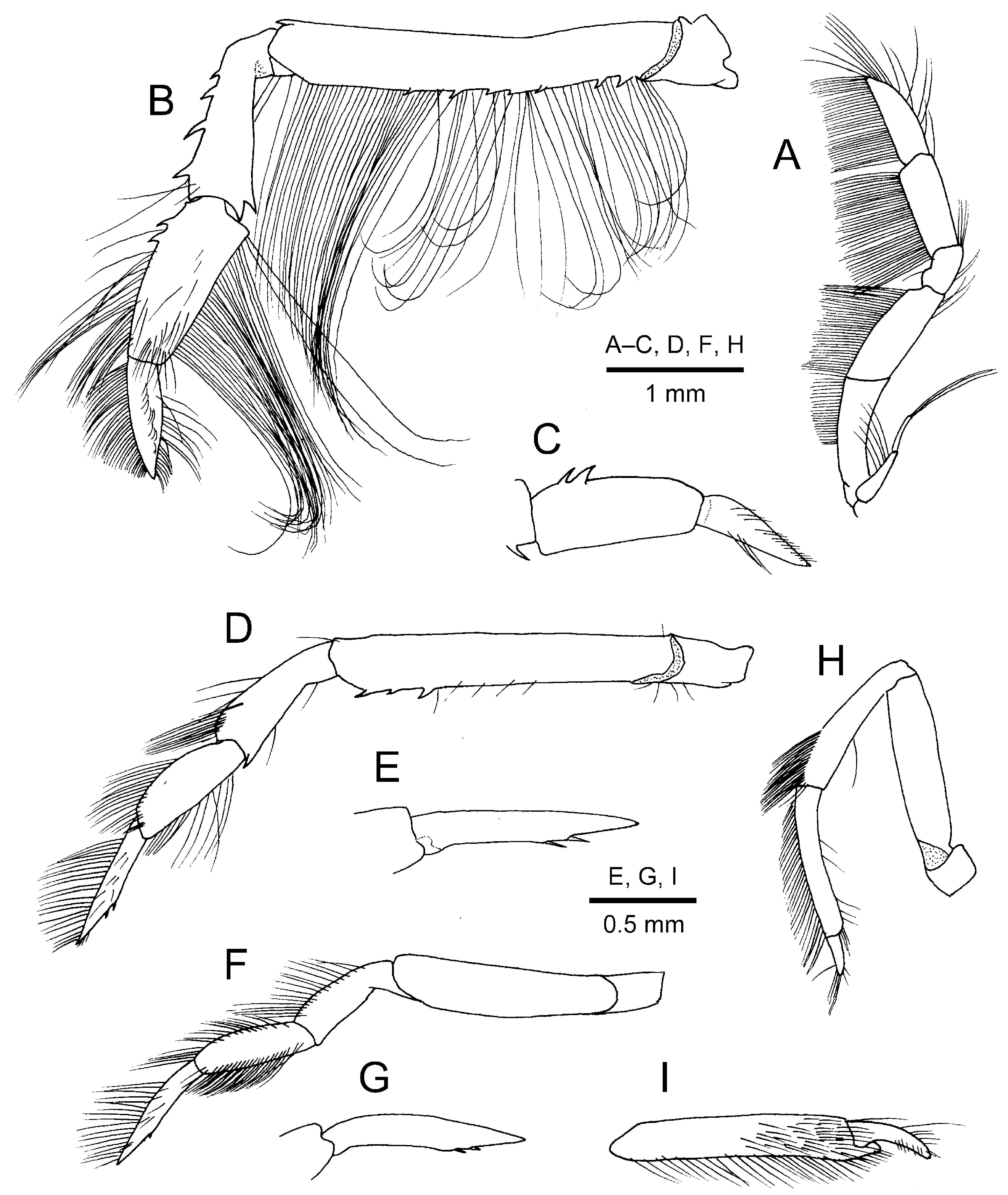
Mouthparts not dissected, though external observation made. Mandible without tooth on anterior margin of incisor process. Maxillule and maxilla without distinctive features. Maxilliped 1 without epipod. Maxilliped 2 without distinctive features. Maxilliped 3 ( Fig. 3 View FIGURE 3 A) with endopod moderately stout, extending as far as distal end of antennal peduncle; ischium to dactylus with thick long setae on lower (flexor) margins, none armed with spines or teeth; dactylus nearly straight, slightly shorter than propodus, terminating in rounded tip; exopod short, tapering distally, flagellum shorter than basal article, with long terminal setae; epipod small, bud-like.
Pereopod 1 ( Fig. 2 View FIGURE 2 A, B) rather slender. Coxa ( Fig. 1 View FIGURE 1 F) with 1 conspicuous procurved spine on distal margin mesially. Ischium with 1 small procurved spine on lower margin. Merus with row of small spines and row of long setae on lower margin; upper margin nearly straight, armed 1 subdistal spine and 1 smaller spine located at proximal one-fourth; with slender subdistal spine. Carpus cup-shaped, slightly more than half length of palm; upper surface with 2 rows of spines noticeably increasing in size distally (distomesial spine strongest) and sparse moderately long setae; lateral surface with row of minute denticles adjacent to lower margin; mesial face unarmed, distomesial margin with 1 strong spine slightly smaller than distomesial spine; lower distal angle also with 1 strong spine subequal in length to distomesial spine. Palm about 3.3 times as long as high; upper margin with row of strong spines and sparse long setae over entire length (distalmost spine subdistal); dactylar condyle on lateral face little developed; mesial face with numerous small to moderately strong spines arranged in 3 longitudinal rows and 1 prominent spine somewhat proximal to base of fixed finger and 1 smaller spine adjacent to lower margin; lower margin slightly concave, with 2 moderately strong spines proximal to midlength and long setae particularly numerous in proximal one-fourth; fixed finger small, slightly curved, upper margin with few spinules proximally; 1 small subacute spine just superior to base of fixed finger on lateral side. Dactylus about 0.6 times as long as palm, slightly curved, terminating in calcareous claw; upper (extensor) margin bearing numerous setae increasing in length proximally, but no conspicuous armature; lateral face with obliquely longitudinal row of setae and row of longer setae adjacent to lower (flexor) margin; lower (flexor) margin faintly denticulate proximally.
Pereopods 2–5 relatively slender; setation typical of genus. Pereopod 2 ( Fig. 3 View FIGURE 3 B, C) reaching base of dactylus of pereopod 1; coxa ( Fig. 1 View FIGURE 1 F) bearing some spinules on mesial face; ischium unarmed; merus with small subdistal spine on upper margin and row of small spines on lower margin in proximal two-thirds; carpus widened distally, upper margin with row of 4 spines increasing in size distally and 1 small subdistal spine on lower margin; propodus subrectangular, subequal in length to carpus, narrowing distally, upper margin with 1 (right) or 2 (left) proximal spines, lower proximal margin angular; dactylus tapering to minute corneous tip, about 0.7 times as long as propodus. Pereopod 3 ( Fig. 3 View FIGURE 3 D, E) with coxa ( Fig. 1 View FIGURE 1 F) with some spinules on mesial face; ischium unarmed; merus with 3 small spines on lower margin in distal one-third, upper margin unarmed; carpus widened distally, upper margin unarmed, lower margin with 1 small subdistal spine; propodus with slightly convex upper margin; dactylus slightly longer than propodus, terminating in acute tip, lower (flexor) margin slightly sinuous, with 2 closely set spiniform setae distal to midlength. Pereopod 4 ( Fig. 3 View FIGURE 3 F, G) with coxa ( Fig. 1 View FIGURE 1 F) bearing 1 spinule on mesial face; merus to propodus unarmed; dactylus subequal in length to propodus, terminating in acute tip, upper margin slightly convex, lower margin slightly sinuous, with 3 minute spiniform setae distal to midlength. Pereopod 5 ( Fig. 3 View FIGURE 3 H, I) semichelate, articles unarmed; coxa ( Fig. 1 View FIGURE 1 F) with gonopore on either side; propodus slightly curved, with short fixed finger; dactylus curved near base, terminating in blunt tip.
Arthrobranchs of type C (cf. Ngoc-Ho 1981), composed of deeply divided, narrow lamellae on either side of rachis.
Pleopods 1 absent. Pleopods 2–5 (cf. Fig. 1 View FIGURE 1 G) generally similar, with endopods small and slender, less than 0.3 length of respective exopod; exopods relatively slender.
Uropod ( Fig. 1 View FIGURE 1 E, H) with both endopod and exopods exceeding posterior margin of telson. Protopod with small spine on posterior margin. Endopod elongate suboval, 2.2 times longer than wide, inner margin fairly convex, outer margin nearly straight except for distal part, distal margin with long bristle-like setae; upper face with 1 median carina. Exopod elongate suboval, 1.2 times as long as endopod, 2.6 times as long as wide; upper surface with 2 longitudinal carina.
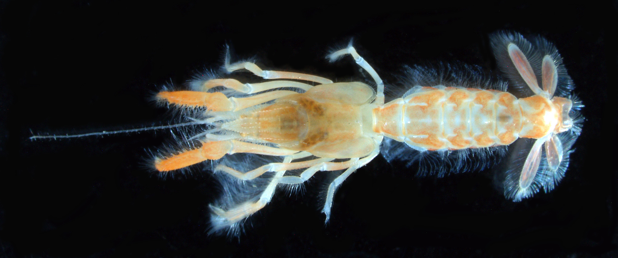
Colouration in life. Body generally light orange-brown, pleon mottled. Cornea of eye yellowish brown. Pereopod 1 with merus pale brown, carpus and chela orange-brown; pereopods 2–5 pale brown or translucent. Uropods with tinge of red. See Fig. 4 View FIGURE 4 .
Remarks. I n spite of the small size (cl 3.2 mm), the holotype has paired gonopores on the pereopod 5 coxae; pleopods 1 are absent. The pereopod 1 palm is slender with the armature being generally well-developed, thus perhaps male characteristics are not fully differentiated in the holotype.
The present new species appears closest to Gebiacantha reunionensis , so far known only from La Réunion, western Indian Ocean ( Ngoc-Ho 1989; Sakai 2006). Shared characters include: rostrum flattened dorsoventrally, far overreaching distal corneal margins; gastric lateral ridge of carapace terminating anteriorly in small spiniform tubercle; anterolateral margin of carapace with two or more spinules; postorbital region of carapace with scattered spinules; cervical groove with row of spinules; telson slightly longer than wide, with 1 minute spiniform seta on each lateral margin, posterior margin medially concave; article 1 of antennular peduncle with row of spines on lower margin; articles 2 and 4 of antennal peduncle with row of spines on lower margin; palm of pereopod 1 with numerous spines on inner face, arranged in irregular longitudinal rows and 1 prominent spine on lower margin proximal to fixed finger. Gebiacantha sagamiensis n. sp. differs from G. reunionensis in the following particulars: (1) the rostrum reaches nearly to the distal margin of article 4 of the antennal peduncle in the new species ( Fig. 1 View FIGURE 1 A), rather than reaching to the midlength of article 4 of the antennal peduncle in G. reunionensis (cf. Ngoc-Ho 1989: figs 1a, 2d, j); (2) the ventral surface of the rostrum is devoid of even a trace of a denticle or spine in G. sagamiensis n. sp., there is usually one distinct rostral ventral spine (or a trace of denticle in juveniles) in G. reunionensis (cf. Ngoc-Ho 1989: fig. 1a, 2d, i); (3) the pleomere 6 is longer than wide with the greatest width posterior to the midlength in G. sagamiensis n. sp. (Fig.), whereas almost as long as wide with parallel lateral margins in G. reunionensis (cf. Ngoc-Ho 1989: fig. 1h, 2n); (4) article 2 of the antennal peduncle is unarmed on the upper margin in G. sagamiensis n. sp., rather than armed with two or three spinules in G. reunionensis (cf. Ngoc-Ho 1989: fig. 1a, d, j); (5) the lower distal spine on the pereopod 1 carpus is prominent, being subequal to the inner distal spine, in G.
sagamiensis View in CoL n. sp., whereas it is distinctly weaker than that in G. reunionensis View in CoL (cf. Ngoc-Ho 1989: fig. 1d, 2a, f, g, k, l, m); (6) the upper lateral spines on the pereopod 1 carpus are generally better developed in G. sagamiensis View in CoL n. sp. than in G. reunionensis View in CoL (cf. Fig. versus Ngoc-Ho 1989: fig. 1c, 2a, f, l); (7) the pereopod 1 merus is armed with a small spine on the upper margin proximally in G. sagamiensis View in CoL n. sp., but such a spine is absent in G. reunionensis View in CoL (cf. Ngoc-Ho 1989: fig. 1c, 2f, l).
Comments on the generic assignment of the present new species are necessary. The close relative of the present new species, G. reunionensis , has been recently transferred to Paragebicula Sakai, 2006 by Sakai (2015) along with other species theretofore assigned to other upogebiid genera. However, Sakai’s (2015) action was based only on the superficial similarity in the uropodal shape (Sakai said that in species assigned to Paragebicula , the uropodal exopod is elongate and pear-shaped), although the shape of the exopod is actually quite variable in those species assigned to Paragebicula (see Sakai 2015: figs. 1H, 2E, 3C, 5C, 6C, 7D, 9C). Furthermore, if the uropodal shape alone is considered, Mantisgebia Sakai, 2006 is also closely similar to Paragebicula , although Sakai (2006) classified Mantisgebia and Paragebicula into two separate subfamilies ( Upogebiinae and Neogebiculinae , respectively). Poore (2008) already pointed out that new genera proposed by Sakai (2006) were diagnosed without much justification. Indeed, it is difficult to distinguish between Mantisgebia and Paragebicula sensu Sakai, 2015 as separate genera with characters offered by Sakai (2006, 2011, 2015). Consequently, I do not follow Sakai’s (2015) classification, and return back “ Paragebicula ” reunionensis to Gebiacantha . Although K. Sakai has not accepted Gebiacantha as a valid genus ( Sakai & Türkay 1995, 2014; Sakai 2006; 2015), the present new species closely fits the generic diagnosis of Gebiacantha provided by Ngoc-Ho (1989; 2003) except for the absence of rostral ventral spine(s): gastric region with lateral ridges; linea thalassinica depressed anteriorly in groove subparallel to cervical groove, extending to posterior border of carapace; rostrum bordered with tubercles; anterolateral margin of carapace with two or more spines; posterior margin of telson medially concave; mandible without acute anterior tooth on incisor process; maxilliped 1 without epipod; maxilliped 3 with small, bud-like epipod; pereopod 1 subchelate, carpus and propodus with numerous spines, lower border of propodus with one strong spine proximal to fixed finger, fixed finger short, not exceeding half-length of dactylus; coxae of pereopods 1–4 with spinules; uropodal exopod longer than telson. In fact, variation in the development of the rostral ventral spine in G. reunionensis was already mentioned by Ngoc-Ho (1989). Consequently the present new species is assigned to Gebiacantha for the time being. Although future study may eventually reveal that Gebiacantha is not monophyletic, but for the time being, it is advisable to follow the classification based on more thorough character analysis. Gebiacantha is now represented by the following 19 species: G. acanthochela ( Sakai, 1967) , G. acutispina (de Saint Laurent & Ngoc-Ho, 1979), G. albengai Ngoc-Ho, 2005, G. arabica Ngoc-Ho, 1989, G. bermudensis ( Williams, 1993) , G. ceratophora (De Man, 1905) , G. dampieri Ngoc-Ho, 2008, G. lagonensis Ngoc- Ho, 1989, G. laurentae Ngoc-Ho, 1989, G. lifuensis Ngoc-Ho, 1994a, G. monoceros (De Man, 1905) , G. multispinosa Ngoc-Ho, 1994a, G. plantae ( Sakai, 1982) , G. poorei Ngoc-Ho, 1994b, G. priochela Sakai, 1993 , G. reunionensis Ngoc-Ho, 1989, G. richeri Ngoc-Ho, 1989, G. sagamiensis n. sp., and G. talismani ( Bouvier, 1915) (type species) ( Ngoc-Ho 1989, 1994a, 1994b, 2003, 2005; 2008; Sakai 1993).
The other species of Gebiacantha known from Japanese waters is G. acanthochela . Gebiacantha acanthochela is easily distinguished from the present new species in having two conspicuous rostral ventral spines, one pair of prominent spines on the posterior carapace just posterior to the cervical groove and the much broader rami of the uropod (cf. Sakai 1967). Mantisgebia kyusyuensis ( Yokoya, 1933) , described from waters around Kyushu, Japan, exhibits some degree of similarity to the present new species in the elongate rostrum and the deeply concave posterior margin of the telson ( Yokoya 1933), although Sakai (2006) noted that the generic assignment of Yokoya’s taxon was only provisional. Indeed, Yokoya’s taxon is represented only by the rather brief type description, and adequate assessment of its diagnostic characters remains difficult. Nevertheless, the unarmed cervical ridge and lower margins of the antennular and antennal peduncles and the much broader rami of the uropod, clearly illustrated by Yokoya (1933), indicate that Yokoya’s taxon is not conspecific with the present new species. Mantisgebia multispinosa Liu & Liu, 2013 , described from the South China Sea, off Nansha Islands, also exhibits substantial similarity to G. reunionensis and the present new species in the general spination of the carapace and of the antennular and antennal peduncle, the deeply concave posterior margin of the telson and the shape of the uropod. Nevertheless, M. multispinosa differs from the latter two species in the absence of a minute spiniform seta on each lateral margin of the telson and the lack of spines on the mesial face of the pereopod 1 palm ( Liu & Liu 2013).
Upogebiids represent deeply burrowing infauna, particularly abundant in coastal shallow waters ( Itani 2004; Dworschak et al. 2012), although many species have been described also from the sublittoral zone (e.g., Sakai 2006, 2011, 2015; Liu & Liu 2010, 2013), of which the vast majority is rare. It is interesting to note that no species of Upogebiidae have been recorded from sublittoral depths in Sagami Bay, although investigations of the benthic fauna have been long continued from the late 19th century ( Takeda et al. 2006; Fujita & Akasaka 2007; Nakano et al. 2015). The discovery of the present new species highlights the difficulty of inventory of the deeply burrowing infauna inhabiting the sublittoral or deeper zone in spite of its existence.
Etymology. The specific name refers to Sagami Bay, the location embracing the type locality.
No known copyright restrictions apply. See Agosti, D., Egloff, W., 2009. Taxonomic information exchange and copyright: the Plazi approach. BMC Research Notes 2009, 2:53 for further explanation.
|
Kingdom |
|
|
Phylum |
|
|
Class |
|
|
Order |
|
|
Family |
|
|
Genus |