
Tripartiella obtusa Ergens & Lom, 1970
|
publication ID |
https://doi.org/10.11646/zootaxa.3681.2.6 |
|
publication LSID |
lsid:zoobank.org:pub:C6752FC1-4A02-4DA6-8798-3B864561C21D |
|
DOI |
https://doi.org/10.5281/zenodo.6158931 |
|
persistent identifier |
https://treatment.plazi.org/id/653B5414-FF8C-FF99-75F2-B268FA87F8E9 |
|
treatment provided by |
Plazi |
|
scientific name |
Tripartiella obtusa Ergens & Lom, 1970 |
| status |
|
Tripartiella obtusa Ergens & Lom, 1970
Host: Rhinogobio typus
Locality: Qu County, Sichuan
Site: Gills
Date of sampling: April, 2012
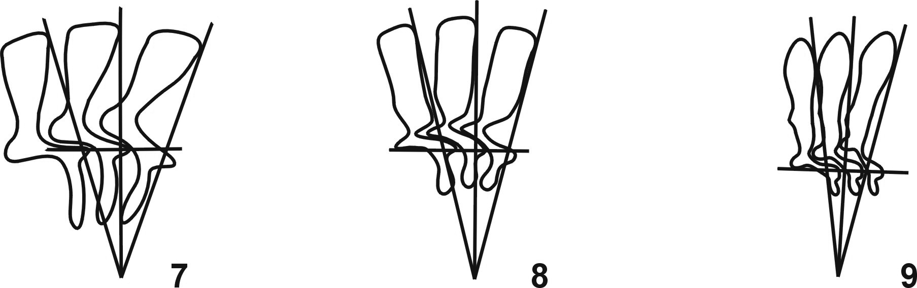
Description ( Figs. 1, 2 View FIGURES 1 – 6 , 7 View FIGURES 7 – 9 ). The following morphological description was based on 25 specimens measured (n = 25). Small to medium freshwate r Tripartiella with a compressed body, diameter 21.5–30.5 (25.6 ± 2.7); adhesive disc 17.0–23.5 (20.5 ± 1.9); width of border membrane 1.5–3.0 (2.1 ± 0.2); diameter of denticle ring 10.0–13.0 (11.5 ± 0.9); number of denticles 21–24; number of radial pins per denticle usually 4–5; span of denticle 5.5–8.0 (6.8 ± 0.7); length of denticle 2.5–4.0 (3.0 ± 0.2); blade length 2.5–4.0 (3.1 ± 0.4), generally elongated and slanted obliquely backwards; distal blade surface straight, in parallel to border membrane, and a little higher than the sharp tangent point; anterior and posterior surfaces straight, but not in parallel with each other with anterior surface blade apophysis present, prominent just touching the Y +1 axis; blade apophysis absent or invisible. Central part relatively developed with a sharp point fitting loosely into preceding denticle and far away from the Y-1 axis. Shapes of the central part above and below the X-axis not symmetrical and 1.2–2.2 (1.5 ± 0.2) in width. Ray connection inconspicuous and barely distinguishable from the ray. Ray relatively long when compared to other Tripartiella species, and tip of ray directed slightly forward; ray apophysis absent and length of ray was 1.5–2.5 (1.9 ± 0.3); ratio between denticle above and below X axis less than two. Macronucleus U-shaped, external diameter 16.5–24.0 (18.2 ± 1.2) and internal diameter 12.0–18.5 (14.8 ± 1.5). Micronucleus elliptical, 1.5–2.5 (2.0 ± 0.5) in length and 1.0–2.0 (1.6 ± 0.5) in width, situated in + Y position. Adoral ciliary spiral turns about 180° - 230° around peristomial disc.
Remarks. In 1970, Tripartiella obtusa was originally found from gills of Gobio gobio by Ergens & Lom, the Czechoslovakia scientists ( Ergens & Lom, 1970). Later, it was found regularly but exclusively on the gills of the same host from Czechoslovakia by Lom and Haldar (1977). In terms of body size, it is very close to Tripartiella copiosa Lom, 1959 , but these two species can be easily distinguished by the morphological features of the denticles, especially the shape of denticles ( Lom, 1959). The morphology and morphometric data of the present population from the gills of Rhinogobio typus are well within the range of the original description by Lom & Haldar (1977). This finding has established a new host record, Rhinogobio typus , for Tripartiella obtusa , and this is also the first report in Asia, which extends the known host and geographic range for Tripartiella obtusa .

No known copyright restrictions apply. See Agosti, D., Egloff, W., 2009. Taxonomic information exchange and copyright: the Plazi approach. BMC Research Notes 2009, 2:53 for further explanation.
|
Kingdom |
|
|
Phylum |
|
|
Class |
|
|
Order |
|
|
Family |
|
|
Genus |