
Sporadotragus parvidens (Gaudry, 1861)
|
publication ID |
https://doi.org/10.4202/app.2012.0129 |
|
DOI |
https://doi.org/10.5281/zenodo.10989539 |
|
persistent identifier |
https://treatment.plazi.org/id/6471BC2B-C223-FF9A-FFEA-F89001B31523 |
|
treatment provided by |
Felipe |
|
scientific name |
Sporadotragus parvidens (Gaudry, 1861) |
| status |
|
Sporadotragus parvidens (Gaudry, 1861)
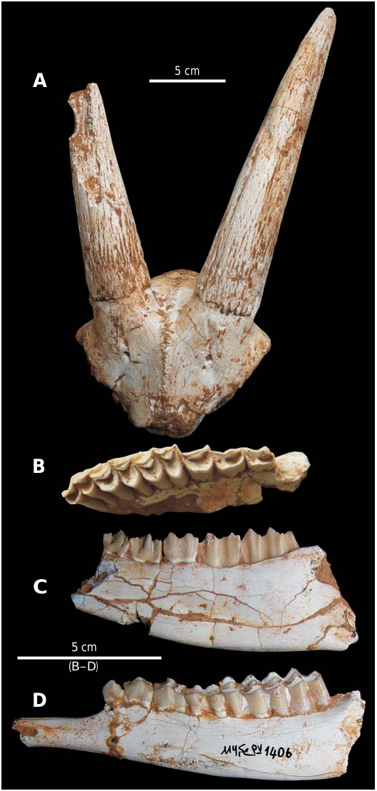
Material.— MYŞE PV- 2502, partial skull; MYŞE PV- 1573, frontlet preserving the basal part of the horn cores; MYŞE PV- 1300, partial left horn core; MYŞE PV- 1522, right upper tooth row with P2–M3; MYŞE PV- 1412. right M3; MYŞE PV- 1423, left M3; MYŞE PV- 1533, left M1; MYŞE PV- 1511, right mandibular body with p3–m3; MYŞE PV- 2561, right mandibular body with p2–m2; MYŞE PV- 1429, right mandibular body with p4–m2; MYŞE PV- 2556, right mandibular body with m1–m3; MYŞE PV- 1630, left mandibular body with p3–m3; MYŞE PV- 1407, PV- 1574, left mandibular body with p2–m2; MYŞE PV- 2569, left mandibular body with p4– m3; MYŞE PV- 1406, PV- 2559, left mandibular ramus with p2–m3; MYŞE PV- 1311, left mandibular ramus with p4–m2. All from Şerefköy-2, Turkey, Late Turolian (Late Miocene).
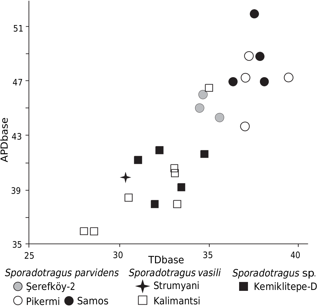
Description. —This species is represented by at least 7 individuals.The frontals form a 105–115° angle along the sagittal plane and appear moderately pneumatised above the orbits. The supraorbital foramen (doubled on the left side in both MYŞE PV- 1573 and PV- 2502) is small and round, placed well below the pedicle, and opens directly into the orbit ( Fig. 6A View Fig ). The interfrontal suture is complex in outline and slightly pinched between the horn cores ( Fig 6A View Fig ). The frontoparietal suture is also complex, Y-shaped, and runs very close to the base of the horn core. The postcornual fossa is shallow and round. As preserved, the horn core is long (maximum length: 210 mm along the anterior surface) and gently curved posteriorly. It furthermore bears thin and discontinuous grooves on its surface and shows no evidence of torsion ( Fig. 6A View Fig ). In MYŞE PV- 1300, a weak anterior keel runs along its basal portion. The cross section of the horn core is elliptical at the base, with its maximum transverse diameter located posteriorly ( TD × 100/APD at the base = 75.3–76.4, n = 3; Table 3). The angle between the greatest anteroposterior diameter of the horn core base and the sagittal plane ranges from 42 to 50°. Towards the tip, the cross section of the horn core remains elliptical, but becomes symmetrical ( TD × 100/APD at 7 cm above the base = 72.9–78.3, n = 3; Table 3).
The upper premolars are moderately long compared to the molars, with a premolar/molar ratio 69.7% (n = 1) ( Fig. 6C View Fig , SOM 3: Table 1 View Table 1 ). P2 and P3 have a strong anterolabial cone and bear a fossa unequally divided by a central fold ( Fig. 6B View Fig ). The anterior style of P3 and P4 is also strong, but the anterolabial cone of P4 is weak. The upper molars have a strongly developed paracone, parastyle and mesostyle, a weak metacone, and no basal pillars (entostyle) ( Fig. 6B View Fig ). A metaconule fold is present on M2.
The lower premolar row is moderately short compared to the molars, with a premolar/molar ratio of 58.3–62.4%, n = 2 ( Fig. 6C, D View Fig , SOM 3: Table 2 View Table 2 ). The p3 and p4 have a strong anterior stylid and a barely developed anterior conid ( Fig. 6C View Fig ). The mesolingual conid is simple and elongated on p3, but rounded towards the base and slightly curved anteriorly on p4. On both p3 and p4, the posterolingual conid fuses with the posterior stylid during early wear and the posterolabial conid is well developed ( Fig. 6C View Fig ). Both the metastylid and especially the entostylid of the lower molars are well marked during early wear. There is no anterior cingulid. A low ectostylid is present on m1 ( Fig. 6D View Fig ).
Remarks.—Both the size and the overall horn core and dental morphology of the material from Şerefköy-2 match those of Sporadotragus , the taxonomic status of which was recently revised by Geraads et al. (2006) and Kostopoulos (2009a). The specimens from Şerefköy-2 have larger horn cores than Sporadotragus vasili Geraads, Spassov, and Kovachev, 2006 from the SW Bulgarian localities of Kalimantsi and Strumyani ( Geraads et al. 2006, 2011; Fig. 7 View Fig ), which furthermore differs from the present material in having a fused interfrontal suture, as well as a less curved horn core with a flat medial and a wide anterior surface, strong longitudinal grooves, and an anteromedial keel ( Geraads et al. 2006). By contrast, the specimens from Şerefköy-2 have several features in common with Sporadotragus parvidens , mainly known from Pikermi, Samos ( Greece) and Kemiklitepe-D ( Turkey) ( Bouvrain 1994; Kostopoulos 2009a). These include a small supraorbital foramen not located inside a pit, a complex and slightly raised interfrontal suture, and a Y-shaped frontoparietal suture. In addition, both have a horn core that is long, sub-cylindrical and gently (but markedly) curved posteriorly, bears irregular grooves, and shows no evidence of torsion or keels. The flexion of the frontals along the sagittal plane appears slightly stronger in the material from Şerefköy-2 (105–115°) than in Sp. parvidens from Samos (100–105°); however, this, as well as other minor differences (such as somewhat longer horn cores in some specimens from Samos or the degree of p4 molarisation) may well reflect intraspecific variation.
Stratigraphic and geographic range.—Upper Miocene; Aegean region.
No known copyright restrictions apply. See Agosti, D., Egloff, W., 2009. Taxonomic information exchange and copyright: the Plazi approach. BMC Research Notes 2009, 2:53 for further explanation.
|
Kingdom |
|
|
Phylum |
|
|
Class |
|
|
Order |
|
|
Family |
|
|
Genus |