
Skoufotragus cf. Sk. schlosseri (Andree, 1926), 2009
|
publication ID |
https://doi.org/10.4202/app.2012.0129 |
|
DOI |
https://doi.org/10.5281/zenodo.10989541 |
|
persistent identifier |
https://treatment.plazi.org/id/6471BC2B-C221-FF96-FFEA-FE35030916BD |
|
treatment provided by |
Felipe |
|
scientific name |
Skoufotragus cf. Sk. schlosseri (Andree, 1926) |
| status |
|
Skoufotragus cf. Sk. schlosseri (Andree, 1926)
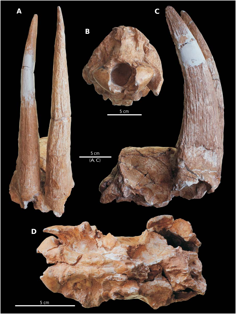
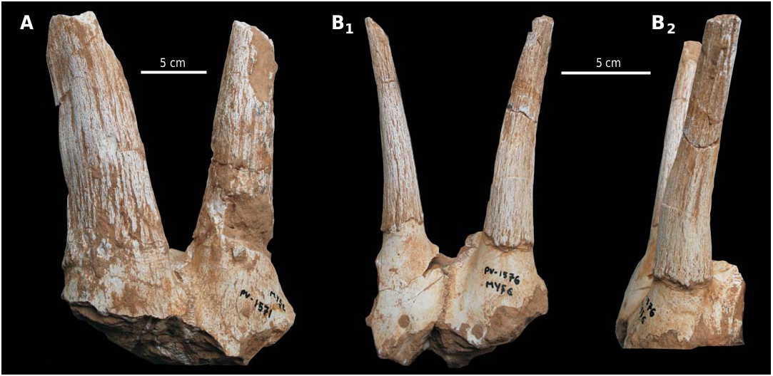
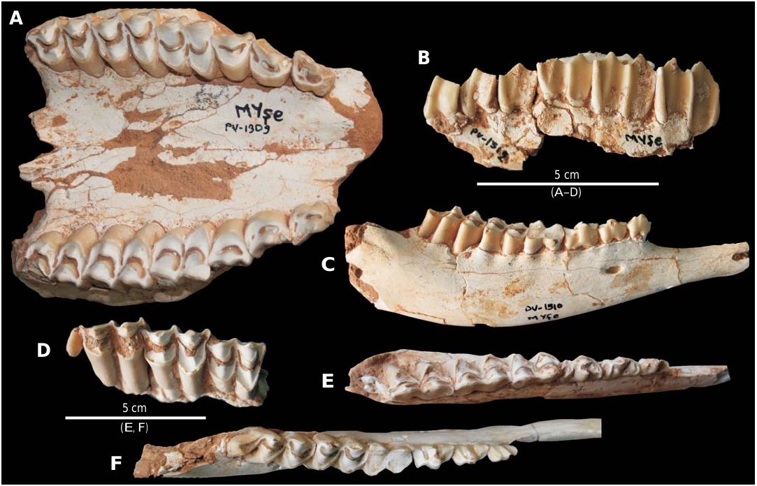
Figs. 8–10 View Fig View Fig View Fig .
Material. —MYŞE PV-547, partial cranium with horn cores; MYŞE PV-1571, 1576, female frontlet; MYŞE PV-1570, left female horn core; MYŞE PV-1575, right horn core; MYŞE PV-1579, partial left female horn core; MYŞE PV-2606, partial female horn core; MYŞE PV-1309, palate; MYŞE PV-1512, PV-1516, PV-2570, right upper tooth row with P2–M3; MYŞE PV-1513, PV-1514, PV-1515, right upper tooth row with P3–M3; MYŞE PV-1622, right upper tooth row with M1–M3; MYŞE PV-1491, right upper tooth row with M2– M3; MYŞE PV-1523, right M3; MYŞE PV-1520, PV-1521, left upper tooth row with M2–M3; MYŞE PV-1519, left upper tooth row with P2–M2; MYŞE PV-1525, left upper tooth row with P4–M1; MYŞE PV-2571, left upper tooth row with P3–M3; MYŞE PV-1410, left upper tooth row with M1–M3; MYŞE PV-1517, left M2–M3; MYŞE PV-1312, left upper tooth row with P2–P4; MYŞE PV-1315, left M3; MYŞE PV-1434, PV-1436, PV-1313, M1 or M2; MYŞE PV-1532, left P2; MYŞE PV-1510, PV-1542, PV-2560, right mandibular body with p2–m3; MYŞE PV-1541, right mandibular body with p3–m3; MYŞE PV-2551, right mandibular body with p4–m2; MYŞE PV-2568, right mandibular body with m1–m3; MYŞE PV-1546, left mandibular body with m2–m3; MYŞE PV-1543, PV-1544, PV-2554, PV-2566, left mandibular ramus with p2–m3; MYŞE PV-1540, left mandibular ramus with p4–m3; MYŞE PV-2001, left mandibular ramus with p3–m1; MYŞE PV-1156, left mandibular ramus with m2–m3.All from Şerefköy-2, Turkey, Late Turolian (Late Miocene).
Description. —This is by far the most abundant bovid found at Şerefköy-2 and represented by at least 10 individuals. The opisthocranium is high, narrow, dolichocephalic (sensu Bosscha-Erdbrink 1978) and has a straight dorsal profile in lateral view ( Fig. 8C View Fig , Table 4 View Table 4 ). The temporal lines are moderately developed and run parallel to each other posteriorly. In posterior view, the occiput is triangular and bears a sharp occipital crest ( Fig. 8B, C View Fig ) that ends dorsally in a strong protuberance surrounded by deep scars. The nuchal crest is well developed. The mastoid faces posterolaterally and the paroccipital process is large and flattened. In lateral view, the occipital condyles project posteroventrally, thus forming a very acute angle with the occipital level. The basioccipital is long, relatively narrow and bears a shallow, narrow longitudinal groove ( Fig. 8D View Fig ). The sharp and prominent (crestlike) posterior tuberosities of the basioccipital are oriented perpendicular to the sagittal plane, whereas the weak anterior tuberosities are oriented anteroposteriorly. The oval foramen faces laterally. The preserved outline of the auditory bulla indicates that it was large and bulbous.
The frontal contains large sinuses, one of which occupies the pedicle and even reaches the base of the horn core. There is no postcornual fossa. The horn core, inserted above the orbit, is sabre-like without keels or torsion ( Figs. 8A, C View Fig , 9A View Fig ). It is moderately long, moderately curved posteriorly in lateral view and strongly compressed mediolaterally along its entire length (TD × 100/APD at the base: 40–57, n = 5; Table 4 View Table 4 ). In cross section, the horn core forms an elongated ellipse, which becomes narrower towards the tip. A deep furrow occasionally runs along the upper half of the posterior surface, and, in combination with the strong mediolateral compression, may give the impression of a distal posterior keel.
Two frontlets and three horn core specimens (MYŞE PV-1570, PV-1571, PV-1576, PV-1579, and PV-2606; Fig. 9 View Fig ) likely represent female individuals. The supraorbital foramen is large and round, and placed far anterior to the pedicle. The interfrontal suture is open and simple in outline. The horn core is thin and long (~ 140 mm) and inserted above the back of the orbit. It is far removed from its counterpart, weakly curved posteriorly and barely twisted homonymously ( Fig. 9B View Fig ). In cross section, the horn core is elliptical at the base but becomes more compressed mediolaterally towards the tip ( Table 4 View Table 4 ). In anterior view, the divergence of the horn cores is weak up to their mid-height, but stronger above.
The premolars are moderately long compared to the molars, with upper and lower premolar/molar ratios of 59.6– 65.6% (n = 5) and 60.3–66.6% (n = 4), respectively ( Fig. 10 View Fig , SOM 3: Tables 1 View Table 1 , 2 View Table 2 ). The upper molars have strong styles, bear a fossetta (central islet) and lack entostyles ( Fig. 10A, B, D View Fig ). P2 is bilobed lingually, whereas P3 has a trapezoidal occlusal outline. Both have a strong anterolabial cone ( Fig. 10A View Fig ). The protocone of M1 protrudes lingually. The metastyle of M3 is strong and, in one specimen ( MYŞE PV-1622 ), flares distally ( Fig. 10D View Fig ). The mandibular body is shallow and long ( Fig. 10C View Fig ). On the labial face, a second mental foramen appears below p3 ( Fig. 10C View Fig ). The p2 is simple without an anterior conid, but with a strong mesolingual conid and anterior stylid ( Fig. 10E, F View Fig ). The p3 has a well-developed anterior conid and stylid, which become fused together with wear. The mesolingual conid of p3 is oriented parallel to the posterolingual conid, which in turn fuses with the weaker and posterolingually directed posterior stylid during early wear ( Fig. 10E, F View Fig ). The p4 resembles p3, but has an anteroposteriorly developed mesolingual conid ( Fig. 10E View Fig ). The lower molars have a strong meta- and mesostylids, but both elements disappear with wear. There is no anterior cingulid. A basal pillar appears on 5 out of 13 m 1s, and 3 out of 13 m 2s. The third lobe of m3 is labially displaced and has an elongated, semicircular occlusal outline; it bears a strong posterolingual stylid on the upper half of the crown.
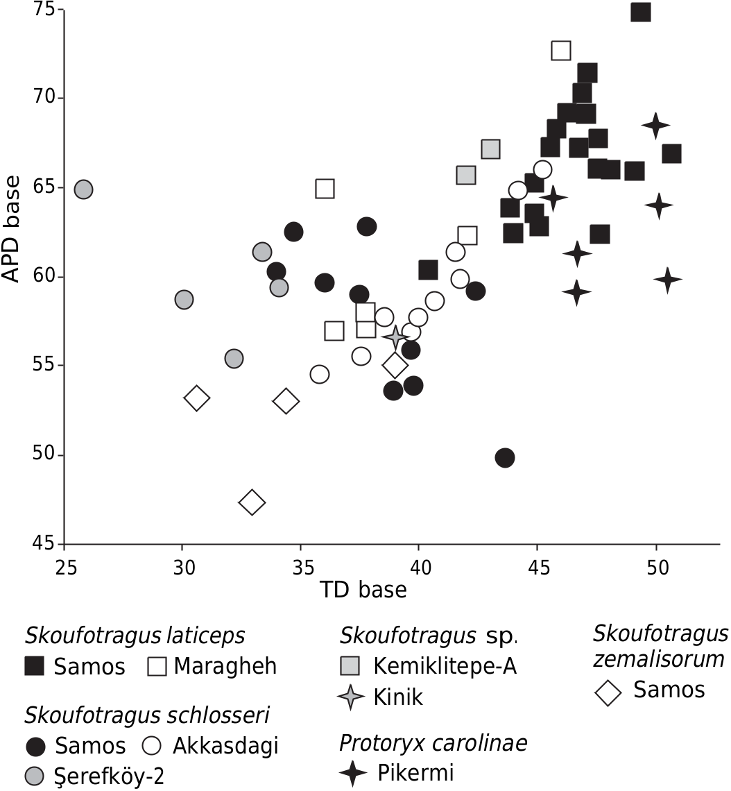
Remarks.—Protoryxoid bovids appear in the Eastern Mediterranean area as early as the Late Astaracian (e.g., Gentry 2000). Although they remained rare during the Vallesian, they strongly radiated and dispersed during the Turolian, especially in Anatolia and adjacent territories. They include small to medium-sized antelopes of caprine/hippotragine cranial appearance, but their taxonomy and evolutionary relationships remain debated. Kostopoulos (2009a) partly revised the Turolian protoryxoids from SE Europe assigned to the so-called “ Protoryx / Pachytragus complex” and recognised two distinct genera: Protoryx Major, 1891 and Skoufotragus Kostopoulos, 2009 (= partim Pachytragus Schlosser, 1904). The Şerefköy- 2 specimens resemble Skoufotragus in their dental morphology and in having (i) a narrow, long braincase with a straight dorsal profile and parallel sides, (ii) a triangular occiput and (iii) a sabre-like, mediolaterally compressed and uprightly inserted horn core ( Kostopoulos 2009a: 364). Skoufotragus is known from the Turkish Late Miocene assemblages of Kinik ( Protoryx sp. of Köhler 1987), Kemiklitepe-A ( Protoryx laticeps of Bouvrain 1994), and Akkaşdağı ( Pachytragus crassicornis of Kostopoulos2005), as well as from Samos, Greece and Maragheh, Iran ( Kostopoulos 2009a, Kostopoulos and Bernor 2011).
Skoufotragus
laticeps
Samos Maragheh Kemiklitepe-A
zemalisorum Kinik
Samos
Samos Akkasdagi
Şerefköy-2
Pikermi
The Şerefköy- 2 specimens differ from Skoufotragus laticeps from Samos, Kemiklitepe-A and Maragheh in having a narrower braincase, as well as a shorter dorsal parietal sector, less developed anterior tuberosities of the basioccipital, shorter, more slender, and more mediolaterally compressed horn cores, and, on average, smaller tooth rows (but with a similar premolar/molar ratio) ( Figs. 11 View Fig , 12). They are more similar to Sk. zemalisorum from Samos in terms of their cranial proportions, but differ in having a narrower braincase (61.7 mm vs. 70–78 mm; Kostopoulos 2009a) and a more anteroposteriorly expanded horn core ( Figs. 11 View Fig , 12). Except for a somewhat longer lower premolar row (relative to the molars), the Şerefköy- 2 specimens closely resemble Sk. schlosseri from Samos and Akkaşdağı in their size and morphology ( Figs. 11 View Fig , 12), although the material from Samos Q5) is characterised by slightly longer and more divergent horn cores bearing an anterior keel (the latter seems to be more common in short-horned individuals; cf. Kostopoulos 2005: 777).
Female individuals of Skoufotragus are rare, but Gentry 1971: 252, pl. 3: 3) interpreted AMNH 20687 as a female individual of Sk. laticeps . The frontlet MYŞE PV-1576 from Şerefköy-2 is very similar to this specimen, suggesting that females of Sk. schlosseri were likely horned.
No known copyright restrictions apply. See Agosti, D., Egloff, W., 2009. Taxonomic information exchange and copyright: the Plazi approach. BMC Research Notes 2009, 2:53 for further explanation.