
Adelosgryllus ferratilis, Merlo & Castro-Souza & Junta & Ferreira, 2022
|
publication ID |
https://doi.org/ 10.11646/zootaxa.5133.1.4 |
|
publication LSID |
lsid:zoobank.org:pub:31F5A638-2922-41DC-BEA4-0988B49BE6C4 |
|
DOI |
https://doi.org/10.5281/zenodo.6521006 |
|
persistent identifier |
https://treatment.plazi.org/id/635AA513-9516-AD4D-FF17-FF18FC16FE96 |
|
treatment provided by |
Plazi |
|
scientific name |
Adelosgryllus ferratilis |
| status |
sp. nov. |
Adelosgryllus ferratilis View in CoL n. sp.
( Figures 2–6 View FIGURES 2–6 , 7–14 View FIGURES 7–14 , 15–17 View FIGURES 15–17 , 18–19 View FIGURES 18–19 , 20-24 View FIGURES 20–24 , Table 2 View TABLE 2 )
Material examined. Holotype ♂, code ISLA 66166, Brazil, Pará state, municipality of Curionópolis , SL-110 cave (5º 57’ 32.351” S; 49º 37’ 49.592” W), 16.i.2012, CARSTE leg GoogleMaps . Holotype condition: right tegmen and legs were detached, and maintained in holotype’s tube. Paratypes, 1 ♂, 03.xi.2012 ( ISLA 66154), CARSTE leg. SL-94 cave (5º 57’ 6.291” S; 49º 37’ 56.475” W) and GoogleMaps 1 ♂, 16.i.2012 ( ISLA 66165), CARSTE leg. SL-109 (5º 57’ 32.350” S; 49º 37’ 49.591” W), all specimens collected in same municipality of holotype GoogleMaps .
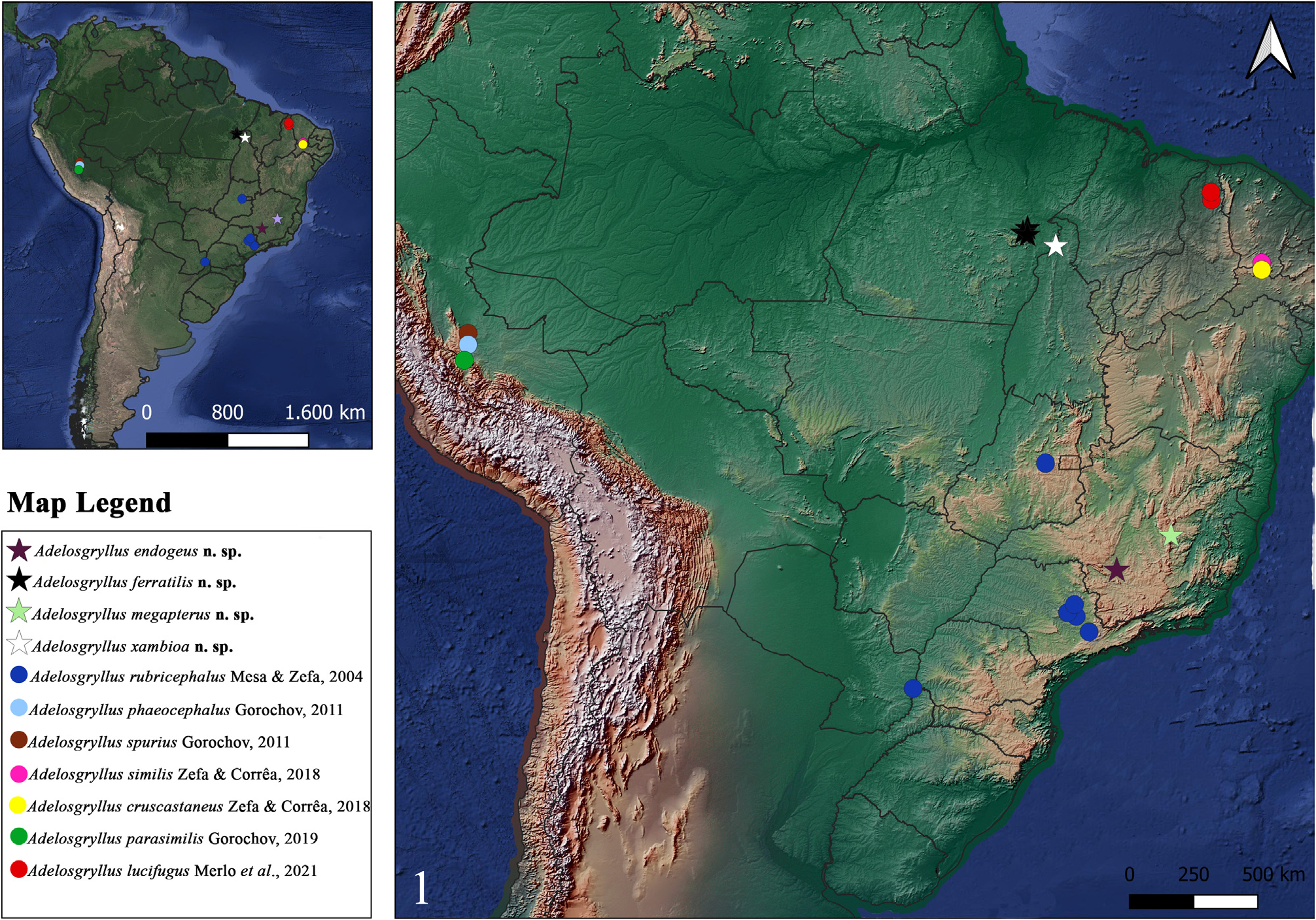
Distribution. Known for six caves: SL-79 (05º 57’ 53.438” S; 49º 38’ 19.786” W) (photographic register), SL-82 (05º 57’ 30.961” S; 49º 38’ 14.866” W) (photographic register), SL-94 (5º 57’ 6.291” S; 49º 37’ 56.475” W), SL-109 (5º 57’ 32.350” S; 49º 37’ 49.591” W), SL-110 (5º 57’ 32.351” S; 49º 37’ 49.592” W) and SL-121 (05º 55’ 44.369” S; 49º 40’ 30.007” W (photographic register) ( Fig. 1 View FIGURE 1 , dark stars), well sampled only on SL-94, SL-109 and SL-110, all localities distributed in the municipality of Curionópolis, Pará state, Brazil.
Etymology. The specific epithet ferratilis refers to the presence of this species in iron caves.
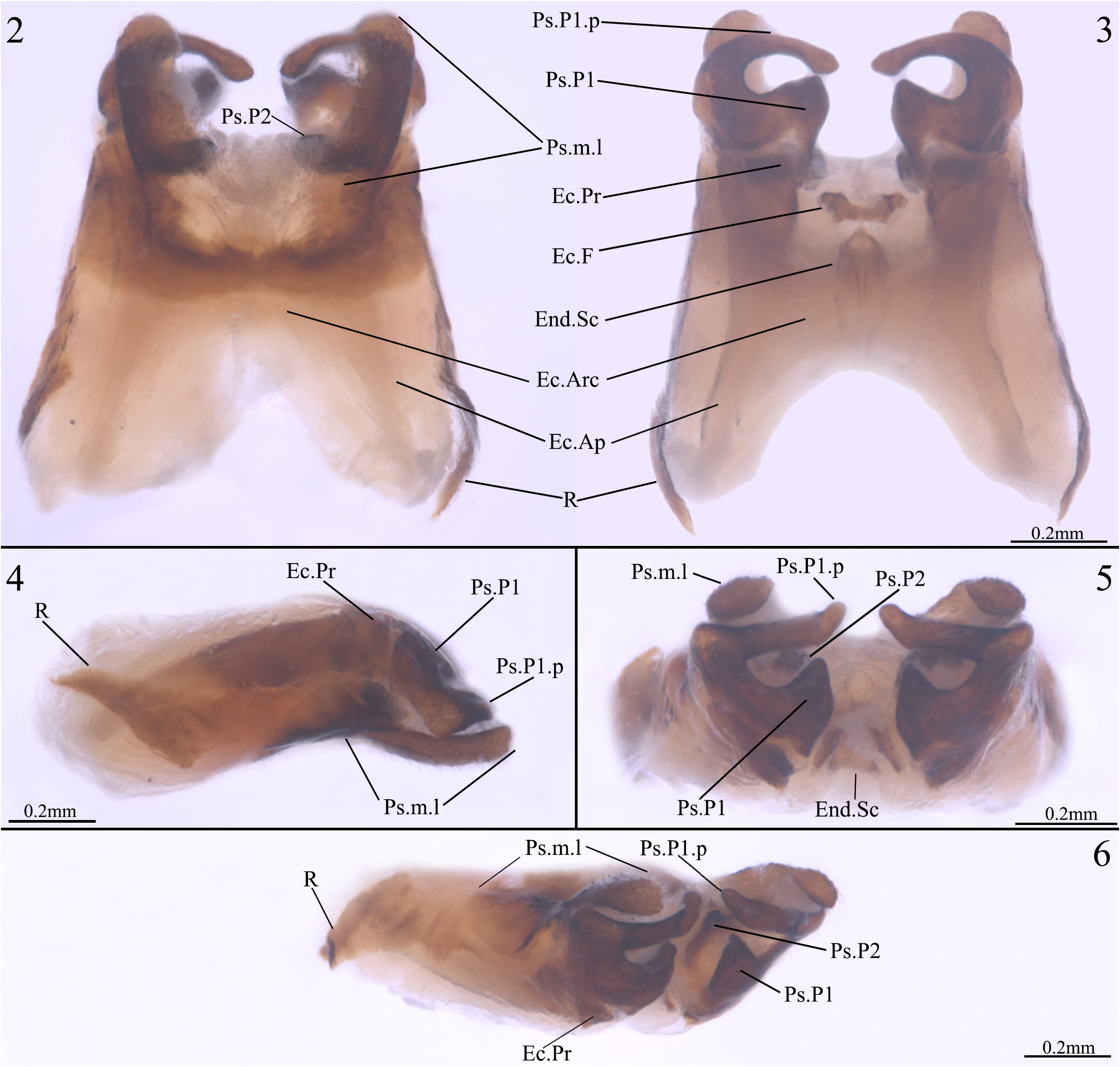
Diagnosis. Combination of the following characteristics: paramere 1 well developed, C-shaped (very similar to A. spurius and A. lucifugus ), inner basal margin acuminated, apex dilated and curved inward (Ps.P1.p, Figs 2–6 View FIGURES 2–6 ); rami elongated (very similar to A. spurius and A. lucifugus ), narrow and well sclerotized slightly curved inside and apex triangular shaped (R, Figs 2, 3, 4 and 6 View FIGURES 2–6 ); ectophallic fold well sclerotized, with the lateral border directed outward, central part slightly convex at apex and linear shaped bottom borders (Ec.F, Fig. 3 View FIGURES 2–6 ); endophallus circularshaped and vertically elongated, with short ventral crest (End.Sc), similar to A. lucifugus , but more sclerotized and slightly developed (End.Sc), connected to ectophallic fold by an inverted V-shaped membrane (End.Sc, Figs 3 and 5 View FIGURES 2–6 ).
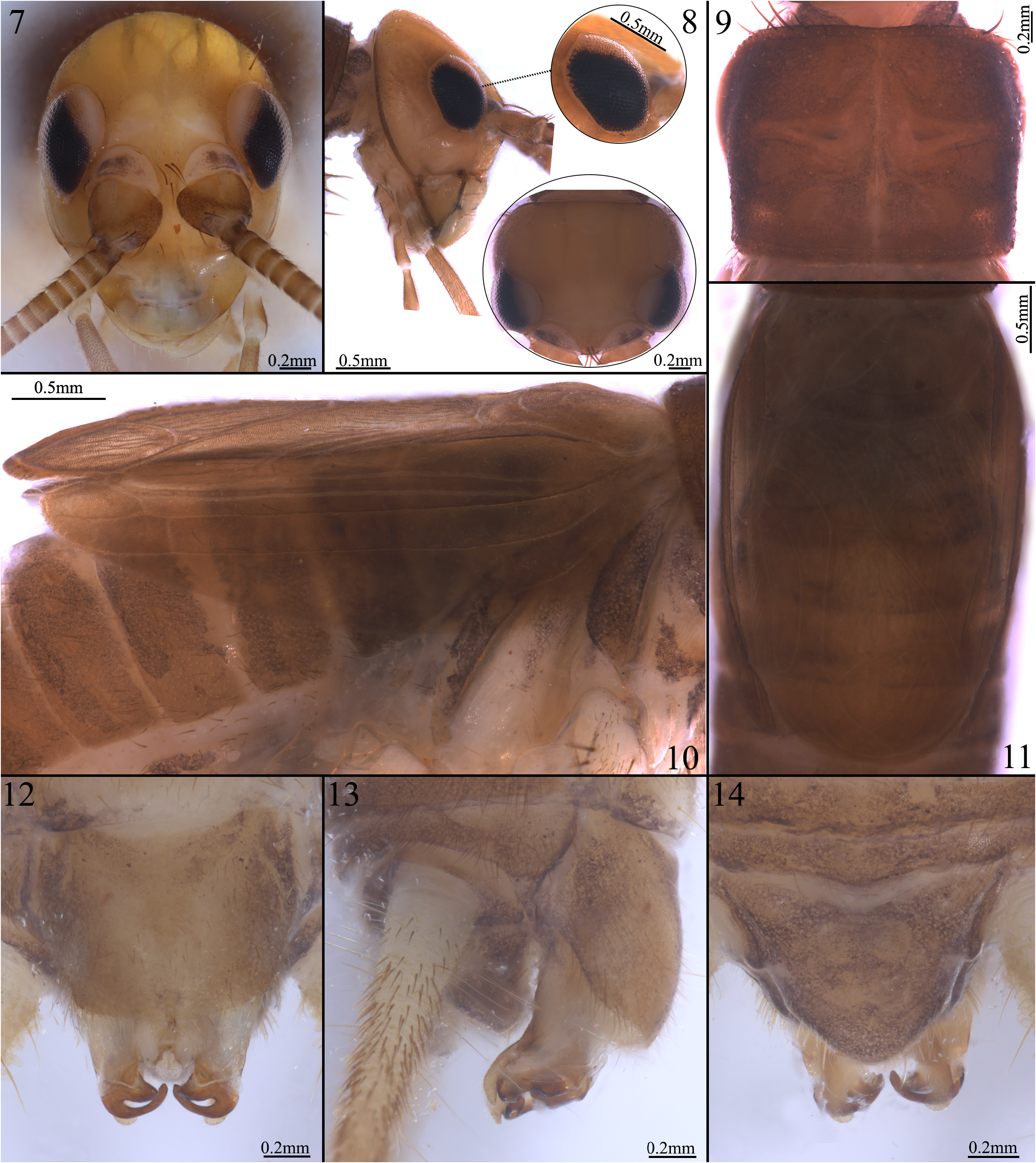
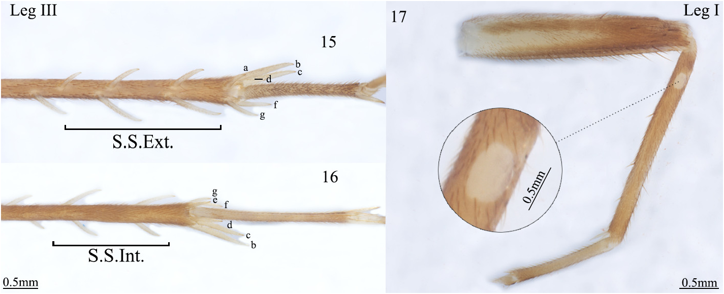
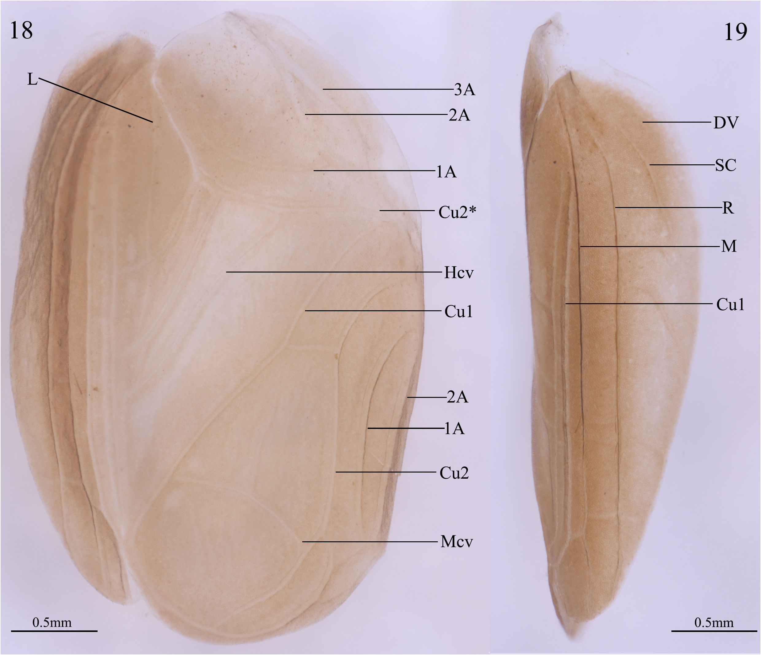
Description, male holotype. General Coloration. Body light brown and head slightly yellowish, possibly discoloration occurred after fixation in ethanol 70% ( Fig. 7—14 View FIGURES 7–14 ); Head. slightly pubescent with long bristles between the scapes ( Fig. 7 View FIGURES 7–14 ), around the eyes and on the posterior and occiput margins of the head almost no bristles are evident (apparently lost after fixation in ethanol 70%), occiput region slightly darkened behind the eyes ( Fig. 8 View FIGURES 7–14 ); Eyes. compound eyes black and with depigmented upper region near the scape insertion ( Fig. 8 View FIGURES 7–14 ); ocelli absent ( Fig. 7 View FIGURES 7–14 ); Mouthparts. clypeus and labrum whitish, mandibles dark outline ( Figs 7 and 8 View FIGURES 7–14 ); maxillary and labial palps yellowish and whitish brown between the articulations ( Figs 7 and 8 View FIGURES 7–14 , picture shows only the first three); maxillary palp slightly pubescent, elongated, with five articulations; first and second palpomeres similar in size and shorter than the others; third and fourth similar in size and shorter than fifth; fifth palpomere longest of all, claviform shape, dilated in distal portion ( Figs 7 and 8 View FIGURES 7–14 , picture shows only the first three); labial palps less pubescent than maxillary palps, with three articulations of increasing size, third palpomere claviform shape ( Figs 7 and 8 View FIGURES 7–14 ); Antennae. scape pubescent, dorsal portion whitish brown and with some slightly darkened spots, oval and dilated shape, inner distal portion with long bristles ( Fig. 7 View FIGURES 7–14 ); pedicel dark brown with whitish regions on outer face, narrow, cylindrical and slightly compressed on median portion; antennomeres with dark brown base and whitish distal region, slightly pubescent, twice as short than pedicel ( Figs 7 and 8 View FIGURES 7–14 ). Thorax. pronotum pubescent with few long bristles on the edges (with lost bristles after fixation in ethanol 70%), darkened brown towards the extremities and whitish in the medial portion, marked with a whitish vertical and horizontal median band, and two small symmetrical white spots near the proximal portion, dorsal disc wider than long, lateral lobe rounded ( Fig. 9 View FIGURES 7–14 ). Legs. Leg I: femur proximal part whitish becoming brownish distally, tibia brownish and with two subequal apical spurs, tibia with an oval auditory tympanum at inner side, first tarsomere twice as large than second and third together, second tarsomere with one quarter of the third tarsomere length, all tarsomeres brownish between the articulations, pre-tarso broken ( Fig. 17 View FIGURES 15–17 ). Leg II: similar to leg I, with tibial apical spurs longer than leg I. Leg III: same appearance as legs I and II, femur developed, whitish towards proximal region, articulation between femur and tibia with a reddish-brown color with black spots at basilateral inner and outer regions, distal portion brownish; tibia slightly brownish, with three inner (SS Int., Fig. 16 View FIGURES 15–17 ) and three outer subapical spurs (SS Ext., Fig. 15 View FIGURES 15–17 ), and four inner (d, e, f, and g, Fig. 16 View FIGURES 15–17 ) and three outer apical spurs (a, b, and c, Fig. 15 View FIGURES 15–17 ), first tarsomere developed with two apical spurs, inner slightly bigger than outer ( Figs 15 and 16 View FIGURES 15–17 ). Right tegmen. Light brown, covering the first four abdominal tergites ( Figs 10 and 11 View FIGURES 7–14 ). Lateral field (in lateral view, Figs 10 View FIGURES 7–14 and 19 View FIGURES 18–19 ): diagonal vein (DV) poorly marked in its distal region; subcostal vein (SC) well marked, reaching a third of the lateral field, with one branch on the lateral margin, the branch connects with R vein in the medial region of the wing length; subcostal (SC), radial (R) and medial (M) veins parallelly distributed in the lateral field; R with a median branch, curved to the distal direction of the wing; between the parallel veins M and R can be seen some cross-vein poorly marked (three or more); Field (in ventral view, Fig. 18 View FIGURES 18–19 ): anal area, chordal area, harp area and the mirror area well developed; anal region with veins anal 1 (1A), anal 2 (2A) and anal 3 (3A) poorly marked, 1A more narrow than 2A and 3A; chordal area with veins 1A, 2A and curbital 2 (Cu2) well marked; Cu2* modified in stridulatory file; harp with a median-longitudinal vein (L), and three crossed veins (Hcv), forming four well demarked cells, connecting Cu2* towards the lateral field, the first two external veins crossing L; mirror triangular oval, with one crossed vein (Mcv) connected to a distal cross vein in the opposite orientation, Mcv with a poorly marked vertical upper branch, one cell distal is present below the Cu2; stridulatory file with 92 teeth. Abdomen. tergites pubescent, light brown and slightly darkened dorsally ( Figs 10 and 11 View FIGURES 7–14 ); sternites pubescent, slightly whiter than the tergites and with a pattern of dark spots above the light brown spot in the most central region of each sternite ( Fig. 10 View FIGURES 7–14 ); subgenital plate pubescent, light brown and slightly darkened towards the side edges, distal and side margin with long bristles, quadrangular shape, distal central region with a slightly indentation ( Fig. 12 View FIGURES 7–14 ); supra-anal plate pubescent, darkened throughout the structure than subgenital plate, trapezoidal shaped, with small lateral projections, distal portion rounded and with long bristles ( Fig. 14 View FIGURES 7–14 ); cerci light brown and whitish at the base ( Fig. 13 View FIGURES 7–14 ).
Observations in Paratypes. Male phallic sclerites (paratype ISLA 66165, Figs 2–6 View FIGURES 2–6 ) Pseudepiphallic: pseudepiphallic median lophy claviform and thin, apex rounded and slightly curved inward, with bristles (Ps.m.l, Figs 2, 4, 5 and 6 View FIGURES 2–6 ); paramere 1 well developed, C-shaped (very similar to A. spurius and A. lucifugus ), inner basal margin acuminated, apex dilated and curved inward (Ps.P1.p, Figs 2–6 View FIGURES 2–6 ); paramere 2 connected to paramere 1 by membranous tissue, slightly more bulging and sclerotized than A. spurius and A. lucifugus (Ps.P2, Figs 5 and 6 View FIGURES 2–6 ); rami elongated (very similar to A. spurius and A. lucifugus ), narrow and well sclerotized slightly curved inside and triangular shaped at the tip (R, Figs 2, 3, 4 and 6 View FIGURES 2–6 ). Ectophallic invagination: ectophallic sclerite H-shaped shortened (similar to A. spurius and A. lucifugus ) ( Figs 2 and 3 View FIGURES 2–6 ), apex of posterior projections quadrangular-shaped in ventral view, dilated and well sclerotized, following the shape of the internal basal margin of pseudepiphallic paramere and connected to the Ps.P1 by membranous tissue (Ec.Pr, Figs 2–4 and 6 View FIGURES 2–6 ); ectophallic arc slightly longer and wider than A. spurius and A. lucifugus (Ec.Arc, Figs 2 and 3 View FIGURES 2–6 ); ectophallic fold well sclerotized, with the lateral border towards outside, central part slightly convex at the top and linear shaped bottom borders (Ec.F, Fig. 3 View FIGURES 2–6 ). Endophallus: circular-shaped and vertically elongated, with a short ventral crest (End.Sc), similar to A. lucifugus , but more sclerotized and slightly developed (End.Sc), connected to the ectophallic fold by an inverted V-shaped membrane (End.Sc, Fig. 3 and 5 View FIGURES 2–6 ).
Ecological Remarks
Specimens of Adelosgryllus ferratilis n. sp. were found in Curionópolis municipality, Pará state, Brazil. The caves where specimens were observed are located in an area regionally known as “Serra Leste”. This area presents several iron-ore caves, but also some quartzite caves. The species was observed in several caves in the area, especially those ferruginous. Such caves are usually small (few dozens of meters) and several cavities only comprises rock shelters, without aphotic areas. The main organic resources observed in those caves is the bat guano and vegetable organic debris, deposited by wind or water. Although the area was originally covered by a tropical rainforest (Amazon rainforest), it is currently highly impacted, by both the replacement of the forests by pastures and by mining activities. The populations of A. ferratilis n. sp. are usually small inside the caves, so that the species may use the caves only sporadically. However, since the external environments were not sampled, it is currently impossible to establish the actual distribution of this species ( Figs 20–24 View FIGURES 20–24 ).

No known copyright restrictions apply. See Agosti, D., Egloff, W., 2009. Taxonomic information exchange and copyright: the Plazi approach. BMC Research Notes 2009, 2:53 for further explanation.
|
Kingdom |
|
|
Phylum |
|
|
Class |
|
|
Order |
|
|
Family |
|
|
Genus |