
Fulvius borgesi, Frédéric, Chérot, Jordi, Ribes & Jacek, Gorczyca, 2006
|
publication ID |
https://doi.org/10.5281/zenodo.172195 |
|
DOI |
https://doi.org/10.5281/zenodo.6263645 |
|
persistent identifier |
https://treatment.plazi.org/id/5F313850-BC67-FFF3-8943-FD33FD0EB69E |
|
treatment provided by |
Plazi |
|
scientific name |
Fulvius borgesi |
| status |
sp. nov. |
Fulvius borgesi View in CoL n. sp.
Holotype (ɗ): AÇORES, Terceira, site S3B, Porto Judeu, São Sebastião (42 m.), UTM 4909664278171, 0108.x.2003, P.A.V. Borges (Arruda Furtado collection, Universidade dos Açores, Azores Island). Paratypes: 5ɗɗ, 1Ψ: AÇORES, Terceira, Porto Judeu, São Sebastião (42 m.), UTM 490966 4278171, 0108.x.2003, P.A.V. B o rg e s (Arruda Furtado collection, Universidade dos Açores, Azores Island; Silesian University, Bankowa, Poland; Museo Nacional de Ciencias Naturales, Madrid, Spain, Museu de Zoologia, Barcelona, Spain and Institut royal des Sciences naturelles de Belgique, Brussels, Belgium); 1Ψ: AÇORES, Terceira, São Sebastião, PortoNovo, 0108.x.2003 (Coll. J. Ribes). All in banana plantation at low altitude («em pomares»; Borges pers. com.).
Description
Male (paratype): total length: 3.36; total width: 1.16; head length (dorsal view): 0.55; head width across eyes: 0.55; vertex width: 0.22; length of antennal segments: I: 0.57; II: 1.17; III: 0.57; IV: 0.63; length of pronotum: 0.46; posterior width of pronotum: 1.10; length of (left) cuneus: 0.55; width of (left) cuneus: 0.35.
Female (paratype): total length: 3.26 (membrane damaged); total width: 1.33; head length (dorsal view): 1.02; head width across eyes: 0.53; vertex width: 0.26; length of antennal segments: I: 0.43; II: 0.96; III: (damaged); IV: (damaged); length of pronotum: 0.46; posterior width of pronotum: 1.14; length of (left) cuneus: 0.54; width of (left) cuneus: 0.31.
General body color black with whitish areas. Head black, eyes dark brown, their inner margin red brown. First antennal segment brown, second antennal segment black with a brown ring at base, distally clear yellow, third and fourth segments black to dark brown. Rostrum yellow brown, reaching middle of abdomen. Pronotum and scutellum black, with short, narrow, suberect white pilosity, the scutellum with a medial carina. Mesoscutum uncovered, with lateral fossae. Hemelytra black to dark brown, with short, narrow, suberect, white pilosity. Median parts of exocoria and coria, basal part of cunei, and apical part of clavii yellow to whitish. Membrane dark brown. Procoxae red brown. Meso and metacoxae dark brown basally, yellow apically. Femora brown. Tibiae basally brown, distally yellow, with numerous spinulae. Tarsi yellow.
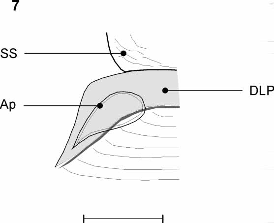
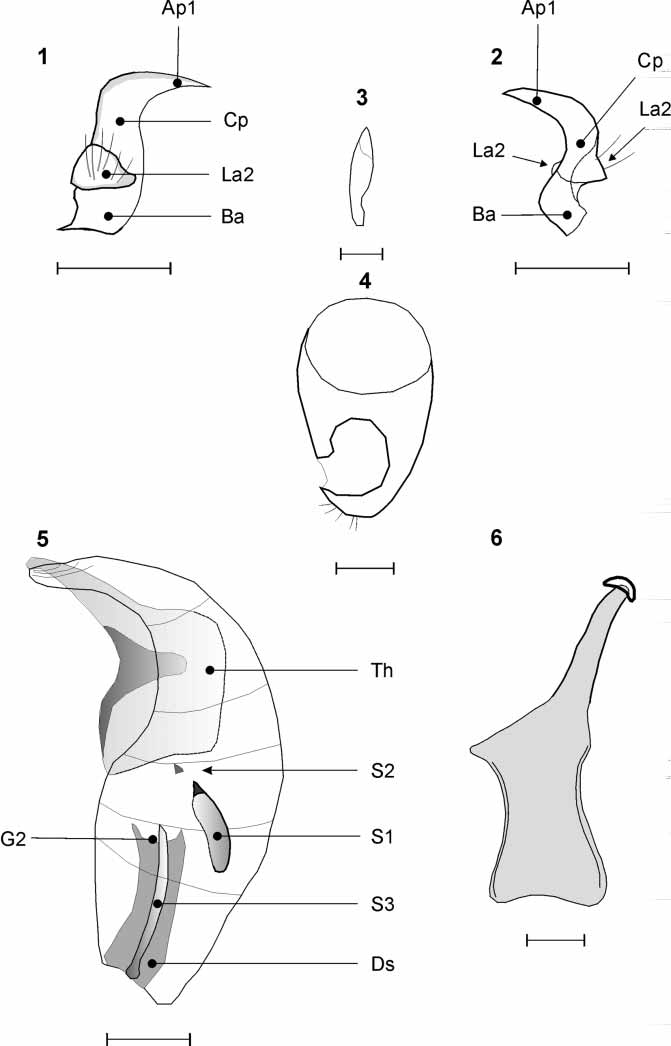
Genital structures. Left paramere stout, curved; base and body (Ba and Cp, Figs 1–2 View FIGURES 1 – 6 ) wide; primary apophysis (Ap1) pointed; ventral surface with a large secondary lobe (La2) bearing numerous setae. Right paramere ( Fig. 3 View FIGURES 1 – 6 ) very small, apex of primary apophysis blunted. Endophallus ( Figs 5–6 View FIGURES 1 – 6 ) complex, with a sclerotized field on the lobe surface ( Fig. 5 View FIGURES 1 – 6 , S1) and two sclerites (S2 and S3, Fig. 5 View FIGURES 1 – 6 ), one pointed, above the secondary gonopore and apex of ductus seminis (G2 and Ds respectively) and the second triangular, more posterior. Theca ( Fig. 5 View FIGURES 1 – 6 , Th. and Fig 6 View FIGURES 1 – 6 ) very large. Seminal depository large, lacking glandular circumference, its connection to median part of vagina wide. Parietovaginal rings ( Fig. 7 View FIGURE 7 , Ap) relatively small, narrow, obviously separated, their anterior margins convex, their posterior margins concave, their outer margins acute, devoid of any prolongation, and their inner margins (ImAp) convex. No medial sclerites. Dorsolabiate plate (DLP) large, anterior and posterior margins slightly thickened. Ventrolabiate plate absent. Dorsal wall very slightly sclerotized. Vaginal projection sensu Rosenzweig (1997) and MiRs sensu Chérot (2002) absent. Vermiform gland indistinct. Lateral oviducts large, not conflating in a common dorsal sac, partially hiding in dorsal view the parietovaginal rings. Posterior wall membranous.
No known copyright restrictions apply. See Agosti, D., Egloff, W., 2009. Taxonomic information exchange and copyright: the Plazi approach. BMC Research Notes 2009, 2:53 for further explanation.