
Russula quercina J. J. Zhou & R. Q. Ji, 2022
|
publication ID |
https://doi.org/ 10.11646/phytotaxa.549.1.6 |
|
DOI |
https://doi.org/10.5281/zenodo.6605371 |
|
persistent identifier |
https://treatment.plazi.org/id/5D3AE467-FFC8-636C-2C9A-FF487AE83030 |
|
treatment provided by |
Plazi |
|
scientific name |
Russula quercina J. J. Zhou & R. Q. Ji |
| status |
sp. nov. |
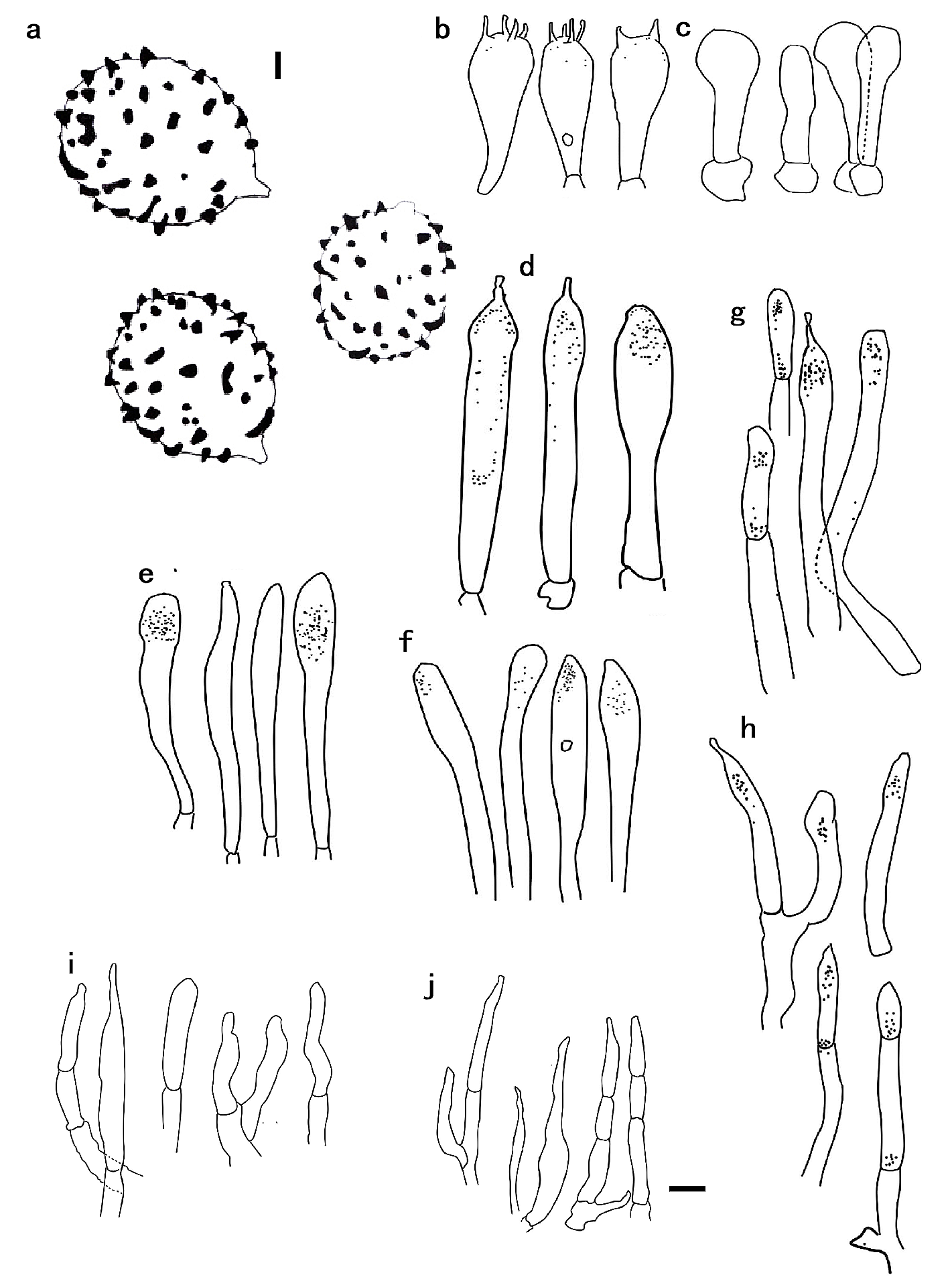
Russula quercina J. J. Zhou & R. Q. Ji View in CoL , sp. nov., ( Figs. 2–4 View FIGURE 2 View FIGURE 3 View FIGURE 4 )
MycoBank: MB840897
Etymology:—The name refers that the holotype host genus is the Quercus .
Holotype:— CHINA, Heilongjiang Province, Wudalianchi County, Wudalianchi Scenic Area , Quercus mongolica forest, 126˚11’ E, 48˚37’ N, asl. 303 m, Peng-Jie Xing & Yang Xu, 12 August 2018 (holotype HMJAU49121 View Materials , ITS: MZ 571933 View Materials ).
Diagnosis:— Russula quercina is morphologically similar to R. integriformis , but is distinguished by its association with Quercus mongolica whereas R. integriformis is associated with Picea . In addition, R. quercina has caulocystidia and longer pleurocystidia while R. integriformis has 47–70 × 9–12 μm size pleurocystidia and 25–32 × 8–11 μm size basidia.
Description:— Basidioma medium-sized. Pileus 34–64 mm broad, hemispheric to convex at first, and then gradually flattens and becomes slightly concave. The edge is raised and dented toward the middle when mature, coral pink to light coral red (xiii 5ʹ d–5ʹ b) when juvenile, xanthine orange to coral pink (iii 13ʹ i–xiii 5ʹ d) with age, and pompeian red to acajou red (xiii 3ʹʹ i–1ʹʹ i) when dried; center flesh is ocher to apricot buff (xiv 9ʹʹ b–11ʹʹ b) when juvenile, vinaceous rufous to cinnamon rufous (xiv 7ʹʹ i–11ʹʹ i) with age, and eugenia red to coral red (xiii 1ʹʹ–5ʹʹ) when dried; viscid when wet and glabrous, smooth, and non-pruinose when dried; margin obtuse, very slightly striate when old, cracks absent, cuticle dry, peeling to almost 1/3 of radius. Context 1.0– 1.5 cm thick from the stipe top to the pileus center, brittle, white (iii) when juvenile, naples yellow (xvi 19ʹ d) with age, distinctly yellowing when dry or bruised. Taste is mild and odor is indistinct. Lamellae 2.2–5.0 mm broad, equal in length, no lamellulae, adnate to almost free, forked, fragile, pale yellow-orange (iii 15f) first, pale orange-yellow (iii 17f) when mature or dry. Stipe is clavate, enlarged towards the base, 3–6.4 × 0.7–1.6 cm, smooth, solid but irregularly hollowing when old, white (iii) when juvenile, becoming baryta yellow (iv 21f), wax yellow (xvi 21), or primuline yellow (xvi 19) towards the base when old or dry. Spore print yellow.
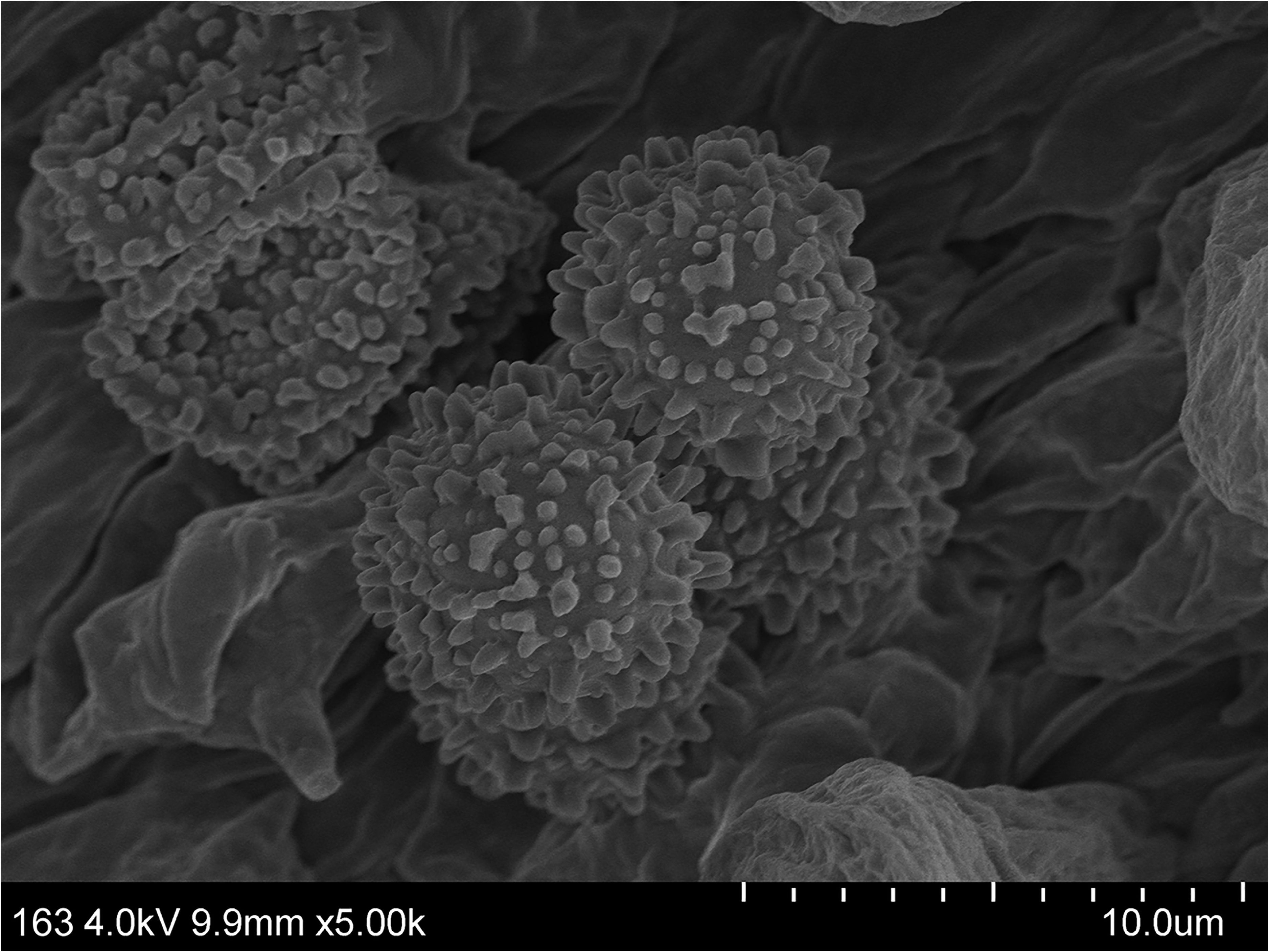
Basidiospores [80/4/4] (6) 6.7–9.5 (10) × (4) 5.2–7.2 (8) μm, Q = (1.09) 1.14–1.28 (1.33), Q av = 1.20 ± 0.05, broadly ellipsoid, rarely subglobose; ornamentation amyloid, mostly isolated, rarely linked by fine lines and do not form a mesh, conic to cylindrical warts, 0.3–1.0 μm in height, suprahilar spot moderately large, amyloids. Basidia are (40) 44–57 (60) × 13–14 (15) μm, clavate to subclavate, broadly tapered towards the base, 4-spored, sometimes twospored, hyaline in KOH, occasionally contain a large droplet; sterigmata 3–5 μm. Pleurocystidia 75–90 × 9–15 μm, emergent, fusiform, clavate to subclavate, often have a subacute tip or mucronate apex and sometimes have a frayed small appendage and dense crystal inclusions. Cheilocystidia 50–61 × 7–13 μm and mostly similar to pleurocystidia. Pileipellis two-layered, 100–125 μm thick, gelatinous, epicutis ixotrichoderm; hyphae 3–4 μm in diameter, septate, branched, erect to suberect, tangled, hyaline in KOH; terminal cells are thin-walled and cylindrical with obtuse, undifferentiated, and often tapered ends. Hyphal terminations near the pileus margin are (27) 50‒70 (90) × (4) 5‒7 (10) μm, mainly unbranched, often apically flexuous; terminal cells are usually distinctly longer and thin-walled; terminal cells are apically attenuated or subcylindrical and obtuse and constricted on septa. Hyphal terminations near the pileus centre are (20) 37‒69 (90) × (3) 5‒7 (9) μm, and the terminal elements are similar to the hyphal terminations of the pileus margin. Pileocystidia 64–92 × 4–6 μm, dispersed, numerous on the surface, scattered, clavate, subclavate to cylindrical, with crystalline and granulate contents. Stipitipellis mostly composed of interwoven branched elongated hyphae, hyphae that are 2–4 μm in diam, and hyaline with inflated cells; caulocystidia 72–100 × 6–9 μm, dispersed, rare, clavate. Clamp connections are absent in all tissues.
Habitat and distribution:—Solitary on soil under Q. mongolica from Heilongjiang, Northeast China.
Additional specimens examined for taxa in this study:— CHINA, Heilongjiang Province, Wudalianchi County, Wudalianchi Scenic Area, in Q. mongolica forest, 126°11’E, 48°37’N, asl. 303 m, 12 August 2018, Peng-Jie Xing & Yang Xu (HMJAU49122, ITS: MZ571934 View Materials ); ibid, 126°12’E, 48°20’N, asl. 301 m, 13 August 2018, Peng-Jie Xing & Yang Xu ( HMJAU49123 View Materials , ITS: MZ 571932 View Materials ) GoogleMaps ; ibid, 126°11’E, 48°23’N, asl. 303 m, 23 August 2018, Peng-Jie Xing & Yang Xu ( HMJAU49124 View Materials ) GoogleMaps

| E |
Royal Botanic Garden Edinburgh |
| N |
Nanjing University |
| MZ |
Museum of the Earth, Polish Academy of Sciences |
No known copyright restrictions apply. See Agosti, D., Egloff, W., 2009. Taxonomic information exchange and copyright: the Plazi approach. BMC Research Notes 2009, 2:53 for further explanation.
|
Kingdom |
|
|
Phylum |
|
|
Class |
|
|
Order |
|
|
Family |
|
|
Genus |