
Dendromonocotyle ukuthena, Vaughan, David, Chisholm, Leslie & Christison, Kevin, 2008
|
publication ID |
https://doi.org/ 10.5281/zenodo.183100 |
|
DOI |
https://doi.org/10.5281/zenodo.5670108 |
|
persistent identifier |
https://treatment.plazi.org/id/5C258787-FF8D-1A65-FF49-FAC2FD78FC20 |
|
treatment provided by |
Plazi |
|
scientific name |
Dendromonocotyle ukuthena |
| status |
sp. nov. |
Dendromonocotyle ukuthena View in CoL n. sp. ( Figs. 2 View FIGURE 2 A, 4C–4E, 4G, 5–7)
Type host: Himantura gerrardi (Gray) .
Additional host: Himantura uarnak (Forsskål) .
Type locality: uShaka Sea World, Durban, South Africa.
Site on host: Dorsal skin surface.
Etymology: This species is named after the Zulu word “ ukuthena ” which is the ritual of becoming-of-age, in recognition of the opening of uShaka Marine World in KwaZulu Natal.
Materials examined: SAMCTA 29455 (holotype), SAMCTA 29456 (18 paratypes), SAMCTA 29457 (paratype with spermatophore), SAMCTA 29458 (paratype with missing distal portion of male copulatory organ), AHC 29290 (5 paratypes), BMNH 2008.6.25.1-5 (5 paratypes).
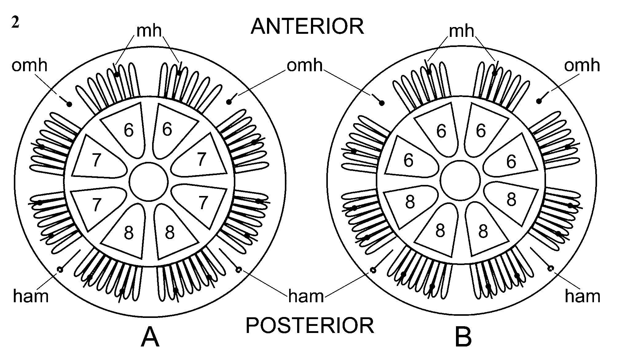
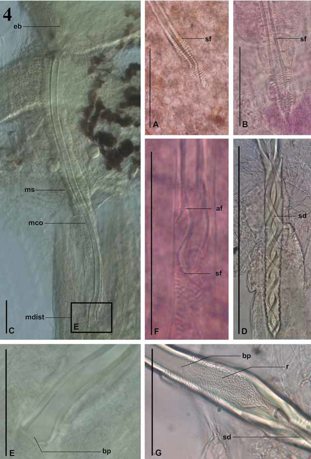
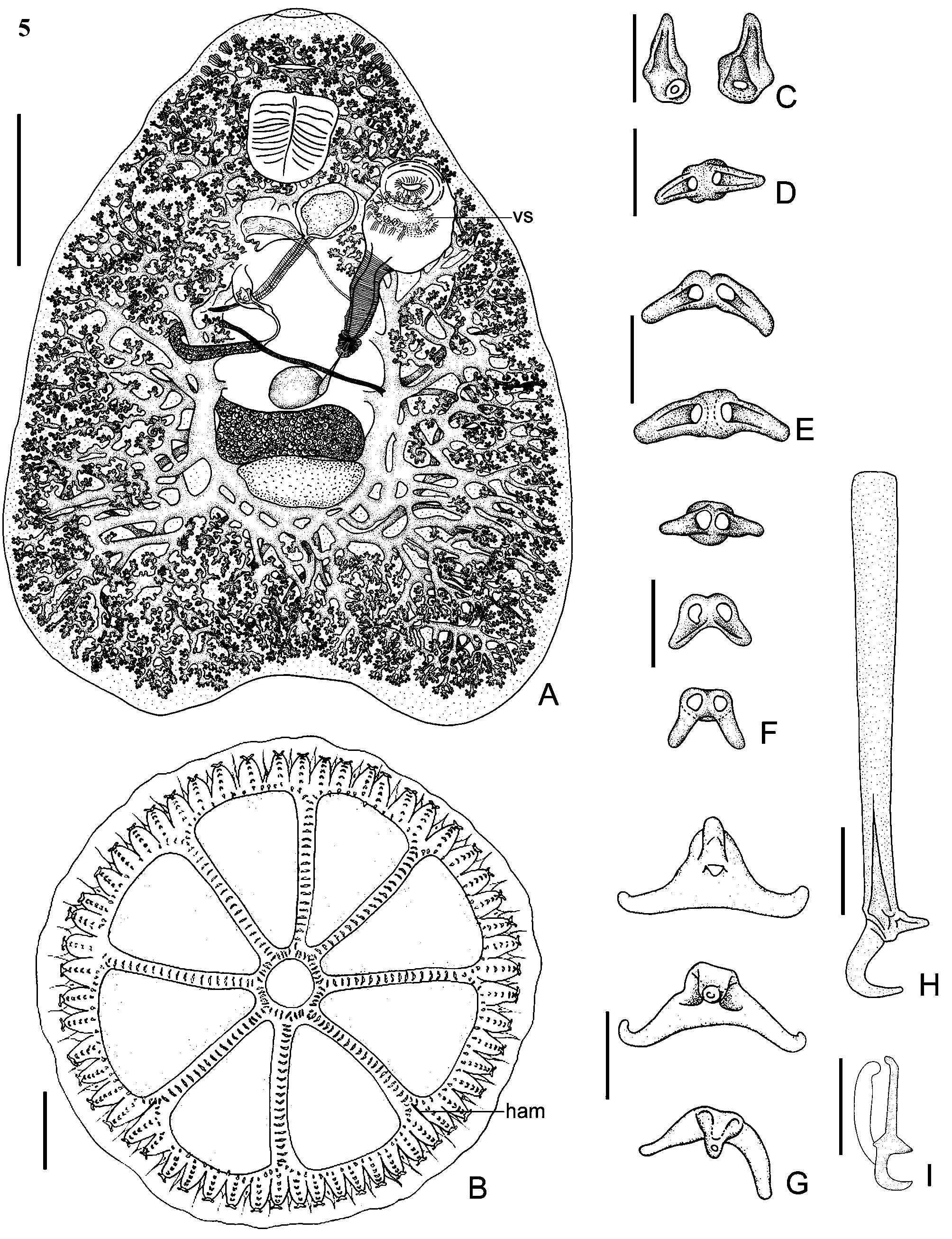
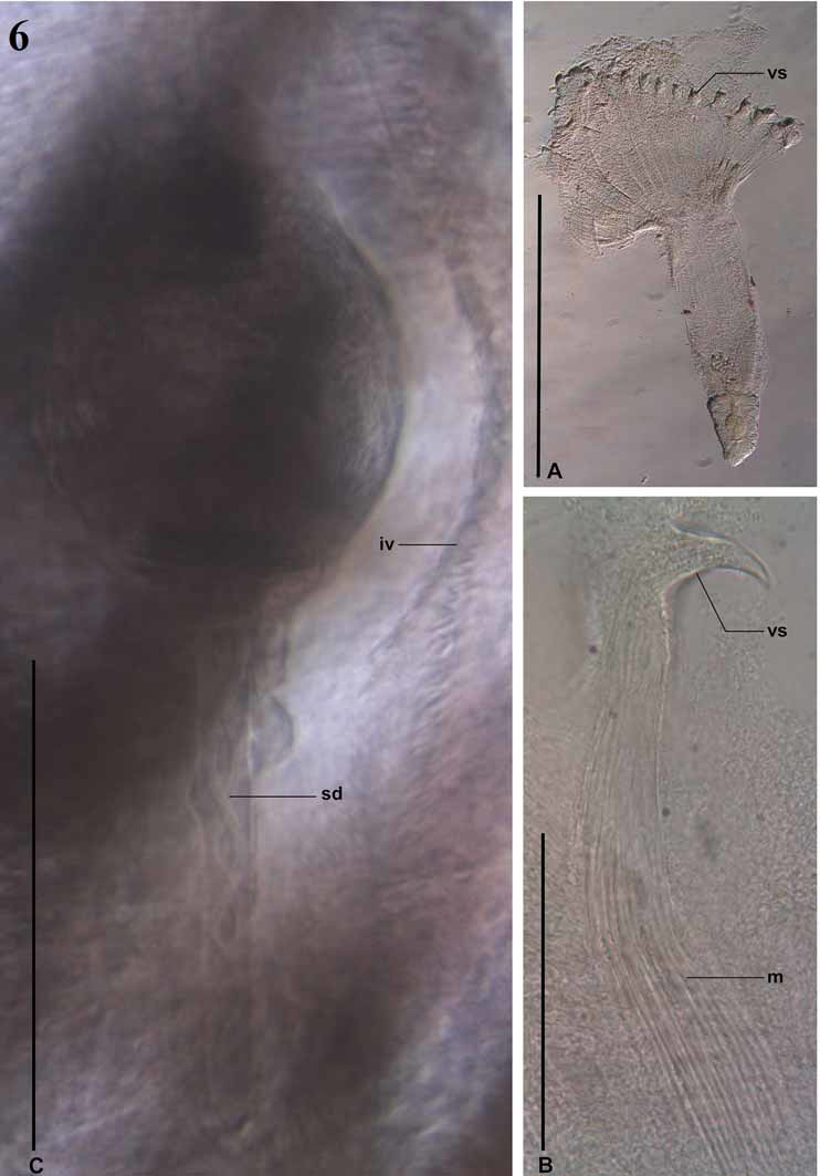
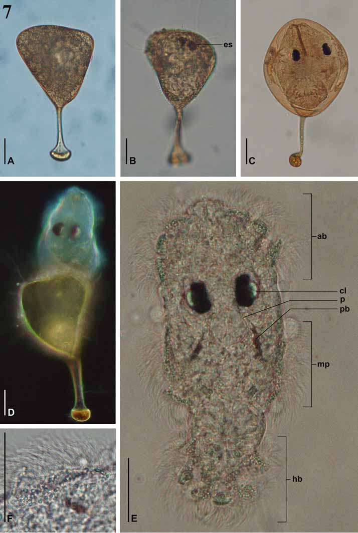
Description. Based on 17 flattened adult specimens. Total body length 1946 ± 750.5 (1250–3290, n = 6). Body-proper length 1957 ± 634.3 (1000–3580, n = 17), and 1522 ± 395.2 (790–2110, n = 17) maximum width ( Fig. 5 View FIGURE 5 A). Circular haptor, 1540 ± 455.8 (810–2110, n = 14) in diameter ( Fig. 5 View FIGURE 5 B), large in proportion to body, with edge of marginal valve extending beyond anterior section of ovary. Haptor with 8 peripheral and 1 central loculus and 56 marginal haptoral papillae, each with between 5 and 6 sclerites including the terminal papillary sclerite ( Figs 5 View FIGURE 5 F, 5G). Outer ring, inner ring and septal sclerites as illustrated ( Figs 5 View FIGURE 5 C–E). Tripartite sclerites at junction of radial septa and inner-ring septum in most specimens. Anterior loculus pair associated each with 6 marginal haptoral papillae, anterolateral and posterolateral loculi each associated with 7 marginal haptoral papillae, and posterior-most loculus pair each associated with 8 marginal haptoral papillae. Hamuli present ( Figs 5 View FIGURE 5 B, 5H), 73 ± 3.7 (68–80, n = 11) long. Fourteen marginal hooklets ( Fig. 5 View FIGURE 5 I), 15 ± 0.5 (14–15, n = 10) long distributed in marginal value between every 4 marginal haptoral papillae ( Figs 2 View FIGURE 2 A, 5B). Mouth anterior to pharynx. Subterminal groove anterior to mouth. Six anterior gland duct openings open laterally on anterior end of worm as illustrated ( Fig. 5 View FIGURE 5 A). Eye spots likely present but only observed in juvenile specimens, obscured by heavily pigmented branches of intestinal caecum. Pharynx 247 ± 75 (110–360, n = 12) long, and 200 ± 53.4 (120–300, n = 13) wide. Dendritic intestinal caecum dense, heavily pigmented. Branches extend from anterior end of body-proper, dorsal and anterior to pharynx, and extend to end of body-proper. Testis 181 ± 28.2 (150–240, n = 11) long, and 365 ± 59.7 (290–450, n = 11). Vas deferens runs dorsal to vagina, ventral to male copulatory organ before swelling to form seminal vesicle. Ejaculatory bulb situated left to centre of body-proper, 171 ± 28.8 (140–220, n = 10) long, and 161 ± 26.4 (110–200, n = 10) wide. Male copulatory organ 486 ± 32 (440–530, n = 10) long, and 11 ± 1.1 (10–14, n = 15) wide; surrounded by muscular sheath, 76 ± 2.5 (75–80, n = 4) wide ( Figs 4 View FIGURE 4 C, 5A). Distal portion of male copulatory organ sclerotised with criss-crossed sperm duct ( Fig. 4 View FIGURE 4 D). Distal portion of male copulatory organ absent in 5 specimens ( Figs 4 View FIGURE 4 C, 4E). Possible breakage point and area of regrowth on distal portion of male copulatory organ seen in a number of specimens ( Fig. 4 View FIGURE 4 G). Ovary ovoid, 154 ± 25.7 (110–190, n = 12) long, 543 ± 57.2 (460–640, n = 12) wide. Ootype 223 ± 15 (210–240, n = 4) long with tetrahedral egg side length 88 ± 5 (80–90, n = 4). Short filament 43 ± 5.8 (40–50, n = 3) extends from 1 pole, ending in blunt, rounded butt ( Fig. 7 View FIGURE 7 A). Muscular vagina distinct 648 ± 23.8 (610–670, n = 5) long. Vaginal pore on left side of body at level of posterior portion of pharynx; leading to vaginal chamber with inner-ring of sclerotised spines embedded in thick musculature ( Figs 5 View FIGURE 5 A, 6A, 6B). Vagina narrows then swells slightly to enter ovoid seminal receptacle 110 ± 12.2 (100–130, n = 5) long, and 145 ± 39.3 (100–190, n = 5) wide. Duct from seminal receptacle to oviduct not seen. Transverse vitelline duct inconspicuous, running just anterior to ovarian loop, crossing over proximal portion of vagina. A spermatophore 230 (n = 1) long, and 60 (n = 1) wide, with distal portion resembling the distal tip of the male copulatory organ present in 1 specimen (SAMCTA 29457) ( Fig. 6 View FIGURE 6 C).
Egg hatching. Eggs of D. ukuthena are laid singly. Eggs incubated in a LD 12:12 light regime at 23°C contained larvae with eyes ( Fig. 7 View FIGURE 7 B) after 5 days. Eggs incubated at 26°C and 30°C contained larvae with eyes after 3 days. Spontaneous hatching ( Fig. 7 View FIGURE 7 D) commenced at 14, 5 and 4 days after laying at 23, 26 and 30°C, respectively.
Description of oncomiracidium. Observations based on 2 freshly hatched, living oncomiracidia. Larva 168 ± 4.2 (165–171, n = 2) long and 89 ± 1.4 (88–90, n = 2) wide. Ciliated cells in 3 zones; 1 anterior band, 2 median patches (1 on each side of body) and 1 haptoral band ( Fig. 7 View FIGURE 7 E). Refringent droplets in ciliated epidermal cells ( Fig. 7 View FIGURE 7 F).
Two pairs of pigmented eyes with crystalline lenses arranged as shown ( Fig. 7 View FIGURE 7 E); pairs very close together. Light brown pigment in body distributed in 2 short distinct longitudinal bands on either side of body from level just posterior pharynx ( Fig. 7 View FIGURE 7 E). Mouth not observed. Pharynx muscular, 20 ± 3.1 (17–24, n = 2) in diameter. Glands associated with pharynx not observed. Gut immediately posterior to pharynx, bilobed. Two anteromedian gland cells containing granular secretion just anterior to pharynx; single duct from each gland cell runs anteriorly opening at anterior end. Glands containing needle-like secretion present on either side of pharynx. Protonephridial system not investigated.
Haptor with 14 hooklets, each of similar shape and size, 12 ± 0.4 (12–13, n = 5) long with domus. Hamuli not observed.
Remarks. Dendromonocotyle ukuthena can be distinguished from all other species of Dendromonocotyle by the morphology of the vagina, which possesses a ring of spines embedded within the thick musculature near the vaginal pore ( Figs 5 View FIGURE 5 A, 6A, 6B). Dendromonocotyle taeniurae is the only other species with spines associated with the vaginal pore but the 2 species are clearly differentiated by the morphology of the distal portion of the male copulatory organ. In addition, D. taeniurae lacks hamuli.
A spermatophore was present in the vagina of 1 specimen. The distal end of the spermatophore resembles the distal end of the male copulatory organ (c.f. Figs 6 View FIGURE 6 C and 4D) complete with the criss-crossed sperm duct. We also observed that the distal part of the male copulatory organ was missing in 5 of the 17 specimens examined. It is possible that the distal end of the male copulatory organ breaks off during mating to form the spermatophore (see Discussion).
Dendromonocotyle ukuthena was found on the same ray specimens as D. citrosa at uShaka. Sea World, Durban.
Eyespots were observed in the larvae and juvenile D. ukuthena but were obscured by the darkly pigmented intestinal caeca in adult specimens.
No known copyright restrictions apply. See Agosti, D., Egloff, W., 2009. Taxonomic information exchange and copyright: the Plazi approach. BMC Research Notes 2009, 2:53 for further explanation.