
Stenopsocus formosanus Banks
|
publication ID |
https://doi.org/ 10.11646/zootaxa.4057.2.2 |
|
publication LSID |
lsid:zoobank.org:pub:89E858EB-2E3B-47FC-B2F0-35B2A3ED6EBB |
|
DOI |
https://doi.org/10.5281/zenodo.6122751 |
|
persistent identifier |
https://treatment.plazi.org/id/5764C334-1248-FFF3-A4B7-FEF73682FE14 |
|
treatment provided by |
Plazi |
|
scientific name |
Stenopsocus formosanus Banks |
| status |
|
Stenopsocus formosanus Banks View in CoL
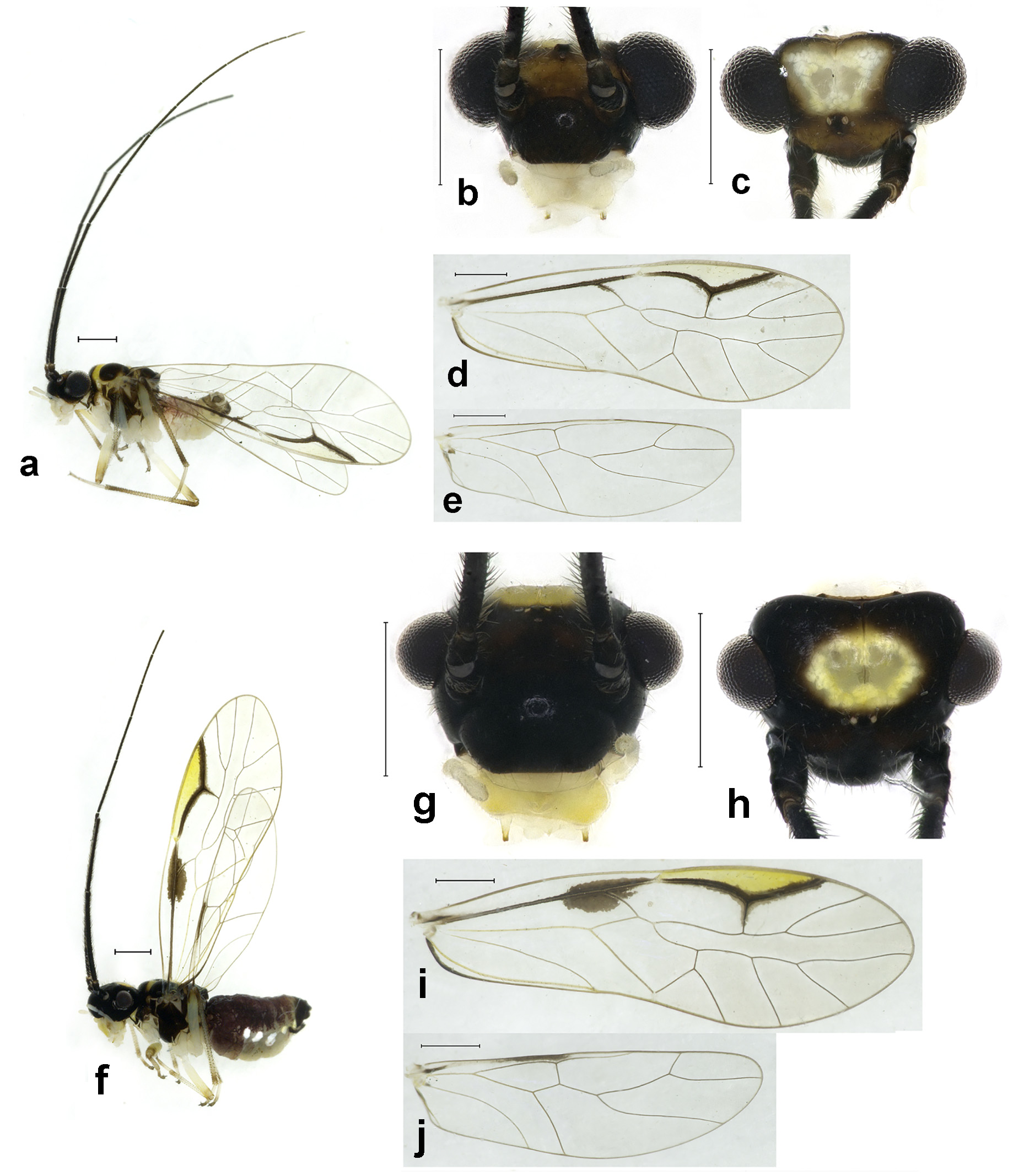
( Figs. 11–12 View FIGURE 11 View FIGURE 12 )
Stenopsocus formosanus Banks, 1937: 259 View in CoL . Type locality: China ( Taiwan: Hassenzan, Arisan).
Diagnosis. This species is characterized by the yellowish pterostigma with narrow brown stripe on the entire R1, and blackish brown antenna. Female forewing has a blackish brown marking on the point of R and Rs, male forewing has a narrow brown stripe along R.
Adult male. Body ( Fig. 11 View FIGURE 11 a) length 2.41 mm, length from postclypeus to wing tip 4.83 mm. IO: 0.37 mm, d: 0.31 mm, IO/d=1.19, f1: 1.01 mm, f2: 0.91 mm, f3: 0.76 mm, FWL: 3.87 mm, FWW: 1.39 mm, HWL: 2.89 mm, HWW: 0.95 mm, t1: 0.39 mm, t2: 0.13 mm.
Colour (in alcohol). Head ( Figs. 11 View FIGURE 11 b, c) dark brown, vertex with a yellowish area, which is variable in shape, a broad brown stripe present between eyes cross ocellar area, frontal area yellowish brown. Antenna dark brown. Postclypeus blackish brown, labrum laterally much paler, apex of maxillary palpus pale brown, remaining segments of maxillary palpus whitish.
Thorax dark brown. Leg yellowish, with coxae, tibiae, apices of 1st tarsomeres, and entire 2nd tarsomeres brown; hind leg mostly brown, with trochanter, middle of tibia, and entire 2nd tarsomere whitish. Abdomen yellowish, 1–4 segments with pale purplish brown terga. Genital segments brownish.
Forewings ( Fig. 11 View FIGURE 11 d) transparent. R and R1 dark brown, anterior margin of pterostigma yellowish, other veins brown. R with an extremely narrow brown marking; pterostigma yellowish. Hindwing ( Fig. 11 View FIGURE 11 e) immaculate.
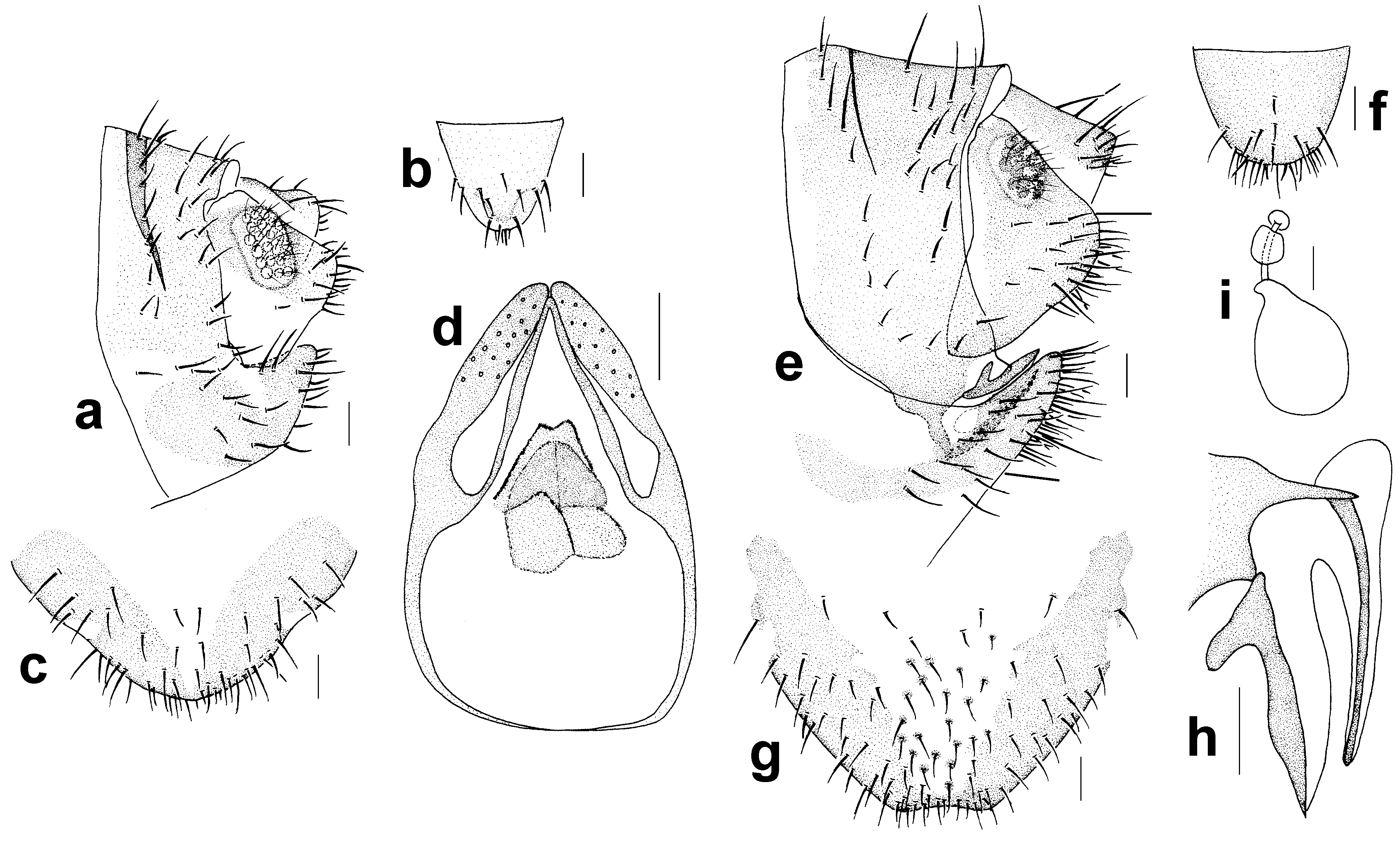
Genitalia ( Figs. 12 View FIGURE 12 a–d) strongly sclerotized. Epiproct ( Fig. 12 View FIGURE 12 b) subtrapezoidal with a round apex. Paraproct semi-sclerotized, with 32 trichobothria. Endophallus ( Fig. 12 View FIGURE 12 d) strongly sclerotized, external parameres robust, with some punctures on broadened apex, not exceeding apex of aedeagal arch; aedeagal arch narrow. Hypandrium ( Fig. 12 View FIGURE 12 c) strongly sclerotized.
Adult female. Body ( Fig. 11 View FIGURE 11 f) length 3.11 mm, length from postclypeus to wing tip 4.34 mm. IO: 0.55 mm, d: 0.20 mm, IO/d=2.75, f1: 1.24 mm, f2: 1.04 mm, f3: 0.77 mm, FWL: 4.26 mm, FWW: 1.41 mm, HWL: 3.02 mm, HWW: 0.92 mm, t1: 0.31 mm, t2: 0.10 mm.
Colour generally similar to male, but slightly darker. Head ( Figs. 11 View FIGURE 11 g, h) with a yellowish area on vertex. Forewing ( Fig. 11 View FIGURE 11 i) with a distinct dark brown marking around branching point of R and Rs, entire R1 with narrow dark brown marking; a brown marking present between Sc and R on hindwing ( Fig. 11 View FIGURE 11 j). Abdomen purplish brown, lateral 3–7 segments with white markings; genital segments blackish brown.
Genitalia ( Figs. 12 View FIGURE 12 e–i) strongly sclerotized. Epiproct ( Fig. 12 View FIGURE 12 f) subtrapezoidal with round apex. Paraproct ( Fig. 12 View FIGURE 12 e) with 19 trichobothria. Gonapophyses ( Fig. 12 View FIGURE 12 h) with external valve reduced, fused with dorsal valve, middle of dorsal valve slightly broadened, ventral valve narrowly elongate, with acute apex. Subgenital plate ( Fig. 12 View FIGURE 12 g) with sclerotized area separated into two parts, a narrow sclerotized stripe along margin of subgenital plate to connect two parts.
Specimens examined. CHINA ( TAIWAN): Hualien, Pi-lu Sacred Tree (2150 m), 5 males, 5 females, 6.vi.2013, Liang Feiyang.
Distribution. China ( Taiwan, Zhejiang).
Remarks. S. formosanus differs from S. externus by the entire dark brown antenna, entire R1 with dark brown marking and the male half-sclerotized praproct.
No known copyright restrictions apply. See Agosti, D., Egloff, W., 2009. Taxonomic information exchange and copyright: the Plazi approach. BMC Research Notes 2009, 2:53 for further explanation.
|
Kingdom |
|
|
Phylum |
|
|
Class |
|
|
Order |
|
|
Family |
|
|
Genus |
Stenopsocus formosanus Banks
| Liang, Feiyang, Dai, Yuting, Yue, Lu, Li, Fasheng & Liu, Xingyue 2015 |
Stenopsocus formosanus
| Banks 1937: 259 |