
Lernanthropus microlamini Hewitt, 1968
|
publication ID |
https://doi.org/ 10.11646/zootaxa.4736.1.1 |
|
publication LSID |
lsid:zoobank.org:pub:970D7D36-6D8C-4463-B9EA-D3B8E191BE72 |
|
DOI |
https://doi.org/10.5281/zenodo.3671101 |
|
persistent identifier |
https://treatment.plazi.org/id/554BDB52-7354-FFF7-5FC9-FAA92D2EFED6 |
|
treatment provided by |
Plazi |
|
scientific name |
Lernanthropus microlamini Hewitt, 1968 |
| status |
|
Lernanthropus microlamini Hewitt, 1968
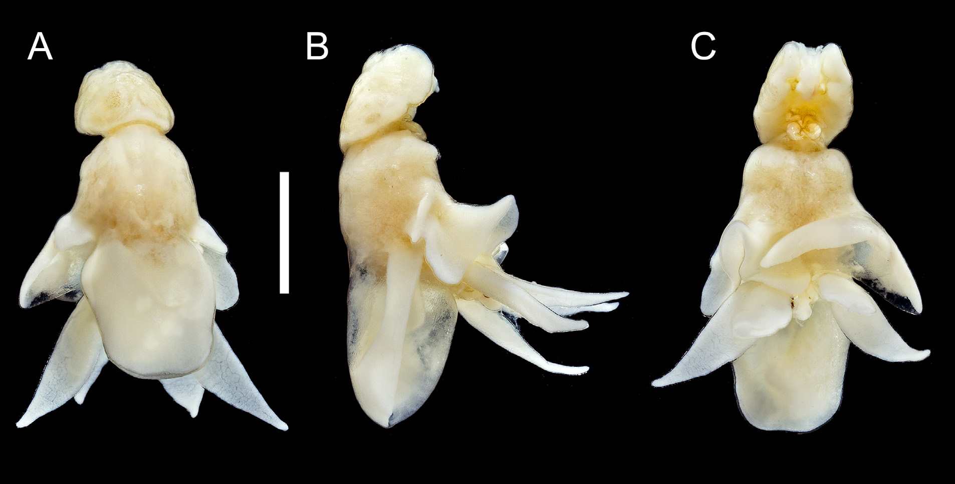
( Figs. 25–26 View FIGURE 25 View FIGURE 26 )
Material examined: 2♀♀ 3♂♂ from gills of Hyperoglyphe antarctica (Carmichael, 1819) , Adelaide , South Australia; 10 May 2007; collected by K.S. Hutson ; 1♀ and 1♂ NHMUK Reg. No. 2007.948–949 and 1♀ and 2♂♂ SAMA Reg. No. C6900 . 1♂ from gills H. antarctica, Coffs Harbour , New South Wales; 27 October 1981; collected by K. Rohde ; NHMUK Reg. No. 1984.90 .
Differential diagnosis: Cephalothorax longer than wide with lateral margins parallel anteriorly; frontal margin weakly concave. Trunk about 2.5 times longer than cephalothorax ( Fig. 25 View FIGURE 25 A–C); anterior part (second and third pedigerous somites) becoming wider posteriorly, posterior part (fourth pedigerous somite) covered by subrectangular dorsal trunk plate with rounded corners and with weakly convex lateral and posterior margins. Urosome comprising fifth pedigerous somite, genital complex and abdomen, all fused. Paired caudal rami small; each ramus about twice as long as width at base; completely concealed beneath dorsal trunk plate. Parabasal flagellum cylindrical. Leg 3 forming large, ventrally-directed, fleshy lamella, extending posterolaterally from side of body by almost one third of length: third legs separate but overlapping slightly in midline. Leg 4 bilobate ( Fig. 25 View FIGURE 25 A–C); inner and outer lobes foliaceous, tapering distally, outer lobe longer than inner and protruding posterolaterally from beneath lateral margin of dorsal trunk plate. Leg 5 represented by laterally-directed cylindrical lobe about twice as long as wide, armed with basal seta on dorsal surface. Body length of ♀ 8.45 mm.
Description of male. Body smaller than female, body length 2.74 mm. Cephalothorax large, comprising about 40% of total body length, broadest at middle, with angular lateral margins; frontal area of cephalothorax carrying antennules and antennae, defined by slight indentation ( Fig. 26A View FIGURE 26 ). Trunk slender comprising second to fourth pedigerous somites fused to urosome. Urosome comprising fifth pedigerous somite, genital somite and abdomen, all fused. Genital somite with weakly convex lateral margins, wider than abdomen. Caudal rami ( Fig. 26B View FIGURE 26 ) about 2.1 times longer than wide, as in female.
Antennule indistinctly 6-segmented ( Fig. 26C View FIGURE 26 ) with irregular cuticular thickenings, setal formula: 1, 3, 1, 2, 1, 12 + 2 ae. Parabasal flagellum ( Fig. 26D View FIGURE 26 ) comprising broad base and short, curved distal part. Antenna ( Fig. 26E View FIGURE 26 ) comprising massive corpus and distal subchela; corpus armed with papilliform process medially; subchela armed with rounded knob-like process proximally, curved spine on concave margin, plus distal tooth-like process; small process present in articulation between corpus and subchela; surface integument of claw striated. Postantennal process directed anteriorly, lacking ornamentation ( Fig. 26F View FIGURE 26 ). Mandible ( Fig. 26G View FIGURE 26 ) and maxillule ( Fig. 26H View FIGURE 26 ) similar to those of female. Maxilla with simple subdistal process and additional spinular ornamentation distally on basis ( Fig. 26I View FIGURE 26 ). Maxilliped ( Fig. 26J View FIGURE 26 ) comprising robust corpus with small papilliform process on myxal surface, and distal subchela armed with 2 rounded processes; surface of subchela striated.
Leg 1 with protopod distinct from somite; members of leg pair joined by intercoxal sclerite ( Fig. 26K View FIGURE 26 ). Each leg biramous with outer seta and short, stout inner spine on basis; exopod 1-segmented, armed with 5 robust terminal spines and ornamented with spinules distally; endopod 1-segmented, armed with long terminal seta about 1.2 times longer than segment, and ornamented with patch of spinules distally. Leg 2 ( Fig. 26L View FIGURE 26 ) lacking trace of intercoxal sclerite: protopod with outer seta; exopod indistinctly articulated at base, armed with 3 small distal spines and larger curved element; endopod well defined basally, armed with single apical seta just shorter than segment, without ornamentation. Leg 3 ( Fig. 26A View FIGURE 26 ) uniramous, comprising tapering cylindrical exopodal process protruding ventrolaterally from trunk; outer protopodal seta not observed. Leg 4 ( Fig. 26A View FIGURE 26 ) biramous, comprising 2 long cylindrical processes, with outer protopodal seta on common base; exopodal lobe longer than endopodal lobe. Leg 5 reduced to rounded lobe bearing naked seta subapically ( Fig. 26M View FIGURE 26 ).
Distribution: This species was established by Hewitt (1968) based on the description of a single female collected from Seriolella brama (Günther, 1860) caught “presumably in the region of Wellington ”, New Zealand ac- cording to Hewitt (1968). The discovery of this species on Hyperoglyphe antarctica in Australia extends its known range to include the southeastern sector of the Australian coast from Adelaide in the south to Coffs Harbour on the east coast. Both H. antarctica and S. brama belong to the Centrolophidae and this parasite appears restricted to hosts belonging to this family. This is the first published report of L. microlamini from Australian waters.
Remarks: This is a large species. The body length of the holotype female was 9.92 mm and the female from South Australia was 8.45 mm. Found here for the first time, the male was 2.74 mm in length. Hewitt (1968) pointed to similarities between his L. microlamini and L. trifoliatus Bassett-Smith, 1898 (now synonymized with L. polynemi Richiardi, 1881 (see Piasecki & Hayward, 2002)), but the shape of the dorsal trunk plate differs: it narrows posteriorly in the former but becomes wider and more rounded posteriorly in the latter.

No known copyright restrictions apply. See Agosti, D., Egloff, W., 2009. Taxonomic information exchange and copyright: the Plazi approach. BMC Research Notes 2009, 2:53 for further explanation.
|
Kingdom |
|
|
Phylum |
|
|
Class |
|
|
Order |
|
|
Family |
|
|
Genus |