
Sagum sanguineus (Song, in Song & Chen, 1976 ), 2020
|
publication ID |
https://doi.org/10.11646/zootaxa.4736.1.1 |
|
publication LSID |
lsid:zoobank.org:pub:970D7D36-6D8C-4463-B9EA-D3B8E191BE72 |
|
DOI |
https://doi.org/10.5281/zenodo.3671087 |
|
persistent identifier |
https://treatment.plazi.org/id/554BDB52-7339-FF95-5FC9-FC782AA7F91A |
|
treatment provided by |
Plazi (2020-02-17 07:18:21, last updated 2024-11-26 03:30:40) |
|
scientific name |
Sagum sanguineus (Song, in Song & Chen, 1976 ) |
| status |
comb. nov. |
Sagum sanguineus (Song, in Song & Chen, 1976) n. comb.
( Figs. 47–48 View FIGURE 47 View FIGURE 48 )
Syn: Lernanthropus sanguineus Song, in Song & Chen, 1976
Material examined: 1♀ from Lutjanus johnii (Bloch, 1792) ( OH-M), Lee Point, Outer Harbour, Darwin , Northern Territory, 10 March 2014, collected by D.P. Barton; MAGNT Reg. No. Cr 019251 . 1♀ from Lutjanus johnii, Condor, Melville Island , Northern Territory; 23 August 2012; collected by D.P. Barton; NHMUK Reg. No. 2018.302 .
Differential diagnosis: Cephalothorax about 1.25 times longer than wide with almost linear lateral margins narrowing anteriorly towards short frontal margin ( Fig. 47A View FIGURE 47 ); lateral margins of dorsal cephalothoracic shield projecting ventrally, with strongly convex posterior margin. Anterior part of trunk (second and third pedigerous somites) about 1.7 times wider than long with distinct anterolateral shoulders and with more or less parallel lateral margins; posterior part (fourth pedigerous somite) covered by subcircular dorsal trunk plate. Dorsal trunk plate entirely concealing urosome but with at least distal third of fourth leg lobes extending beyond posterior margin and visible in dorsal view ( Fig. 47A View FIGURE 47 ). Urosome comprising fifth pedigerous somite, genital complex and abdomen, all fused ( Fig. 47B, C View FIGURE 47 ). Genital complex slender, with conspicuous paired gonopores dorsolaterally and with paired copulatory pores located posterolaterally on ventral surface (arrowed in Fig. 47B View FIGURE 47 ); dorsal surface of genital complex ornamented with 2 pairs of sensillae located between gonopores. Egg sacs loosely coiled ( Fig. 47D View FIGURE 47 ) beneath dorsal trunk plate and concealed laterally by third legs. Paired caudal rami elongate ( Fig. 47B View FIGURE 47 ), about 2.7 times longer than maximum width; broadest in proximal-section; tips of caudal rami not reaching middle of elongate fourth leg lobes ( Fig. 47C View FIGURE 47 ). Caudal rami each with 2 dorsal setae, distal dorsal seta located at 30% of length of ramus, small outer seta located at about 60% of ramus length, plus 2 apical setae.
Antennule unsegmented, armed with 6 setae proximally and 10 setae plus 2 aesthetascs around apex ( Fig. 47E View FIGURE 47 ). Parabasal flagellum absent. Antenna ( Fig. 47F View FIGURE 47 ) with robust proximal segment; distal subchela armed with rounded process, small spinous process near medial margin, plus small process in articulation with proximal segment; surface of strongly recurved subchela ornamented with striations and pits. Postantennal process elongate ( Fig. 47G View FIGURE 47 ). Mandible stylet-like with 8 marginal teeth at apex. Maxillule bilobed, smaller lobe tipped with 1 spiniform element; larger lobe tipped with 3 unequal spiniform elements and ornamented with hair-like setules ( Fig. 47H View FIGURE 47 ). Maxilla ( Fig. 47I View FIGURE 47 ) with short tapering syncoxa; basis with apical claw ornamented with marginal rows of denticles, plus blunt subapical process. Maxilliped ( Fig. 47J View FIGURE 47 ) corpus with irregular myxal surface armed with short spine; subchela armed with minute setal vestige on concave margin.
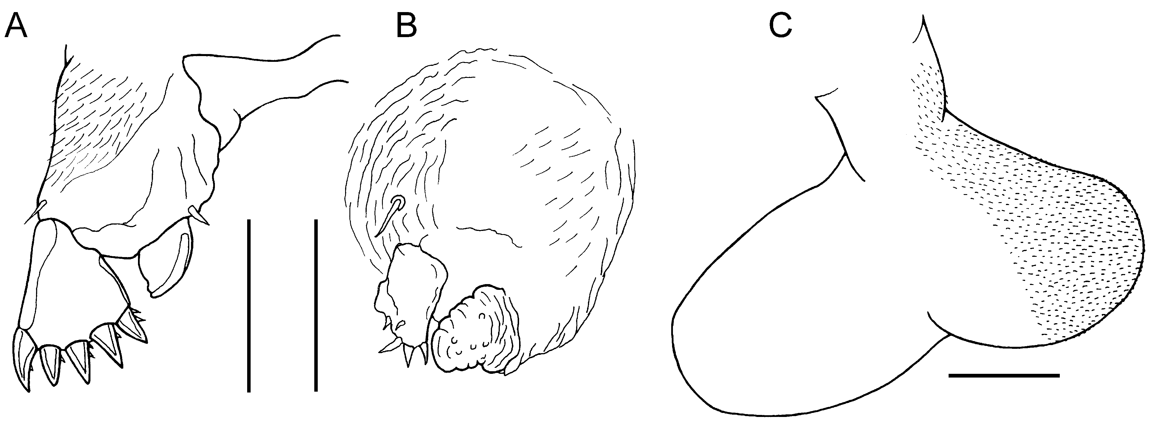
Leg 1 with coxa fused to somite and incompletely separated from basis; members of leg pair joined by intercoxal sclerite ( Fig. 48A View FIGURE 48 ). Each leg biramous with outer seta and short, inner spine on basis; outer surface of coxal part of protopod ornamented with long setules: exopod 1-segmented, armed with 5 robust terminal spines with sparsely denticulate margins; endopod 1-segmented, smoothly tapering to unarmed apex; surface unornamented. Leg 2 ( Fig. 48B View FIGURE 48 ) carried on inflated subspherical prominence, with wrinkled surface, derived from incorporated protopod and armed with outer seta: biramous, with both rami 1-segmented and both with wrinkled cuticular surface; exopod armed with 5 small distal spines; endopod conical with rounded apex, unarmed but with surface papillae. Leg 3 located on ventral surface of third pedigerous somite, forming bilobed fleshy lamella ( Fig. 48C View FIGURE 48 ), large outer lobe held vertically and directed posteriorly, reaching about to middle of dorsal trunk plate; smaller inner lobe about one third length of outer lobe; held vertically and extending posteromedially but legs separate along midline. Leg 4 bilobate ( Fig. 47C View FIGURE 47 ): distinct protopodal part bearing outer basal seta: inner and outer lobes elongate, flattened and tapering slightly from wider base; outer (exopodal) lobe slightly longer than inner (endopodal) lobe: at least distal third of lobes extending beyond posterior margin of dorsal trunk plate. Leg 5 represented by minute papilla carrying short apical seta (arrowed in Fig. 47B View FIGURE 47 ). Body lengths of ♀♀ 3.06 mm and 3.14 mm (based on 2 specimens).
Distribution: This species (as Lernanthropus sanguineus ) was originally established on the basis of two females collected from the gills of Lutjanus sanguineus caught off Sanya, China. The only subsequent record of this copepod, as Sagum sanguineus ( Song, 1976) , was from Vietnamese waters on Lutjanus johnii ( Kazachenko et al., 2014) . This is the first report of this copepod from Australian waters.
Remarks: The presence of loosely coiled egg sacs in the female confirms the validity of the transfer of this species to Sagum informally carried out by Kazachenko et al. (2014). The possession of leaf-like caudal rami, a short and strongly recurved claw on the antenna, and the form of the first leg of the female with its unarmed endopod, are all character states shared by a core group of species within the genus Sagum ( Table 4 View TABLE 4 ). In particular, as mentioned above, this species closely resembles S. lativentris and both are reported here from L. johnii . These two species share even fine details of limb setation but can be readily distinguished by the length of the fourth legs and the position of the distal dorsal caudal seta on the caudal ramus, as discussed above.
Kazachenko, V. N., Kovaleva, N. N., Nguyen, V. T. & Ngo, H. D. (2014) Taxonomic review of the parasitic copepod (Crustacea: Copepoda) fish in Vietnam. Scientific Journal of Dalrybvtuz, 31, 20 - 30.
Song, D. & Chen, G. (1976) Some parasitic copepods from marine fishes of China. Acta Zoologica Sinica, 22, 406 - 424.
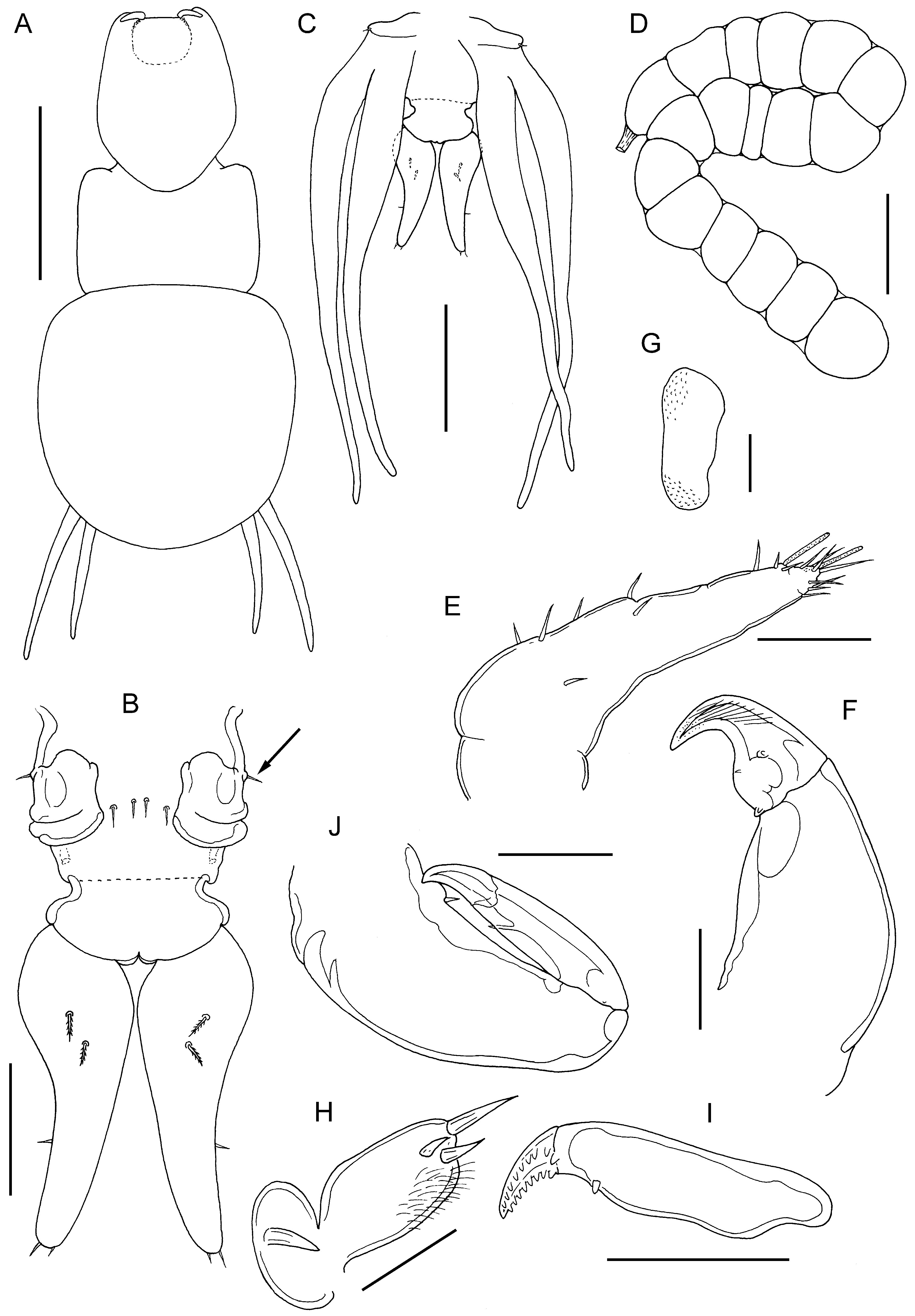
FIGURE 47. Sagum sanguineus (Song, in Song & Chen, 1976) n. comb., adult ♀. A, habitus, dorsal: B, urosome, dorsal view showing vestigial fifth legs (arrowed), paired genital openings and caudal rami; C, posterior end of trunk and urosome, ventral view showing extent of lobes of leg 4 relative to tips of caudal rami; D, egg sac; E, antennule; F, antenna; G, postantennal process; H, maxillule; I, basis of maxilla; J, maxilliped. Scale bars A, 1 mm, B,D, 200 μm, C, 0.5 mm, E,G,H, 50 μm, F,I,J, 100 μm.
No known copyright restrictions apply. See Agosti, D., Egloff, W., 2009. Taxonomic information exchange and copyright: the Plazi approach. BMC Research Notes 2009, 2:53 for further explanation.
|
Kingdom |
|
|
Phylum |
|
|
Class |
|
|
Order |
|
|
Family |
|
|
Genus |
1 (by plazi, 2020-02-17 07:18:21)
2 (by ExternalLinkService, 2020-02-17 07:29:36)
3 (by ExternalLinkService, 2020-02-17 07:49:39)
4 (by ExternalLinkService, 2020-02-17 17:48:26)
5 (by veselin, 2020-02-27 10:06:36)
6 (by ExternalLinkService, 2021-10-20 03:17:43)
7 (by ExternalLinkService, 2021-10-20 05:35:24)
8 (by plazi, 2023-10-31 00:24:02)