
Metapterodectes leptasthenurae, Mironov & González-Acuña, 2011
|
publication ID |
https://doi.org/10.11646/zootaxa.3057.1.1 |
|
DOI |
https://doi.org/10.5281/zenodo.4623144 |
|
persistent identifier |
https://treatment.plazi.org/id/546E87CE-032D-FFA5-FF11-E5E7FF389670 |
|
treatment provided by |
Plazi |
|
scientific name |
Metapterodectes leptasthenurae |
| status |
sp. nov. |
Metapterodectes leptasthenurae sp. n.
( Figs. 23 View FIGURE 23 F–K, 24, 25)
Type material. Male holotype ( ZISP 4671 View Materials ), 7 males and 8 female paratypes from the Plain-mantled Tit-spinetail Leptasthenura aegithaloides (Kittlitz) (Furnariidae) , CHILE: Bío Bío Region, Bío Bío Province, Santa Barbara , 37°39'53"S 72°1'13"W, 11 November 2006, coll. D.A. González-Acuña. GoogleMaps
Type depository. Holotype, 6 male and 7 female paratypes—ZISP, remaining paratypes—DGA.
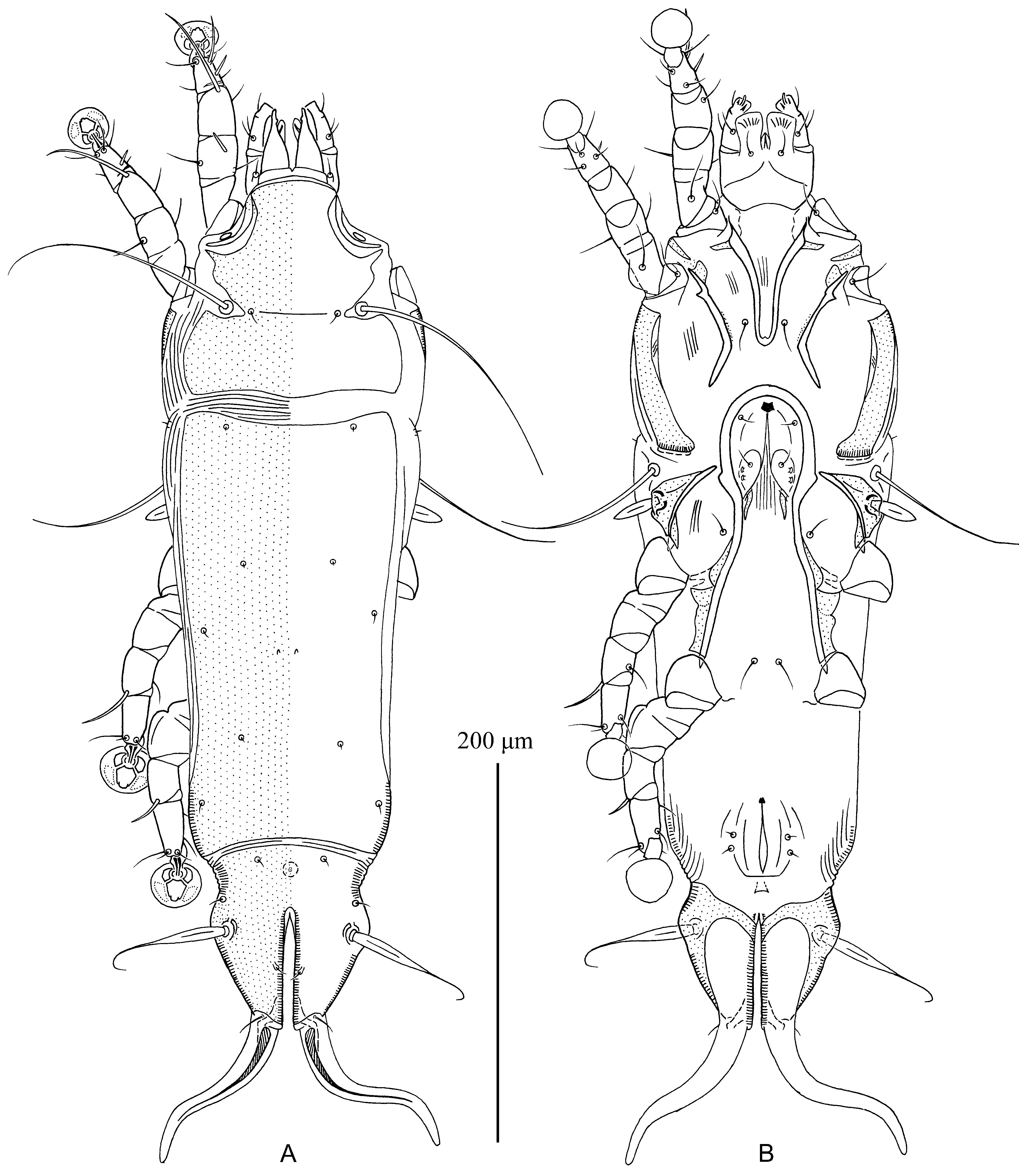
Description. MALE (holotype, range for 7 paratypes in parentheses). Idiosoma, length × width, 325 (315–340) × 130 (124–135), length of hysterosoma 205 (205–220). Prodorsal shield: 108 (105–110) × 102 (99–105), lateral margins entire, posterior margin slightly convex in median part, antero-lateral extensions acute, surface without pattern, scapular setae se separated by 60 (57–65) ( Fig. 24 View FIGURE 24 A). Setae ve absent. Humeral shields absent. Setae cp, c2 situated on soft tegument. Subhumeral setae c3 lanceolate, 22 (20–22) × 8.5 (7–8.5). Hysteronotal shield: greatest length 209 (205–223), width in anterior part 95 (90–100), anterior margin slightly concave, surface without pattern. Distance between prodorsal and hysteronotal shields 15 (15–20). Opisthosomal lobes approximately as long as wide at base; posterior ends of lobes roughly rounded. Terminal cleft shaped as a wide inverted U with divergent branches, 20 (20–22) in length. Supranal concavity present, roughly circular. Setae f2 situated anterior to bases of setae ps2. Setae h1 situated anterior to supranal concavity. Setae h3 spiculiform, 18 (18–21) long; setae ps2 75 (75– 80) long; setae ps1 minute, filiform, about 5 long, situated on margins of terminal cleft approximately at level of setae h2. Distance between bases of dorsal setae: c2:d2 86 (85–90), d2:e2 71 (70–80), e2:h3 47 (45–53), d1:d2 29 (24–30), e1:e2 29 (22–30), h1:ps2 27 (25–30), h2:h2 56 (53–60), h3:h3 38 (36–40), ps2:ps2 69 (65–70).
Epimerites I fused into a V, fused part without lateral extensions ( Fig. 24 View FIGURE 24 B). Coxal fields I, II without extensive sclerotized areas. Rudimentary sclerites rEpIIa absent. Coxal fields II, III open. Coxal fields IV without sclerotized areas. Epimerites IVa absent. Genital arch of moderate size, 22 (20–22) × 42 (42–50); basal sclerite of genital apparatus with widely ovate posterior margin, aedeagus sword-shaped, 60 (60–66) long, almost extending to anterior end of anal opening; genital papillae connected by bases. Genital and adanal shields absent. Anal suckers 13 (13– 14) in diameter, corolla smooth. Opisthoventral shields narrow, inner margins of these shields with angle-shaped extension at level of setae f2; setae ps3 situated on margins of these shields approximately at midlevel of anal suckers. Distance between ventral setae: 3b:3a 13 (7–14), 3a:4a 36 (35–39), 4a:g 36 (36–42), g:ps3 46 (45–52), ps3:ps3 75 (70–77), ps3:h3 25 (25–27).
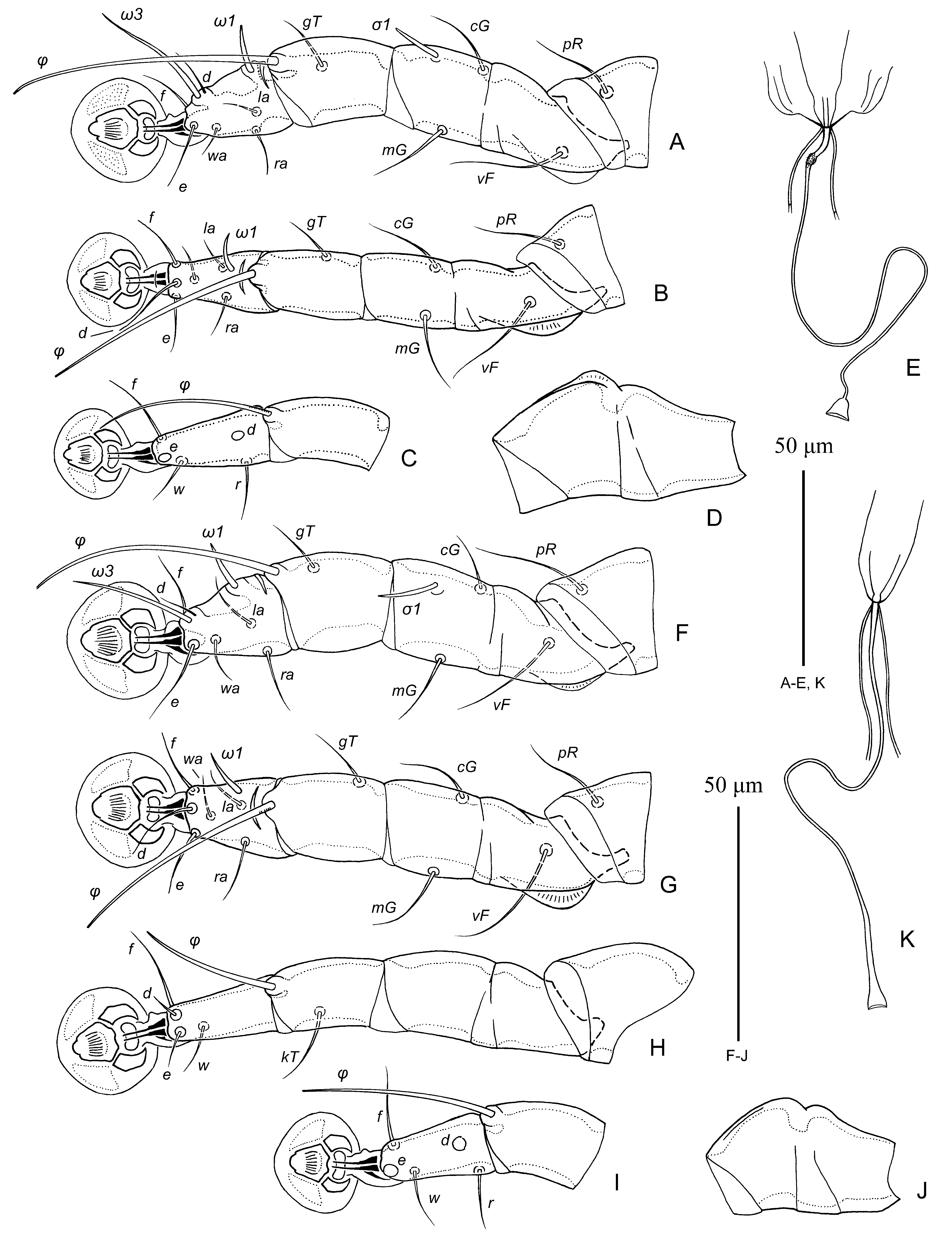
Femora I, II with ventral crests, other segments of legs I, II without processes. Solenidion σ 1 of genu I 13 (12– 13) long, situated at midlevel of segment; genual setae cG I, II and mG I, II filiform ( Figs. 23 View FIGURE 23 G, H). Seta d of tarsus II subequal to corresponding seta f, seta d of tarsus III shorter than corresponding seta f ( Fig. 23 View FIGURE 23 I). Tarsus IV 20 (20–26) long, without apical process; seta d situated in basal half of segment; solenidion φ of tibia IV extending to midlevel of ambulacral disc ( Fig. 23 View FIGURE 23 J).
FEMALE (6 paratypes). Idiosoma, length × width, 450–490 × 135–145, length of hysterosoma 315–348. Prodorsal shield: general form and surface as in male except for angular incisions around bases of setae se, 115–130 × 115–128, setae se separated by 70 ( Fig. 25 View FIGURE 25 A). Setae ve absent. Humeral shields absent. Setae cp, c2 situated on soft tegument. Setae c3 lanceolate, 20–22 × 7–8. Distance between prodorsal and hysteronotal shields 15–22. Anterior and lobar parts of hysteronotal shield separated dorsally by narrow transverse band of soft tegument ( Fig. 25 View FIGURE 25 A). Anterior hysteronotal shields: greatest length 235–255, width at anterior margin 115–122, anterior margin slightly concave, surface without pattern. Length of lobar region 95–106, greatest width 84–90, anterior margin convex. Terminal cleft narrow, parallel-sided, extending slightly beyond level of setae h2, 60–65 long, 5–8 wide at level of lobar apices. Setae h1 on lobar shield, situated close to anterior margin; setae h1 and f 2 in trapezoid arrangement. Setae h2 spindle-like, with filiform apex, length 55–60, greatest width 7–8. Setae ps1 on inner margins of opisthosomal lobes. Setae h3 10–12 long, about 1/8th the length of terminal appendages. Distance between dorsal setae: c2:d2 97–104, d2:e2 100–110, e2:h2 69–75, h2:h3 47–55, d1:d2 35–42, e1:e2 28–36, h1:h2 40–42, h1:h1 38–42, h2:h2 66–73.
Epimerites I fused into a narrow U, without lateral extensions ( Fig. 25 View FIGURE 25 B). Lateral parts of coxal fields I, II without large sclerotized areas. Epimerites IVa absent. Translobar apodemes of opisthosomal lobes present, narrow, not fused to each other anterior to terminal cleft. Epigynum with poorly expressed lateral extensions in posterior part, greatest width 35–57; apodemes of oviporus not fused with epimerites IIIa. Setae ps2 close to corresponding setae ps3, both pairs filiform, situated approximately at midlevel of anal opening and equidistant from midline; distance between pseudanal setae: ps2:ps2 30–33, ps3:ps3 31–30, ps2:ps3 6–8. Primary spermaduct without noticeable enlargement in proximal part and with narrow conical enlargement in distal part; secondary spermaducts 35–40 long ( Fig. 23 View FIGURE 23 K).
Femur I, II with ventral crest, other segments of these legs without processes. Solenidion σ 1 of genu I 10–12 long, situated at midlevel of segment. Genual setae cG I, II and mG I, II filiform. Seta d of tarsus II subequal to corresponding seta f, setae d of tarsi III, IV shorter than corresponding setae f. Genu IV dorsally inflated, with wide longitudinal dorsal crest ( Fig. 23 View FIGURE 23 J), genu III with low longitudinal dorsal crest.
Differential diagnosis. Males of M. leptasthenurae sp. n. are most similar to M. furnarius Mironov, 2008 from Furnarius rufus (Gmelin) ( Furnariidae ) from Brazil (Mirnov et al. 2008) by having narrowly lanceolate setae h3 and the hysteronotal shield without any pattern of lacunae. Metapterodectes leptasthenurae differ from that species and also from M. muticus by having the following features: in males, setae h1 are situated anterior to the level of supranal concavity and the genital papillae are connected by bases; in females, setae ps2, ps3 are filiform, the prodorsal shield has angle-shaped incisions around bases of setae se, and the lobar shield is entire. In males of the two previously known species, setae h1 are situated at the level of supranal concavity, and the bases of the genital papillae are separated; in females, setae ps2, ps3 are represented by small suckers, the prodorsal shield has no incisions surrounding setae se, and the lobar shield is almost completely split by median band of soft tegument into two parts remaining connected just anteriorly.
Etymology. The specific epithet derives from the generic name of the type host and is a noun in the genitive case.

No known copyright restrictions apply. See Agosti, D., Egloff, W., 2009. Taxonomic information exchange and copyright: the Plazi approach. BMC Research Notes 2009, 2:53 for further explanation.
|
Kingdom |
|
|
Phylum |
|
|
Class |
|
|
Order |
|
|
Family |
|
|
Genus |