
Barawertornis tedfordi Rich, 1979
|
publication ID |
https://doi.org/ 10.3853/j.0067-1975.62.2010.1539 |
|
persistent identifier |
https://treatment.plazi.org/id/541D8849-FFCF-B776-FC7C-BFE6A048E1AB |
|
treatment provided by |
Felipe |
|
scientific name |
Barawertornis tedfordi Rich, 1979 |
| status |
|
Barawertornis tedfordi Rich, 1979
Figs 1–4 View Figure 1 View Figure 2 View Figure 3 View Figure 4
Holotype. CPC7341 View Materials , a left femur missing the trochanter femoris and lateral part of the upper shaft.
Referred material. The following specimens were all collected from the Riversleigh World Heritage Area, Lawn Hill National Park, northwestern Queensland, Australia.
Localities are given in parentheses. Femora. QM F30352 (Wayne’s Wok Site), distal end of left femur; QM F39901 View Materials (Upper Site), proximal end of right femur; QM F40231 View Materials (D Site), partial right femur; QM F40232 View Materials (Sticky Beak Site), proximal end of left femur; QM F45056 View Materials (Hiatus Site), right femur missing distal third; QM F45420 View Materials (Hiatus Site), shaft of right femur. Tibiotarsi. QM F30377 (White Hunter Site), distal end of right tibiotarsus; QM F30801 (White Hunter Site), distal end of left tibiotarsus; QM F30802 (White Hunter Site), proximal end of right tibiotarsus; QM F30828 (White Hunter Site), proximal end of left tibiotarsus; QM F45416 View Materials (Hiatus Site), left tibiotarsus missing proximal end; QM F45417 View Materials (Hiatus Site), proximal end of left tibiotarsus; QM F52257 View Materials (Lee Sye’s Outlook Site), proximal half of right tibiotarsus; QM F52260 View Materials (Price Is Right Site), distal end of right tibiotarsus. Tarsometatarsi. QM F 24126 (Neville’s Garden Site), distal end of right tarsometatarsus (trochleae metatarsi III and IV partially preserved); QM F30413 (Dirk’s Towers Site), distal end of left tarsometatarsus (trochleae metatarsi II and III partially preserved); QM F45419 View Materials (Hiatus Site), heavily damaged right tarsometatarsus, broken into proximal and distal ends; QM F40223 View Materials (D Site), distal end of right tarsometatarsus; QM F40239 View Materials (Wayne’s Wok Site), distal end of left tarsometatarsus (trochlea metatarsi IV partially preserved).
The following specimens of vertebrae, described by Boles (2000), were used for character scoring in phylogenetic analyses: QM F24377 (Neville’s Garden Site), atlas-axis vertebral complex; QM F24378 (Neville’s Garden Site), cervical vertebra.
Measurements. See Tables 1–3.
Type locality. D Site (“ BMR Locality 103D” in Rich, 1979), Riversleigh World Heritage Area, Lawn Hill National Park, northwestern Queensland, Australia. Based on biostratigraphic and biocorrelative analyses, D Site forms part of Riversleigh’s “Faunal Zone A”, and as such is interpreted to be Late Oligocene in age ( Archer et al., 1997; Arena, 2004; Travouillon et al., 2006).
Description
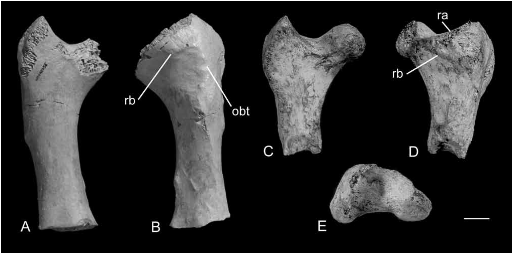
Femur. Description of the femur is based on new specimens, with reference to the holotype specimen CPC7341. The proximal and distal ends of the femur are not well preserved together in any the new specimens. As such, a comprehensive comparison of the proximal end with the distal end cannot be made. In cranial view the trochanter femoris is moderately high, narrow and falcate. It extends slightly laterad of the shaft. The proximal extent of the trochanter femoris and caput femoris are roughly equal. The caput femoris is relatively small in depth compared to Genyornis and Dromornis , with a slightly narrow, elongate collum femoris. The facies articularis antitrochanterica is highly concave both craniocaudally and mediolaterally in a broad U-shape. The fossa trochanteris is shallow. Viewed laterally, the trochanter femoris is largely oval-shaped. Its cranial margin is semicircular and extends slightly craniad of the shaft. The caudal margin of the trochanter femoris is almost straight and does not extend beyond the shaft surface.
In caudal view, a sharp ridge (A) is formed by the caudal border of the facies articularis antitrochanterica merging with the proximal crest of the trochanter femoris ( Fig. 1D View Figure 1 ). Distal to ridge (A) is a second ridge (B), which extends from the caudal face of the trochanter femoris to the caput femoris. Ridge (B) occurs at an angle of about 45 degrees from the lateral shaft margin. In specimen QM F45056 View Materials , ridge (B) trends at an angle of about 75 degrees from the lateral shaft margin. It joins to a broad, proximodistally trending ridge that may be interpreted as the impressiones obturatoriae ( Fig. 1B View Figure 1 ). The length of the impressiones obturatoriae, seen in specimens QM F45056 View Materials and QM F40231 View Materials , is less than half, but slightly more than one-third, of the total shaft length.
The shaft is shallow, craniocaudally compressed, and slender with respect to its length. In the holotype specimen CPC7341 View Materials the minimum width of the shaft is about 40 per cent of the distal end width. In CPC7341 View Materials and the new specimens the minimum shaft width is slightly proximad of its midpoint, with the exception of QM F40231 View Materials —it is slightly distad. The cross-sectional shape of the shaft is roughly trapezoidal to elliptical. Viewed caudally, the medial shaft margin is slightly to moderately curved. The lateral shaft margin is more or less straight. In specimen QM F45056 View Materials the impressiones iliotrochantericae are prominent and broad, terminating proximad of the midpoint of the shaft long axis. The linea intermuscularis cranialis is prominent over the proximal one-third of the femur. It trends proximolaterally and forms an angle of about 5–10 degrees with the long axis of the shaft. The linea intermuscularis caudalis is prominent over the distal half of the femur, appearing narrower and sharper in the new specimens than in the holotype. It trends proximomedially and forms an angle of about 20–25 degrees with the shaft’s long axis .
There is no linea intermusculus caudalis present in specimen QM F30352, but a small protuberance on the caudomedial margin. Lack of the linea intermusculus caudalis, unfused condyles, undeveloped popliteal fossa and porosity of the bone may indicate that this femur belonged to a young bird.
The condyli medialis et lateralis are not well preserved together in the new specimens. Although the condylus medialis is incomplete in specimen QM F40231 View Materials , it appears to have a sharper caudal margin than that in the holotype .
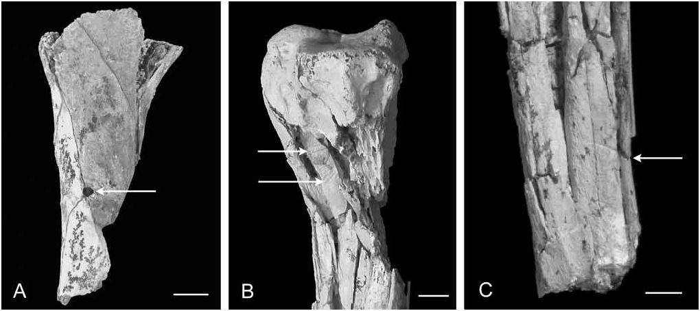
Specimen QMF45420 bears a circular puncture hole of about 5 mm diameter on the cranial surface, mediad of the midline of the shaft ( Fig. 4A View Figure 4 ). This unhealed puncture is associated with several greenstick fractures. Distal to the puncture are two smaller compression marks and a 2 mm long transverse gouge. These injuries indicate that they were formed at or near the time of death, possibly by a predator such as species of Baru ; large, extinct freshwater crocodiles found at Riversleigh (see Geological Setting). Many fossil specimens of the dromornithid Bullockornis planei from the Bullock Creek locality in the Northern Territory bear tooth punctures and point fractures thought to be evidence of crocodile predation (Murray & Vickers-Rich, 2004).
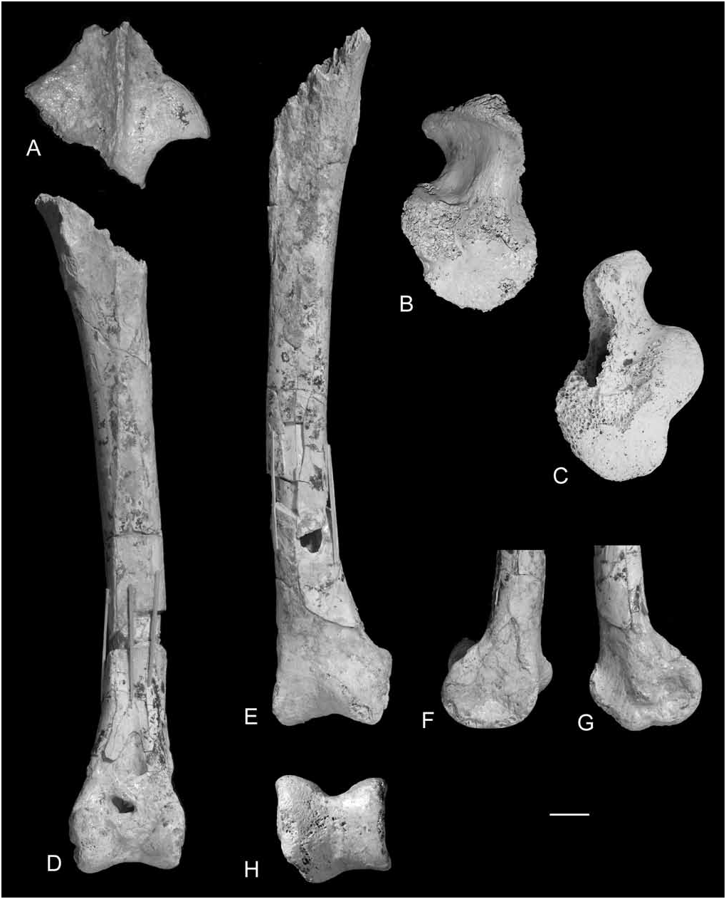
Tibiotarsus. In proximal view, the incisura tibialis lateralis is shallowly concave. The facies articularis lateralis is shallow and dome-like proximally. It extends slightly laterad and is semicircular in outline. The major axis of the facies articularis lateralis trends caudomedially. The facies articularis medialis is broadly convex, semicircular in outline and nearly flat proximally. From what is preserved, the eminentia intercondylaris appears to extend further proximally than the facies articularis lateralis. The fossa retropatellaris is circular and deep. The area interarticularis is moderately broad and concave proximolaterally. This entire surface slants distolaterally. The crista cnemialis lateralis is partially preserved. It is broad and triangular in cranial view, but is missing the proximal section of the crista. Only the base of the crista cnemialis cranialis is preserved. In proximal view the internal angle formed by the cristae cnemialis lateralis et cranialis is acute, approaching 90 degrees. The fossa flexoria is large and pneumatic. The impressio lig. collateralis medialis is distinct, large and elevated. It is deep and triangular in medial aspect.
The shaft is slightly curved mediolaterally. In caudal view, the medial margin of the shaft is more curved than the lateral margin. The caudal shaft surface is nearly planar but is slightly convex at the distal end. Over the proximal one-third, the cranial shaft surface is nearly planar and curves into the medial shaft surface. It smoothly grades into the lateral shaft surface that lies at about 90 degrees to the cranial shaft surface. The cranial and caudal shaft surfaces meet laterally along the crista fibularis at an angle of about 70 degrees in specimen QM F52257 View Materials . In specimen QM F45416 View Materials , these shaft surfaces meet laterally at about 110 degrees. In lateral view, the linea externa musculi peronei is broad and indistinct. Its proximal end is caudad to the crista fibularis. The linea externa musculi peronei trends distocranially and terminates proximad of the condylus lateralis. The linea extensoria is distinct; it trends distomedially away from the long axis of the shaft and intersects the medial shaft margin at about two-thirds down the shaft length. At the proximal and distal ends, the medial shaft surface is slightly convex. The remaining medial surface is nearly planar.
The distal end of the tibiotarsus is broad with respect to shaft width. The trochlea cartilaginis tibialis is moderately deep. The cristae trochleae are near vertical and do not converge proximally. Viewed cranially, the pons supratendineus forms an angle of about 75–85 degrees with the long axis of the shaft. The sulcus extensorius is deep. The distal opening of the canalis extensorius is slightly medial to the mediolateral midpoint of the distal end. The proximal margin of the area intercondylica is concave and forms a broad U-shape. The incisura intercondylaris is very shallow and broadly concave. It does not have an apex and is not V-shaped.
The condyli medialis et lateralis are moderately deep craniocaudally. Viewed cranially, the condyli are about equal in proximal extent. However, the condylus medialis is broader than the condylus lateralis. The proximal and distal margins of the condyli medialis et lateralis are slightly convex. In distal view the condyli appear to slightly diverge cranially, with the condylus medialis projecting further cranially than the condylus lateralis. It also projects further caudally than the condylus lateralis. From what is preserved, the condylus lateralis is roughly parallel with the shaft, whereas the condylus medialis is slightly inclined medially.
Viewed cranially, the condylus lateralis is relatively flat over its proximal half, but convex over its distal half. Its lateral margin is slightly convex and gradually grades into the lateral shaft margin. Viewed laterally, the condylus lateralis is broadly oval in shape. Its cranial margin is semicircular in outline, whereas its caudal margin is almost straight. The condylus lateralis projects a short distance caudad of the shaft but extends much further craniad. The distalmost extension of the condylus lateralis is at or craniad to its craniocaudal midpoint. The depressio epicondylaris lateralis is very shallow. The tuberculum retinaculi m. fibularis is small and slightly raised.
The condylus medialis is very convex over its cranial surface. Although its caudal and medial margins are damaged, the condylus medialis appears broadly oval in medial aspect. Its cranial margin is semicircular in outline. Viewed medially, there is a distinct distal protrusion from the distal margin of the condylus medialis ( Fig. 2G View Figure 2 ), which is apparently absent in published specimens of other dromornithid species. This distal protrusion in Barawertornis , however, may be an artefact of the specimen’s preservation. The depressio epicondylaris medialis is moderately deep. The epicondylus medialis is large, broad and circular. In caudal view the epicondylus medialis projects slightly further medially than the condylus medialis.
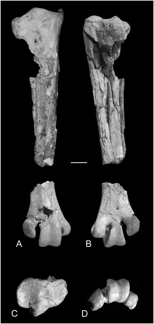
Tarsometatarsus. The proximal end of the tarsometatarsus appears to be relatively shallow compared to the shaft. Viewed proximally, the dorsal border of the proximal end is slightly convex, interrupted by the low eminentia intercotylaris. There is a shallow depression in the area intercotylaris. The hypotarsus is bulbous with a single poorly defined ridge. It is shaped like a scalene triangle, where the lateral side is more elongate than the medial side, and the dorsal side is the longest. Its proximal-most projection is located dorsally. There is a slight indentation in the lateral margin of the hypotarsus. In medial view, the hypotarsus moderately bulges plantarly beyond the shaft margin. The cotylae medialis et lateralis are narrow and roughly symmetrical. The cotyla medialis is moderately excavated, whereas the cotyla medialis is shallower and almost flat.
In dorsal view, the fossa infracotylaris dorsalis is narrow and moderately excavated. The foramina vascularia proximalia are large, deep and adjacent to each other. Distal to these foramina are two low, elongate tuberosities with a broad shallow groove between them; these are perhaps the insertions of the musculus tibialis cranialis. The impressiones retinaculi extensorii is shallow.
In all of the specimens under study, the proximal and middle shaft is not preserved or is heavily damaged. The medial shaft surface on the proximal half is discernable though; it is planar and meets the dorsal surface at about 90 degrees. Over the distal part of the shaft preserved, the dorsal shaft surface reveals a shallow sulcus extensorius extending from the damaged area. The sulcus extensorius appears to trend laterally, before terminating proximad of or level with the foramen vasculare distale. The medial and lateral margins of the sulcus flexorius are more prominent in the new specimens than in the paratype specimen CPC7346 View Materials , described by Rich (1979). It is narrow and terminates proximad of or level with the foramen vasculare distale. In CPC7346 View Materials , the cross-section of the shaft is teardrop-shaped, with the medial section of the shaft more circular in crosssection than the more triangular lateral section. In QM F52259 View Materials , the cross-sectional shape of the shaft is oval, with the shaft dorso-plantarly compressed .
Viewed plantarly, the flare of the distal end is moderate to broad with respect to the shaft width. There is no distinct fossa metatarsi I. The foramen vasculare distale is absent in some specimens of Dromornis stirtoni , and its state is uncertain in Bullockornis planei ( Rich, 1979) . In Barawertornis it is present in specimens QM F40239 View Materials and QM F45419 View Materials but, due to damage, is not preserved in other specimens. This foramen is small and does not open into the incisura intertrochlearis lateralis. The incisura intertrochlearis lateralis is narrow and extends further proximad than incisura intertrochlearis medialis. The surface within the incisura intertrochlearis medialis slopes gradually to a shelf that forms about midway between the dorsal and plantar shaft surfaces.
Trochlea metatarsi II is nearly planar dorsally. Its dorso-plantar plane is tilted plantarly towards the lateral border. In medial view, the dorsal, distal and plantar margins of this trochlea are smoothly rounded and approach a semicircular outline. Its medial face is moderately to deeply excavated.Viewed distally, the medial and lateral margins of trochlea metatarsi II diverge plantarly. The dorsal margin of trochlea metatarsi II is about three-quarters the width of the plantar margin.
Dorsal projection of trochlea metatarsi III with respect to the other two trochleae is moderate to great. The central axis of trochlea metatarsi III is slightly mediad to that of the shaft. Only trochlea metatarsi III bears a trochlear groove on the articular surface. This groove is shallow, and is of same depth dorsally and plantarly. In lateral view, the trochlea metatarsi III is circular in outline and has a large, moderately deep depression on its lateral face. In medial view, this trochlea is oval-shaped and has a small and moderately deep depression. The plantar margin of trochlea metatarsi III projects further beyond the shaft than its dorsal margin. Viewed distally, the medial and lateral margins of this trochlea diverge plantarly, and its plantar margin is about three-quarters of the width of the dorsal margin.
In dorsal view, the articular surface of trochlea metatarsi IV is nearly planar, with its plane tilted plantarly towards the lateral border. The dorsal, distal and planar margins of the trochlea form a semicircle in lateral view. Its most plantar projection is distad of the proximal end of the plantar margin. There is a deep depression in the lateral surface of trochlea metatarsi IV. Viewed distally, the dorsal and plantar margins of this trochlea converge laterally. The medial margin is slightly convex and the lateral margin is highly concave.
There is a series of diagonal gouges present on the posterior surface of a heavily damaged right tarsometatarsus (QMF45419, Fig. 4B View Figure 4 ). Distad to these gouges, another transverse gouge is present ( Fig. 4C View Figure 4 ). These markings may have been inflicted by a predator or a scavenger post-mortem, or animals trampling on the bone.
| QM |
Queensland Museum |
| BMR |
Bureau of Mineral Resources |
No known copyright restrictions apply. See Agosti, D., Egloff, W., 2009. Taxonomic information exchange and copyright: the Plazi approach. BMC Research Notes 2009, 2:53 for further explanation.
|
Kingdom |
|
|
Phylum |
|
|
Class |
|
|
Order |
|
|
Family |
|
|
Genus |