
Ptychogaster (Ptychogaster)
|
publication ID |
https://doi.org/ 10.5252/geodiversitas2021v43a20 |
|
publication LSID |
urn:lsid:zoobank.org:pub:04F82471-9F26-4935-97CE-1A286E08C958 |
|
DOI |
https://doi.org/10.5281/zenodo.5699189 |
|
persistent identifier |
https://treatment.plazi.org/id/523F87B8-FF91-DC57-FC75-FDCAE539F911 |
|
treatment provided by |
Felipe |
|
scientific name |
Ptychogaster (Ptychogaster) |
| status |
|
Ptychogaster (Ptychogaster) sp.
( Figs 2 View FIG ; 3 View FIG )
LOCALITIES. — MWQ1/2001, MWQ2/2003, MWQTC/2001, MWQ4/2018 and MCQ3/2005.
STUDIED MATERIAL. — Czech Republic. South Moravia Region, Mokrá-Quarry, carapace remains ( Fig. 2 View FIG A-R’): Pal. 1300, nuchal and left peripherals 1-3; Pal. 1301, nuchal; Pal. 1302, neural 4; Pal. 1303, neural 5; Pal. 1304, neural 7; Pal. 1305, suprapygal 1; Pal. 1306, suprapygal 2; Pal. 1307, left costal 1; Pal. 1308, right costal 6; Pal. 1309, right costal 6; Pal. 1310, left costal 8; Pal. 1311, right costal 8; Pal. 1312, right peripheral 1; Pal. 1313, left peripheral 7; Pal. 1314, left peripheral 8. Plastral remains ( Fig. 3 View FIG A-R): Pal. 1315, left hypoplastron; Pal. 1316, right hypoplastron; Pal. 1317, right hypoplastron; Pal. 1318, left xiphiplastron.
DESCRIPTION
The ptychogasterid material (Pal. 1300-1318) from Mokrá- Quarry consists of numerous disarticulated plates, which present preserved parts of the almost the entire shell. However, some plates are missing from the carapace (i.e., pygal, costals 2-4 and 7, neurals 1-3 and both medial and posterior peripherals). The plastron is represented only by the hypoplastron and the xiphiplastron. The following description is based on all available material, however, not all preserved plates are depicted in Figures 2 View FIG and 3 View FIG . According to the dimensions of the nuchal plate (Pal. 1300-1301), costals (Pal. 1307-1311) and peripherals (Pal. 1312-1314), the shell would have been relatively large (> 20 cm: Figs 2 View FIG ; 3 View FIG ). Ornamentation of both carapace and plastron external surfaces is absent. As it is typical for Ptychogaster plates, dermal grooves are well developed. Unlike Testudo , neither growth lines nor sutures (i.e., completely fused to each other: see e.g., Fig. 2 View FIG A-D) are preserved, so it is impossible to evaluate them.
The nuchal plate is hexagonal in outline, wider than long (Pal. 1301: Fig. 2 View FIG E-H). It contacts the first pair of peripherals, the first pair of costals and the neural 1. The anterior border possesses a shallow nuchal notch, affecting the nuchal border and the first peripherals (Pal. 1300: Fig. 2A, B View FIG ). The anteroposterolateral sides of the nuchal are rather equal in length, whereas the posterior border is narrow and slightly convex anteriorly ( Fig. 2E, F View FIG ). In lateral view, the nuchal is vaguely curved ( Fig. 2I, J View FIG ). Two transversal thickenings are
developed on the internal surface of this bone ( Fig. 2C, D, 2 View FIG G-L). The cervical scute is present anteriorly, both dorsally and viscerally. It is a relatively large and trapezoidal element that is longer than wide ( Fig. 2E, F View FIG ). The lateral edges of the cervical are slightly curved medially both in dorsal and visceral sides ( Fig. 2A, B View FIG , E-H). The overlap of this scute is less developed on the ventral surface ( Fig. 2G, H View FIG ).
According to the preserved neural plates, an alternating between octagonal and hexagonal plates forms the neural series: neural 4 octagonal (Pal. 1302: Fig. 2M, N View FIG ) and neural 5 and 7 hexagonal with short sides behind (Pal. 1303-1304: Fig. 2 View FIG O-S). A weak medial keel develops on the neural 7 (Pal. 1304: Fig. 2R, S View FIG ) and the suprapygals 1-2 (Pal.1305-1306: Fig. 2T, U View FIG ). In visceral view, remains of the thoracic vertebrae attachments are present in all neural plates ( Fig. 2N, Q, S View FIG ).
The suprapygal plate 1 is trapezoidal with a wider posterior part (Pal. 1305: Fig. 2T View FIG ). The anterior border is concave, whereas the posterior one is slightly convex. It contacts the neural 8 anteriorly, costals 8 laterally and suprapygal 2 posteriorly. Suprapygal 2 is hexagonal, wider than long, and much wider than suprapygal 1 ( Fig. 2T, U View FIG ). It contacts the suprapygal 1 and the posteromedial sides of costals 8 anteriorly, peripherals 11 laterally and pygal posteriorly. The anterolateral sides of the suprapygal 2 are slightly longer anteroposteriorly, compared to the posterolateral ones. The posterior side is vaguely convex anteriorly ( Fig. 2T, U View FIG ).
The vertebral scute series is partially preserved, which is quadrangular and slightly narrower than the costal series. Vertebral 1 contacts the cervical and marginals 1-2 anteriorly ( Fig. 2B, F View FIG ). It seems to be lyre-shaped and covers the lateral corners of the nuchal and costals 1 (Pal. 1300-1301: Fig. 2A, B, E, F View FIG ). According to preserved portion of the vertebral 3, it expanded at least on costals 5 and neural 5 ( Fig. 2X, Y View FIG ). The sulcus between the vertebrals 3-4 is wavy in its medial part, and more specifically in the part that is crossing the neural 5 ( Fig. 2O, P View FIG ). Vertebral 4 likely contacts the vertebral 3 anteriorly, pleurals 3-4 laterally and vertebral 5 posteriorly ( Fig. 2P View FIG , A’, B’, D’, E’). Vertebral 5 is the widest vertebral scute, contacting with the vertebral 4 anteriorly, pleural 4 anterolaterally and marginals 11-12 posteriorly. It expands on costals 8, neural 8, peripheral 12 and pygal, and therefore covers the entire surface of the suprapygals 1-2.
Although not fully preserved costals, the costal plate 1 is much longer than the rest of costal (Pal. 1307: Fig. 2V, W View FIG ). It is trapezoidal and always contacts the peripheral plates 1-3 anterolaterally, nuchal anteromedially and neurals 1-2 medially. The anterior border is sinuous to articulate with the corresponding peripherals. Costal 6 is similar in regards of its shape, being much wider than long (Pal. 1308-9: Fig. 2 View FIG X-B’). The medial side of Pal. 1308 shows a short anteromedial and long posteromedial sides ( Fig. 2 View FIG X-Z). However, the medial side of Pal. 1308 is most likely rounded ( Fig. 2 View FIG A’-C’). Costal 8 is narrow, slightly wider than long, contacting the costal 7 anteriorly, neural 8 and suprapygal 1 medially, and suprapygal 2 and peripherals 11-12 posteriorly (Pal. 1310-11: Fig. 2 View FIG D’-F’). The pleural scutes are not preserved with the exception of the first one. The marginopleural sulcus is entirely situated on peripheral plates, at least in both anterior and posterior part of the carapace ( Fig. 2A, B, E, F, V View FIG , A’, B’, D’, E’).
The peripheral plates 1-3 are longer than wide and slightly trapezoidal ( Fig. 2 View FIG A-D, G’-J’). They are completely fused together (i.e., sutures are not visible) and crossed not only by the intermarginal sulcus, but also by the pleuro-marginal sulcus, unlike in Testudo . Peripherals 1-2 are prominent in anterior direction in Pal. 1300. Peripheral 7, which is partially preserved, is rectangular (Pal. 1313: Fig. 2 View FIG K’-N’). In internal view, it displays a rough elongated area for the cartilaginous union of the inguinal process ( Fig. 2 View FIG M’, N’). Pal. 1313 is the last peripheral involved in the shell bridge and also displays a weak lateral ridge on its external side. Peripheral 8 is rectangular and hosts both the pleuromarginal sulcus and intermarginal sulci (i.e., between marginals 8-9), which are situated far from the costoperipheral suture (Pal. 1314: Fig. 2 View FIG O’-R’). The marginal scute 1 is rectangular, slightly wider than long, whereas marginal 2 is trapezoidal. Marginal 3 is approximately as wide as long. Marginal 8, the only complete scute from the bridge area, is rectangular and higher than wide. The ventral overlap of all preserved marginals is well developed.
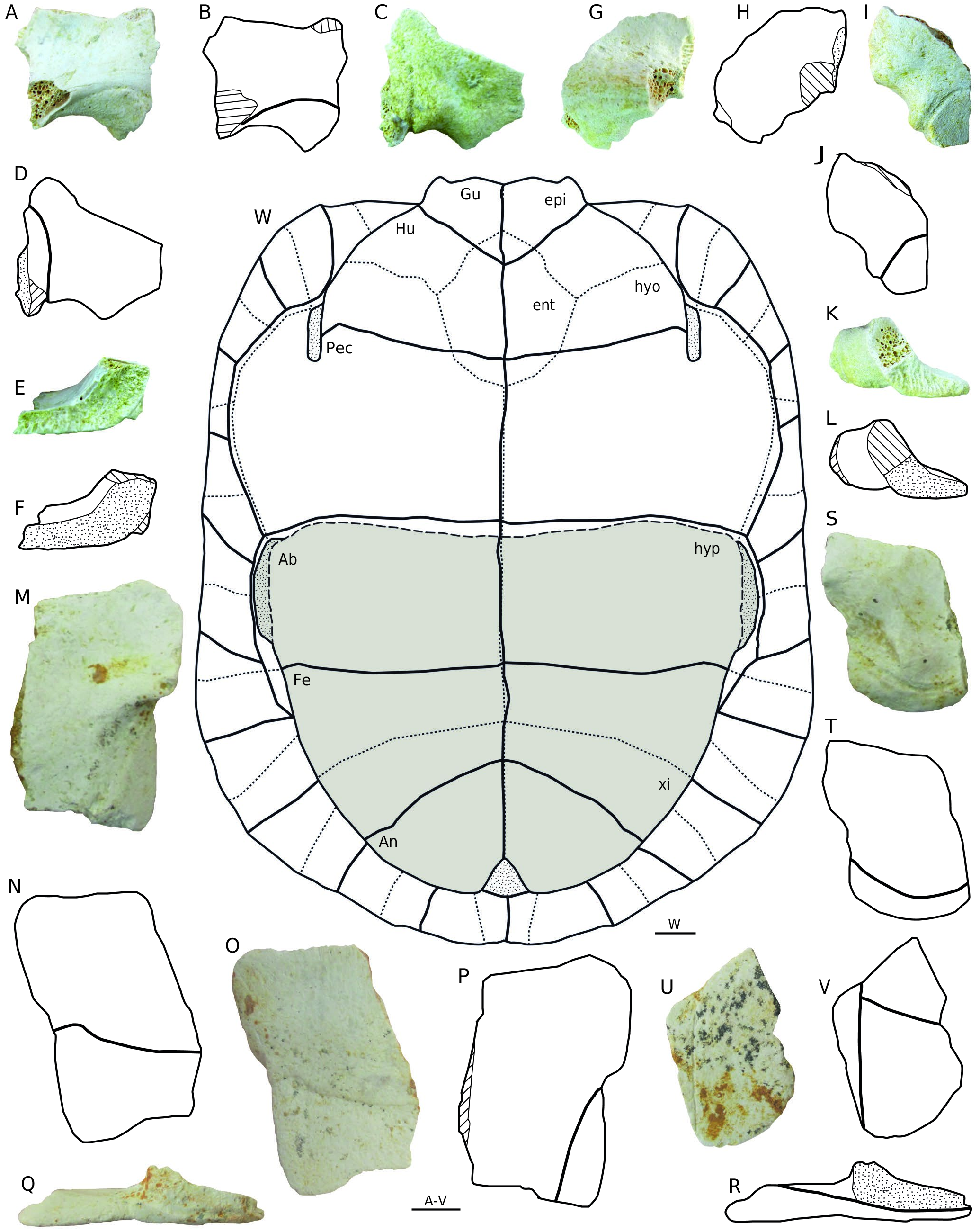
The preserved portion of the hyo-hypoplastral hinge is straight and approximately transversal (Pal. 1377: Fig. 3 View FIG M-P), which is located at the mid of the peripheral 6. The hypoplastron contact anteriorly with the hypoplastron through a hinge and laterally with the peripherals 6-7 through a completely ligamentous union in the inguinal process (Pal. 1315- 17: Fig. 3 View FIG A-R). The visceral overlap of both abdominal and femoral scutes on hypoplastra is well developed ( Fig. 3M, N View FIG ). The xiphiplastron is trapezoidal and its posterior tip is rather rounded. The abdominofemoral sulcus, developed on the ventral surface of each hypoplastron, is concave ( Fig. 2N, O View FIG ). Moreover, the latter does not reach the inguinal process laterally, but is located slightly below it. The femoroanal sulcus is oblique, whereas the anal scute is triangular with rounded lateral borders ( Fig. 2 View FIG S-V). The dorsal overlap of the anal scute is moderately developed ( Fig. 2S, T View FIG ).
REMARKS
Ptychogasteridae is a geoemydid family which includes turtles of medium body size. This clade originated in Europe during the Eocene and its members are characterized mainly by a plastral kinesis ( Lapparent de Broin 2001; Claude 2006). Thus, the anterior part of the plastron is firmly connected to the carapace, whereas the posterior one is movable thanks to a hinge situated between hyo- hypoplastra and peripherals 6. Some studies claimed that ptychogasterids constitute a monophyletic clade diagnosable by several synapomorphies ( Hervet 2004a, b, 2006). However, the inclusion within ptychogasterids of non-kinetic extinct geoemydids genera, such as Merovemys , Clemmydopsis and Hummelemys , has been questioned (see Claude & Tong 2004). A more comprehensive phylogenetic analysis would be required in the future to know what genera belong to subfamily Ptychogasterinae , as well as to further clarify which are the closest extinct relatives of the latter.
As for the genus Ptychogaster View in CoL , it was originally erected by Pomel (1847) from the early Miocene (MN2) of Saint-Gerandle-Puy, France. Two subgenera are currently distinguished within Ptychogaster View in CoL : P. ( Ptychogaster View in CoL ); and P. ( Temnoclemmys ). Ptychogaster (Ptychogaster) emydoides Pomel, 1847 (i.e., type species of subgenus Ptychogaster View in CoL ) has a complex taxonomical and nomenclatural history, with many junior subjective synonyms ( Schäfer 2013; Luján et al. 2014). Although one revision of the genus Ptychogaster View in CoL has recently been performed, the results of this PhD thesis are still unpublished ( Schäfer 2013).

No known copyright restrictions apply. See Agosti, D., Egloff, W., 2009. Taxonomic information exchange and copyright: the Plazi approach. BMC Research Notes 2009, 2:53 for further explanation.
|
Kingdom |
|
|
Phylum |
|
|
Class |
|
|
Order |
|
|
Family |
|
|
Genus |
Ptychogaster (Ptychogaster)
| Luján, Àngel H., Čerňanský, Andrej, Bonilla-Salomón, Isaac, Březina, Jakub & Ivanov, Martin 2021 |
Merovemys
| Hervet 2006 |
Hummelemys
| Hervet 2004 |
Temnoclemmys
| Bergounioux 1957 |
Clemmydopsis
| Boda 1927 |
Ptychogasterinae
| De Stefano 1903 |
Ptychogaster
| Pomel 1847 |
Ptychogaster
| Pomel 1847 |
Ptychogaster
| Pomel 1847 |
Ptychogaster (Ptychogaster) emydoides
| Pomel 1847 |
Ptychogaster
| Pomel 1847 |
Ptychogaster
| Pomel 1847 |