
Guaranidrilus hoeferi, Schmelz, Rüdiger M., Collado, Rut & Römbke, Jörg, 2011
|
publication ID |
https://doi.org/ 10.5281/zenodo.203260 |
|
DOI |
https://doi.org/10.5281/zenodo.5611825 |
|
persistent identifier |
https://treatment.plazi.org/id/520EAA7D-D646-4508-FF40-8759FE84FC26 |
|
treatment provided by |
Plazi |
|
scientific name |
Guaranidrilus hoeferi |
| status |
sp. nov. |
Guaranidrilus hoeferi View in CoL sp. nov.
( Figs 4 View FIGURE 4 , 8 View FIGURE 8 D)
Holotype. UFPR OL-13, adult/subadult specimen, stained whole mount, Guarequeçaba, Itaqui, 25°14'45,3''S, 48°30'19,4''W, 27 m a.s.l., medium old secondary forest on Cambisol [site 38], Sep 2007, leg. P. Heine, R.M. Schmelz.
Paratypes. MZUSP, 6 specimens, Antonina, Cachoeira , stained whole mounts:
MZUSP 1230, 2 specimens, 25°14'38'', 48°40'10'', 140 m a.s.l., old-growth forest on Cambisol [site 13], May 2003, leg. J. Römbke, R. M. Schmelz.
MZUSP 1231, 4 specimens, 25°19'00"S, 48°40'14"W, and 25°19'41"S, 48°40'36"W, 30 and 120 m a.s.l., abandoned pasture and medium old secondary forest on Cambisol [sites 6, 12], Oct 2004. leg. B. Förster, R.M. Schmelz.
UFPR, 44 specimens, Guarequeçaba, Itaqui, stained whole mounts (35) and ethanol-preserved (9):
UFPR OL-14, 5 specimens, same data as holotype.
UFPR OL-15, 13 specimens, 25°15'43.8''S, 48°29'14.8''W, ca. 20 m a.s.l., young forest on Cambisol [site 36], Sep 2007, leg. P.Heine, R. M. Schmelz.
UFPR OL-16, 17 specimens, 25°15'32.7''S, 48°30'31.9''W and 25°16'23.7''S, 48°29'13.7''W, 20 and 31 m a.s.l., respectively, old-growth forest on Cambisol [sites 41, 42], Sep 2007, leg. P.Heine, R. M. Schmelz.
UFPR OL-17, 9 specimens, ethanol-preserved.
Additional material. Fifteen specimens from sites in Itaqui, examined in vivo, not preserved.
Etymology. Named in honour of Hubert Höfer (State Museum of Natural History, Karlsruhe), initiator and German coordinator of the SOLOBIOMA project, in grateful recognition of the tremendous amout of work to launch and to maintain this project, which allowed the discovery and description of so many enchytraeid species.
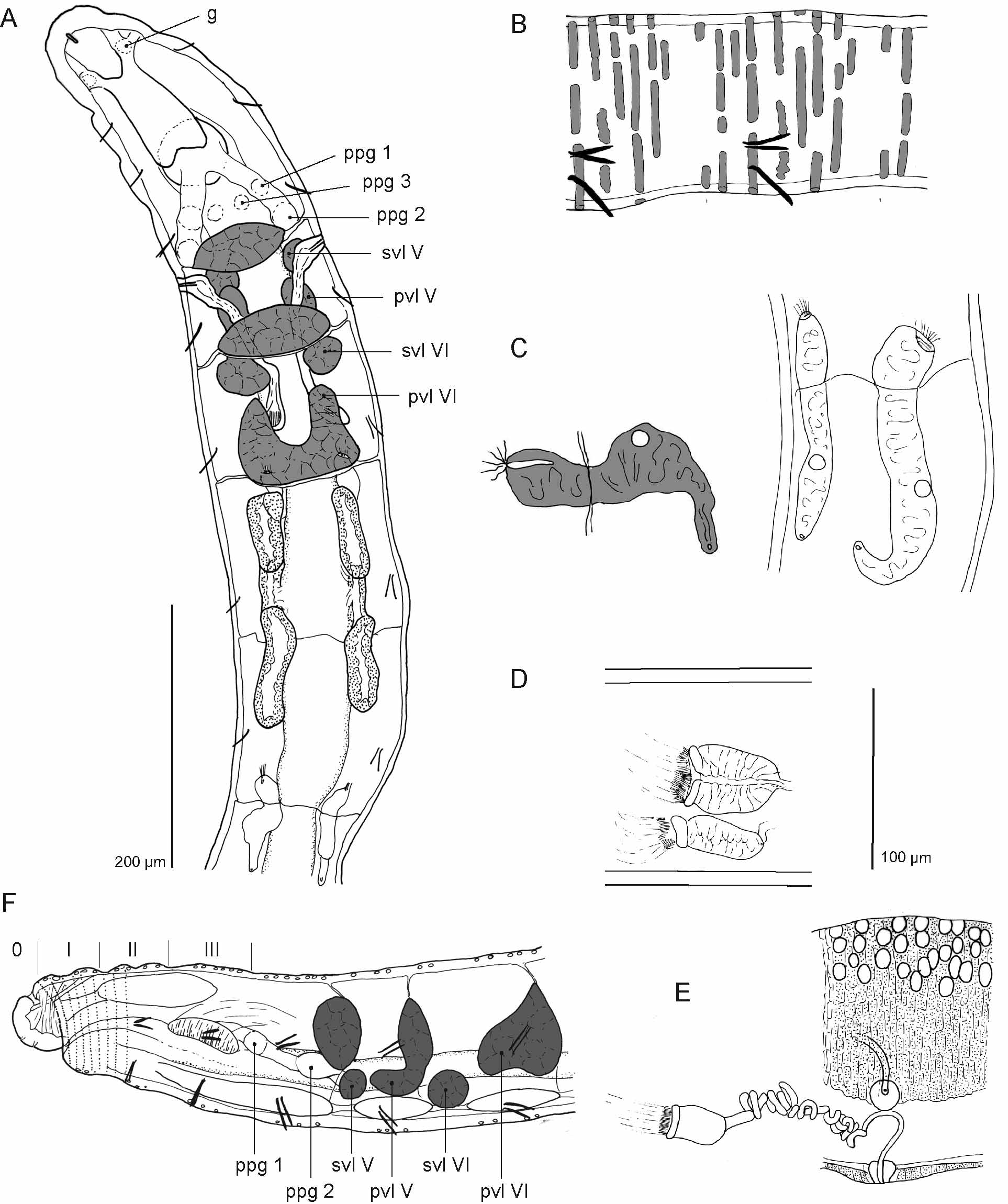
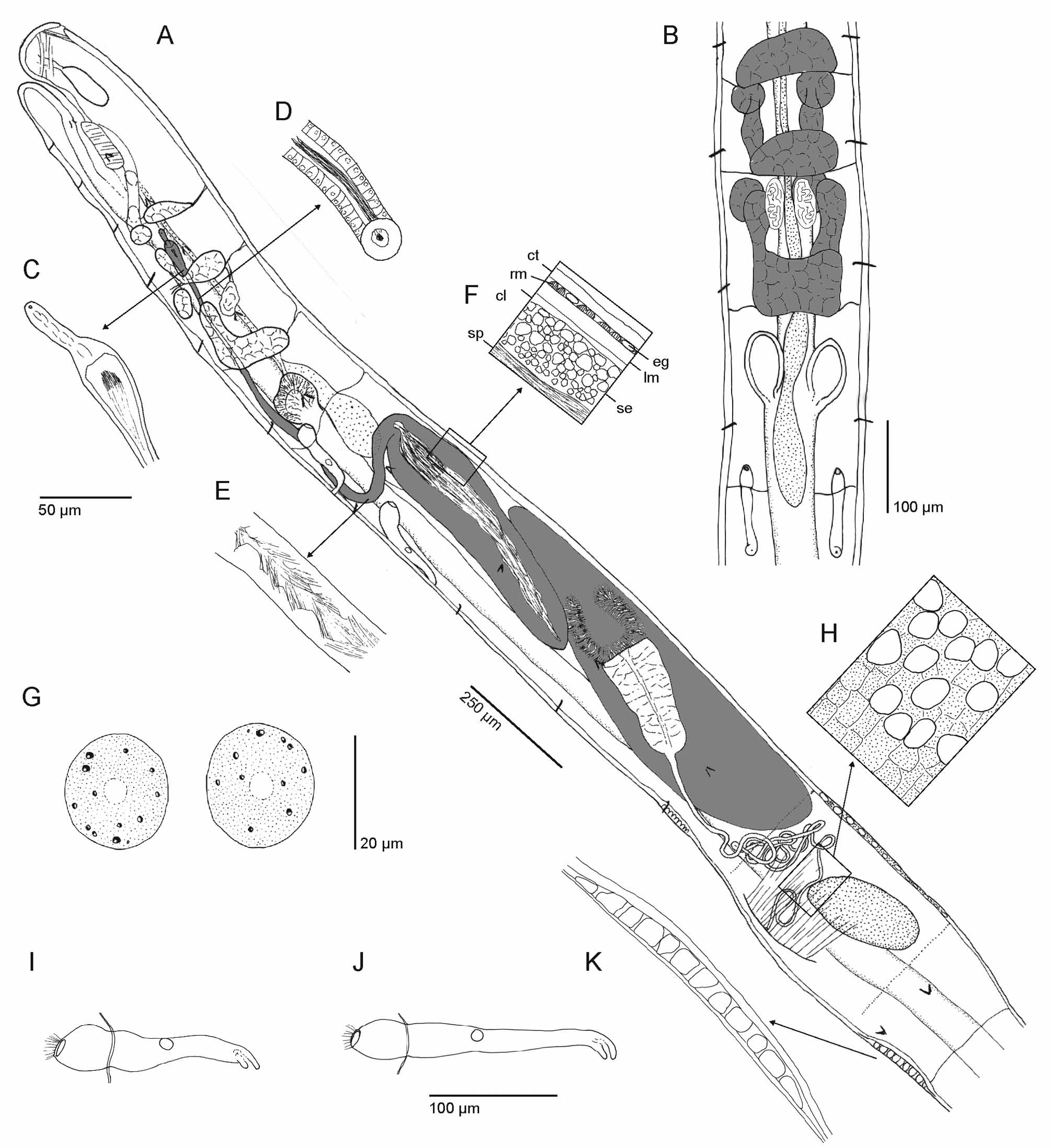
Description. Slow body movements. Body dimensions. Mature specimens about 12–15 mm long in vivo, live diameter ca. 0.2 mm, submatures often distinctly smaller (<8 mm). Segment number 39–48. Chaetae distally straight, with weak proximal hook, not thickened proximally. Preclitellar chaetae small and stout, ca. 22–28 µm long and and 2.5–3 µm thick. Postclitellar chaetae behind clitellum small and inconspicuous, ca. 20 µm long and 2 µm wide; chaetae increasing gradually in size towards posterior end. In caudal segments chaetae stout again, 40–50 µm long and 6 µm thick. Epidermal gland cells inconspicuous in vivo. In whole mounts all epidermis apparently glandular, cells transversely elongate, hyaline, in about 11 rows per segment, almost continuous except mid-ventrally. Clitellum ( Fig. 4 View FIGURE 4 H) saddle-shaped, not developed ventrally; on dorsal half granulocytes and hyalocytes, granulocytes isolated, hyalocytes large, seen as large free interspaces in living specimens; ventro-laterally only granulocytes. On dorsal half cells in indefinite rows or reticulate; latero-ventrally cells in dense rows. In adults accessory glands present, a glandular thickening of the epidermis mid-ventrally in XIII or in XI and XIII, immediately in front of and behind clitellum ( Fig. 4 View FIGURE 4 A,K); glandular field between the ventral chaetae and the respective posterior septum. Head pore on prostomium mid-dorsally.
Prostomium without inner papillae. Pygidium with strongly developed rectal musculature, filling body cavity. Body wall thin (ca. 10–20 µm), ring muscles in one regular layer. Cuticle mostly thick compared to rest of body wall (up to 9 µm), thicker than epidermis + ring muscle layer. Anterior septa not thickened. Brain posteriorly concave; a few perikarya on prostomial nerves, no ganglionic swelling. Pharyngeal glands ( Fig. 4 View FIGURE 4 A,B) in IV–VI, unpaired and fused dorsally; dorsal lobes of V and VI with ventral anterior projections (primary ventral lobes), large in VI, small in V; secondary ventral lobes present in V and VI, spherical. Oesophageal appendages ( Fig. 4 View FIGURE 4 A,B) paired dorso-laterally in anterior half of VI, elongate, with irregular outline. Intestinal diverticula ( Fig. 4 View FIGURE 4 A,B) in VII, as pouches with wide lumen, directed anteriad, i.e. connection with intestine posteriorly; walls with radial striation, possibly folds of inner wall; chloragocytes present on outer surface. Dorsal blood vessel ( Fig. 4 View FIGURE 4 A,B) from 1/ 2 VIII, pulsating. Preclitellar nephridia ( Fig. 4 View FIGURE 4 A,B,I) two pairs, at 7/8 and 8/9. Anteseptale large, with coils of canal, outline spherical in side view, postseptale elongate and narrower than anteseptale, with dorsal vesicle, gradually merging into short and stout efferent duct, no terminal vesicle. Postclitellar nephridia ( Fig. 4 View FIGURE 4 J) of similar shape but slightly longer, anteseptale longer than wide. Nephridia missing at several positions. Coelomocytes ( Fig. 4 View FIGURE 4 G) numerous, darkened in aggregations. Cells flattened, outline broad-oval, length ca. 20–40 µm. A few dark and refractile granules of irregular outline scattered in a finely and regularly vesicular, pale-brown matrix.
Seminal vesicle ( Fig. 4 View FIGURE 4 A) large, extending over up to 5 segments, caused by forward bulge of septum 10/11 and backward bulge of septum 11/12. Spermatozoa very numerous, heads 33 µm long (measured in vivo). Sperm funnel ( Fig. 4 View FIGURE 4 A) longer than body diameter, ca. 2x as long as wide (or longer), tapering distally; collar conspicuous, with free extensions beyond funnel body, often U-shaped, conspicuous by attached sperm. Each extension may have stump-like sub-lobes. Vas deferens ( Fig. 4 View FIGURE 4 A) very long and irregularly coiled, of equal diameter throughout (8 µm in fixed material. Male copulatory organ ( Fig. 4 View FIGURE 4 A) with male pores on body surface, each withdrawn in a longitudinal eversible furrow; both furrows creating a mid-ventral "penial plate" when withdrawn. In all fixed specimens furrows everted into elongate-conical projections; male pores widely separate, distance wider than distance between ventral chaetal bundles of a segment. No male glands around male pore, but epidermis thickened here and also in a field between the pores, the "penial plate". Copulatory muscles ( Fig. 1 View FIGURE 1 A) conspicuous in preserved specimens as a parasagittal series of fine, dorso-ventral strands, arranged in paralles (side view): ventral insertion of muscles at inner wall of papilla, dorsal insertion at inner body wall surface latero-dorsally, slightly above longitudinal row of lateral chaetae. Spermatheca ( Fig. 4 View FIGURE 4 A,C,D,E,F) very large, extending into IX. Ectal duct short, distal part of ampulla conspicuous, thin-walled, with sperm and surrounding free lumen; ental reservoir extending over 3 segments, into IX, here elongately club-shaped, thick-walled; walls with large, non-staining inclusions of irregular outline ( Fig.4 View FIGURE 4 F, "se"); lumen completely filled with spermatozoa, forming a long, compact and iridescent strand. One egg at a time seen, but no specimen with fully developed egg found.
Habitat. G. hoeferi was found at all stages of forest regeneration. It was absent in pastures and only one specimen was found at the agroforestry sites.
Remarks. Among all species present at the study sites, G. hoeferi was instantly distinguished by (1) the peculiar coelomocytes. Further traits that confirmed the identification were: (2) only 2 pairs of anterior nephridia, at 8/9 and 9/10, (3) dorsal blood vessel from VIII, (4) intestinal diverticula confined to VII, mostly directed forwards. With this character combination, specimens of all age stages, including very small juvenile ones, could be identified. The striking gigantism of sperm-related organs is shared by several other Guaranidrilus species that possess oesophageal appendages ( G. atlanticus Christoffersen, 1977 , G. cernosvitovi Healy, 1979 , G. joanae Christoffersen, 1977 , G. m b o i Righi, 1975), and hence is little help in the identification of the species.
G. hoeferi View in CoL is most similar to G. joanae Christoffersen, 1977 View in CoL , another species of the Brazilian Atlantic rain forest, found ca. 250 km up north-east near the city of São Paulo. Both may be indeed be sister species; at least they belong to a common sub-group or clade within the genus. The excellent original description of G. joanae View in CoL allows a detailed comparison. Both species agree in body size, behaviour, all details of pharyngeal glands, oesophageal appendages, intestinal diverticula, and in size and organization of spermathecae and the entire male reproductive system. They differ in four traits: (1) In G. joanae View in CoL , chaetae are enlarged not only in caudal segments but also in the most anterior segments, and here (2) the ental bend is thickened in a claviform manner. (3) Preclitellar nephridia are present not only at 7/8 – 8/9 but also at 6/7, resulting in three preclitellar pairs instead of the two in G. hoeferi View in CoL . (4) Coelomocytes of G. joanae View in CoL appear to lack the conspicuous double texture present in G. hoeferi View in CoL (few coarse granules embedded in a densely and finely vesicular pale-brown matrix), they are described as being "rather inconspicuously granulated" ( Christoffersen 1977: 200). Furthermore segment number is slightly higher (49–55) and clitellar gland cells are irregularly arranged in G. joanae View in CoL , but our specimens were not fully mature. Our specimens were relaxed and Christoffersen's specimens apparently contracted at the time of fixation, which may account for differences in body length (10–11 mm in G. joanae View in CoL ) and shape of preclitellar nephridia (efferent duct bent forwards in G. joanae View in CoL ). Epidermal gland cells are said to be slightly developed in G. joanae ( Christoffersen 1977: 198) View in CoL , but the animals "... were always found enveloped in great amounts of mucus..." (ibid.), which suggests the same situation as in G. h o e f e r i: numerous glands cells, inconspicuous in vivo. In fact even in fixed material the glands are invisible unless interference contrast optics are used. The ectal pore of the spermathecae of G. joanae View in CoL appears to be located ventrally in a figure ( Christoffersen 1977, Fig. 24), but the description gives the usual and probably correct lateral location.
| MZUSP |
Museu de Zoologia da Universidade de Sao Paulo |
No known copyright restrictions apply. See Agosti, D., Egloff, W., 2009. Taxonomic information exchange and copyright: the Plazi approach. BMC Research Notes 2009, 2:53 for further explanation.
|
Kingdom |
|
|
Phylum |
|
|
Class |
|
|
Order |
|
|
Family |
|
|
Genus |
Guaranidrilus hoeferi
| Schmelz, Rüdiger M., Collado, Rut & Römbke, Jörg 2011 |
G. joanae
| Christoffersen 1977 |
G. joanae (
| Christoffersen 1977: 198 |