
Cetacea, Brisson, 1762
|
publication ID |
https://doi.org/ 10.5252/g2009n4a943 |
|
DOI |
https://doi.org/10.5281/zenodo.4688287 |
|
persistent identifier |
https://treatment.plazi.org/id/4D3087DE-FFC5-9B0D-FD14-FF2BFCB6F94B |
|
treatment provided by |
Felipe |
|
scientific name |
Cetacea |
| status |
|
Cetacea
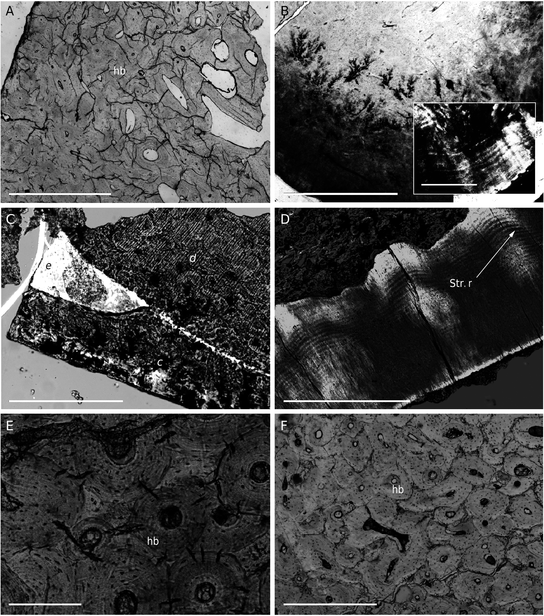
82. “Côte fossile d’un Cétacé, section en travers”. Pencil number: 263. Charles Marchand Préparateur ( Fig. 3E View FIG ).
Description. A cross section in compact bone tissue. The section is irregular in shape, about 8 × 5 mm. It seems that an original external free surface of the bone is preserved. The external cortex is characterized by many osteons in a periosteal tissue containing many Sharpey’s fibers. A few osteons appear to be primary, longitudinally oriented and lying comformably within the periosteal tissue. However, many osteons are secondary, as evidenced by their inconformity with neighbouring structures. Deeper in the bone, most osteons are secondary, although some primary tissue, with Sharpey’s fibers, locally form an “intersticial system” between them. Most secondary osteons are well defined, with a small diameter Haversian canal in the middle. Very few wide open erosion rooms are observed and there is no generalized superposition of several generations of osteons.
Comments. The tissue can be described as “dense Haversian bone” although there are probably more primary osteons than suggested by a quick casual observation and secondary osteon overlaping is not overwhelming. Most secondary osteons would be thus of “first generation”, suggesting a not so mature tissue. As frequent in fossil dense Haversian tissues (especially from Hadrosaurs “ossified tendons”) numerous short radial cracks unite the peripheries of neighbouring secondary osteons, crossing the cementing line limiting each secondary osteon.
83. “Côte fossile d’un Cétacé, section en travers”. Pencil number: 116, ink label: Anat. Comp. 116.
Description. A smaller cross section in compact bone from the same material as above (82) but slightly thinner, allowing sharper observation, but with no preserved peripheral cortex. A few wide open erosion rooms and some “third generation” secondary osteons are observed.
84. Champsodelphis macrogenius ? “terrains de...”. Ink label: Anat. Comp. 1876.
Description. An irregular and ovoid cross section in a tooth 6 × 3 mm. Most of the section is formed of a highly birefringent tissue, surrounding an oval central structure about 2 × 1 mm. The external tissue is entirely non-vascular but densely cellular, and entirely permeated by an extremely large number of thin, slightly undulating and very long radial fibres. Many circumferential lines, closer to each others towards the periphery, are resolved as associated with cracks in the tissue. Although those lines are artefactual, at least in part, they strongly suggest lines of cyclical deposition.
The inner tissues are sharply delineated from the external one by a clear discontinuity. Deeper to this discontinuity, a first thin layer is amorphous, isotropic and featureless. Then a second thicker discontinuity circles the most internal tissue. The latter is highly birefringent and surrounds a tiny, central free pulp cavity. The internal tissue appears mostly globular, especially close to its periphery. Birefringence underlines both radial and circumferential organizations. Successive circumferential regions of relatively more globular or more inotropic mineralizations (sensu Ørvig 1967) seem to alternate. Elongate, very thin radial fibres can be observed, perhaps as numerous, but far less obvious, than in the surrounding cellular tissue.
Comments. Champsodelphis Gervais 1848 in Gervais 1848-1852 was erected on material from the middle to the upper Miocene of Europe ( Simpson 1945). By comparison with available data (e.g., Schmidt & Kiel 1971) we interpret the cellular, non vascular tissue in the periphery as a very thick coat of cementum. Radial fibres there would be the anchoring fibres, akin to Sharpey’s fibres. The inner tissue can be interpreted as orthodentine, although of a highly globular variety. The thin amorphous coating at the periphery remains puzzling. It appears to be natural and well in situ between cementum and dentin. Its optical properties do not suggest enamel, an interpretation which would also be contradicted by its surrounding by the cementum.
No known copyright restrictions apply. See Agosti, D., Egloff, W., 2009. Taxonomic information exchange and copyright: the Plazi approach. BMC Research Notes 2009, 2:53 for further explanation.