
Crucescharellina japonica Silén, 1947a
|
publication ID |
https://doi.org/ 10.11646/zootaxa.5379.1.1 |
|
publication LSID |
lsid:zoobank.org:pub:430102D2-4EAA-41B3-B57F-CC532F929DA3 |
|
DOI |
https://doi.org/10.5281/zenodo.10248949 |
|
persistent identifier |
https://treatment.plazi.org/id/4B6E902E-FFF9-FFC3-FF46-FB8E1E0AFF12 |
|
treatment provided by |
Plazi |
|
scientific name |
Crucescharellina japonica Silén, 1947a |
| status |
|
Crucescharellina japonica Silén, 1947a View in CoL
( Fig. 46 View FIGURE 46 ; Table 41)
Crucescharellina japonica Silén, 1947a: 44 View in CoL , text-figs 28–31, pl. 1, figs 11, 12.
Material examined. Holotype by monotypy UPSZTY 2483 , Goto Islands , Kyushu, Japan; depth 175 m. Leg. Prof. S. Bock 1914.
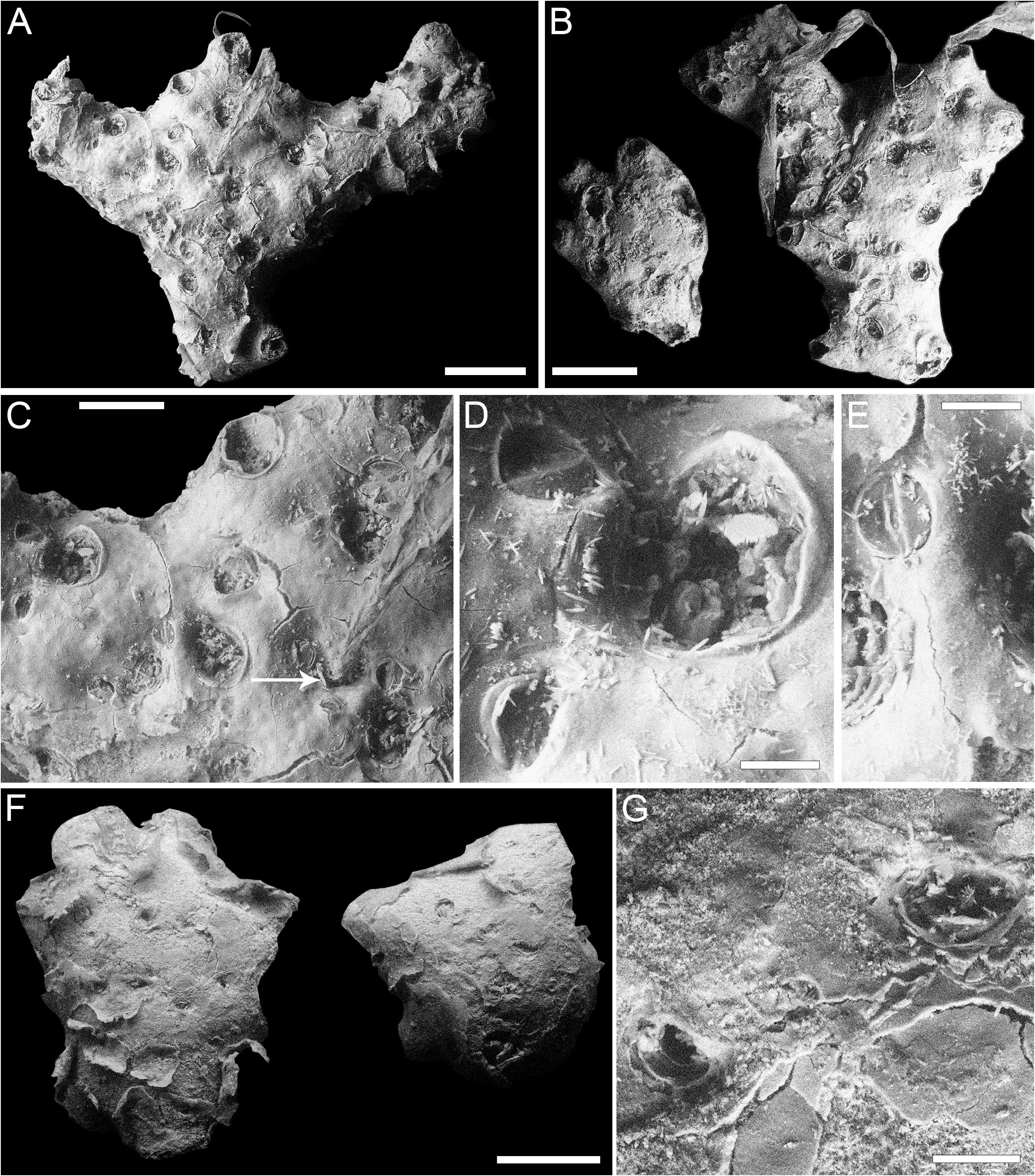
Remarks. Compared to the figures in Silén (1947a, pl. 1, figs 11–12), the preservation state of the specimen has deteriorated preventing an improvement of the description as well as size measurements, except for avicularia ( Table 41). The unique colony available is the designated holotype which is now fragmented in seven parts, five of which are illustrated in Fig. 46 View FIGURE 46 . The rooted colony was originally stellate or cruciform, with five, flat, thick branches with bi- to trilobate tips, developing on the same horizontal plane. The autozooids are opening only on the frontal side, while the dorsal side is occupied by avicularia and kenozooids. The hypothesis is that colonies of this species live with the frontal side facing the substrate ( Silén 1947a; Gordon 1989; Book & Cook 2004). Autozooidal boundaries are indistinct, the subcircular orifice is surrounded by a short peristome forming a pseudosinus proximally ( Fig. 46C View FIGURE 46 ). The frontal adventitious avicularia are subcircular to oval with complete crossbar and semicircular mandibles ( Fig. 46D, E View FIGURE 46 ), similar to those on the dorsal surface ( Fig. 46G View FIGURE 46 ). A single lunoecium (i.e. a crescentic kenozooidal openings) is distinguishable ( Fig. 46C View FIGURE 46 , arrowed).
Ovicells are unknown in the genus,they have not been observed in any of the species attributed to Crucescharellina , such as C. aster Gordon & d'Hondt, 1997 from New Caledonia and New Zealand localities, C. australis Bock & Cook, 2004 from Australia, C. decussis ( Harmer, 1957) from Sulu, Banda and Celebes Seas, and C. jugalis Gordon, 1989 from New Zealand. Crucescharellina japonica was collected at 175 m depth and C. australis at 320 m, while other species in the genus come from much deeper waters, sometimes abyssal: C. aster from 760–1573 m, C. decussis from 535–3112 m, C. jugalis from 1217–1357 m. A single colony, tentatively attributed by Gordon & d'Hondt (1997, p. 73, figs 221–223) to C. japonica , was collected from the Philippines at 640– 668 m. Large spatulate avicularia, lacking in the holotype specimen, were observed and illustrated for this specimen.
No known copyright restrictions apply. See Agosti, D., Egloff, W., 2009. Taxonomic information exchange and copyright: the Plazi approach. BMC Research Notes 2009, 2:53 for further explanation.
|
Kingdom |
|
|
Phylum |
|
|
Class |
|
|
Order |
|
|
Family |
|
|
Genus |
Crucescharellina japonica Silén, 1947a
| Martino, Emanuela Di 2023 |
Crucescharellina japonica Silén, 1947a: 44
| Silen, L. 1947: 44 |