
Amphiblestrum crassispinosum ( Silén, 1954 )
|
publication ID |
https://doi.org/ 10.11646/zootaxa.5379.1.1 |
|
publication LSID |
lsid:zoobank.org:pub:430102D2-4EAA-41B3-B57F-CC532F929DA3 |
|
DOI |
https://doi.org/10.5281/zenodo.10248871 |
|
persistent identifier |
https://treatment.plazi.org/id/4B6E902E-FFA0-FF99-FF46-FC8B1E09FF12 |
|
treatment provided by |
Plazi |
|
scientific name |
Amphiblestrum crassispinosum ( Silén, 1954 ) |
| status |
|
Amphiblestrum crassispinosum ( Silén, 1954) View in CoL
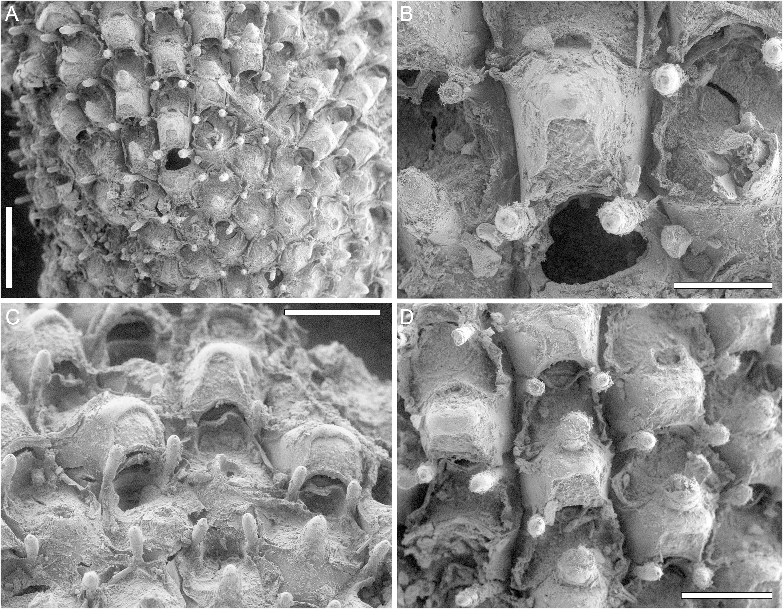
( Fig. 2 View FIGURE 2 ; Table 3)
Rhamphonotus crassispinosus Silén, 1954: 10 , fig. 3.
Material examined. Lectotype (designated here) LUZM 50 a, the largest colony among the syntype material, off Rockingham , south of Garden Island , Western Australia; depth 9–18 m. Leg. Prof. T. Gislén, Australia Expedition 1951–1952, collected 4.1.1952 . Paralectotype: the remaining colony LUZM 50 b.
Description. Colonies encrusting algae, the largest colony 3 × 3.5 mm in size.
Autozooids hexagonal, longer than wide (mean L/ W 1.27), flat, distinct, separated by grooves ( Fig. 2A View FIGURE 2 ). Gymnocyst extensive proximally (120–160 µm), smooth, forming medially a suboral mucro lodging the avicularium and a raised rim at its limit with the adjacent cryptocyst; cryptocyst slightly depressed in respect to the gymnocyst, granular, extended for 50–90 µm.
Opesia bell-shaped with pointed triangular lateral constrictions and slightly concave proximal margin ( Fig. 2B View FIGURE 2 ); a pair of robust, club-shaped spines placed laterally at level with the lateral constrictions, 90–160 µm long and with basal diameter 25–30 µm and tip diameter 35–50 µm, persisting in ovicellate zooids ( Fig. 2C View FIGURE 2 ); a stout, gymnocystal mucro developed suborally and medially, 110–130 µm long and with basal diameter 70–90 µm and tip diameter about 30 µm ( Fig. 2C, D View FIGURE 2 ).
Avicularium adventitious, placed on the suboral mucro, seemingly oval ( Fig. 2D View FIGURE 2 ).
Ovicells squared, prominent, convex frontally and indented distally by the suboral mucro of the next zooid in the row, closed by the operculum ( Fig. 2B–D View FIGURE 2 ); ectooecium smooth, partially calcified, sometimes developing a blunt umbo medially and leaving proximally a semielliptical to bell-shaped fenestra exposing the granular endooecium ( Fig. 2B View FIGURE 2 ).
Remarks. This species, originally described as Ramphonotus , is currently accepted as Amphiblestrum , the main recognized difference between the two genera being the ectooecium, completely calcified in Ramphonotus and partially calcified in Amphiblestrum ( Bishop & Hayward 1989) . However, the boundary between these two genera remains ambiguous. It has been observed, for example, that in colonies of Amphiblestrum , e.g. A. lyrulatum ( Calvet, 1907) , the extent of the ectoooecium calcification can vary, sometimes even appearing uniformly calcified ( López de la Cuadra & García-Gómez 1994; Di Martino et al. 2022, fig. 3).
The ‘prickly’ appearance of this species is typical of taxa encrusting ephemeral, flexible substrates such as algae and seagrass leaves. The presence of thick, long, latero-oral spines and the development of a stout suboral mucro and sometimes of an umbo on the ovicell is likely a protection used to reduce colony damage consequent to friction between fronds ( Di Martino & Rosso 2021).
| T |
Tavera, Department of Geology and Geophysics |
No known copyright restrictions apply. See Agosti, D., Egloff, W., 2009. Taxonomic information exchange and copyright: the Plazi approach. BMC Research Notes 2009, 2:53 for further explanation.
|
Kingdom |
|
|
Phylum |
|
|
Class |
|
|
Order |
|
|
Family |
|
|
Genus |
Amphiblestrum crassispinosum ( Silén, 1954 )
| Martino, Emanuela Di 2023 |
Rhamphonotus crassispinosus Silén, 1954: 10
| Silen, L. 1954: 10 |