
Microporina okadai Silén, 1941
|
publication ID |
https://doi.org/ 10.11646/zootaxa.5379.1.1 |
|
publication LSID |
lsid:zoobank.org:pub:430102D2-4EAA-41B3-B57F-CC532F929DA3 |
|
DOI |
https://doi.org/10.5281/zenodo.10248919 |
|
persistent identifier |
https://treatment.plazi.org/id/4B6E902E-FF9E-FFA0-FF46-FD401F6DFC4E |
|
treatment provided by |
Plazi |
|
scientific name |
Microporina okadai Silén, 1941 |
| status |
|
Microporina okadai Silén, 1941 View in CoL
( Fig. 27 View FIGURE 27 ; Table 25)
Microporina okadai Silén, 1941: 68 View in CoL , figs 79–82, pl. 4, fig. 13, 14. Material examined. Holotype by original designation UPSZTY 2468 , Okinose , Sagami, Japan; depth 150–600 m. Leg. Prof. S. Bock 1914.
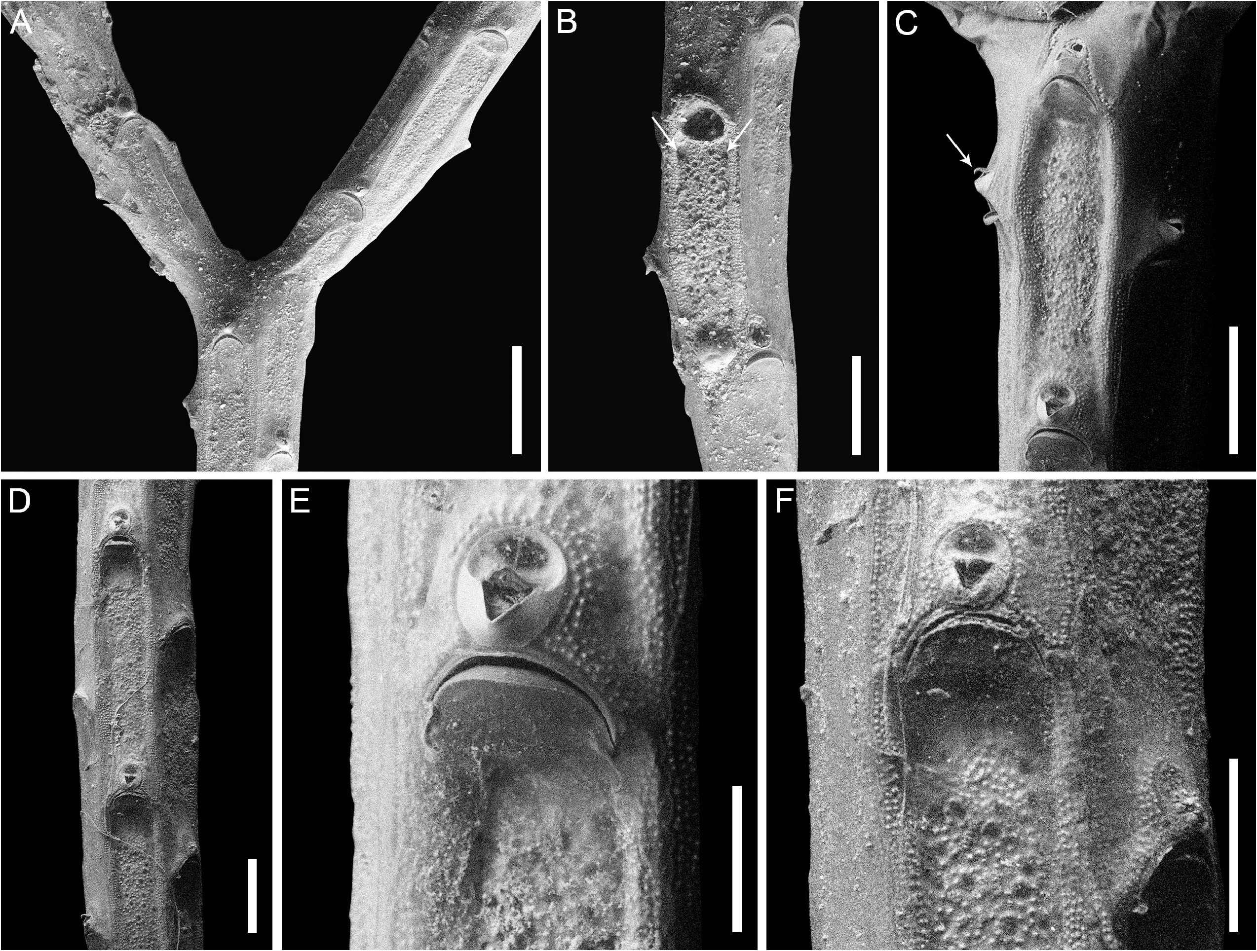
Description. Colony erect, jointed, dichotomously branching ( Fig. 27A View FIGURE 27 ); internodes straight, quadrangular in cross-section, quadriserial ( Fig. 27D View FIGURE 27 ).
Autozooids elongate-rectangular (mean L/ W 3.70), proximal margin concave, distal margin convex, arranged in alternating back-to-back series; cryptocyst forming a raised mural rim (50–60 µm wide), minutely granular with granules (5 µm or less in diameter) regularly aligned forming multiple concentrical rows, and modified to follow the outline of the avicularium at the distal edge ( Fig. 27E View FIGURE 27 ); frontal cryptocyst immersed and flat with sparse granules, slightly coarser than those of the mural rim (8–10 µm), and unevenly distributed circular pseudopores (10–15 µm in diameter) ( Fig. 27B View FIGURE 27 ); a pair of lateral opesiules at a short distance (c. 40–50 µm) from the proximal margin of the orifice ( Fig. 27B View FIGURE 27 , see arrows).
Orifice semielliptical with straight or concave proximal margin, wider than long, outlined by a raised rim ( Fig. 27B View FIGURE 27 ).
Avicularium adventitious, single, placed distally to most autozooids, oval ( Fig. 27D–F View FIGURE 27 ); rostrum acutely triangular, raised, proximally directed; crossbar complete; mandible triangular with downward hooked tip ( Fig. 27C View FIGURE 27 , see arrow).
Ovicells absent.
Remarks. The autozooid illustrated in Fig. 27B View FIGURE 27 was the only one without a frontal membrane, in which lateral symmetrical depressions adjacent to the mural rim and at a short distance from the proximal margin of the orifice were observed. These structures were, therefore, interpreted as the ‘opesiulae’ described and drawn in Silén (1941, fig. 81). SEM images of an internode with well-defined opesiules are available from Arakawa (2016, fig. 9B).
Of the 12 species currently assigned to Microporina more than half are from the Pleistocene to Recent of Japan ( Bock 2023). Based on the species comparison of Arakawa (2023, table 1), among the Japanese Microporina , M. okadai has the narrowest internodes and longest autozooids.
No known copyright restrictions apply. See Agosti, D., Egloff, W., 2009. Taxonomic information exchange and copyright: the Plazi approach. BMC Research Notes 2009, 2:53 for further explanation.
|
Kingdom |
|
|
Phylum |
|
|
Class |
|
|
Order |
|
|
Family |
|
|
Genus |
Microporina okadai Silén, 1941
| Martino, Emanuela Di 2023 |
Microporina okadai Silén, 1941: 68
| Silen, L. 1941: 68 |