
Reticulolaelaps Costa, 1968
|
publication ID |
https://doi.org/10.22073/pja.v8i2.43017 |
|
publication LSID |
lsid:zoobank.org:pub:B1FB2D9B-9080-42EC-93ED-CA6EB2497BF4 |
|
persistent identifier |
https://treatment.plazi.org/id/4B1D9B72-FFED-B843-0577-19C3FBAAF98F |
|
treatment provided by |
Felipe |
|
scientific name |
Reticulolaelaps Costa, 1968 |
| status |
|
Genus Reticulolaelaps Costa, 1968
Reticulolaelaps Costa, 1968: 26 .
Type species: Reticulolaelaps faini Costa, 1968 .
Definition
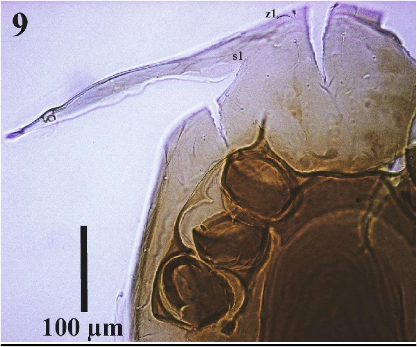
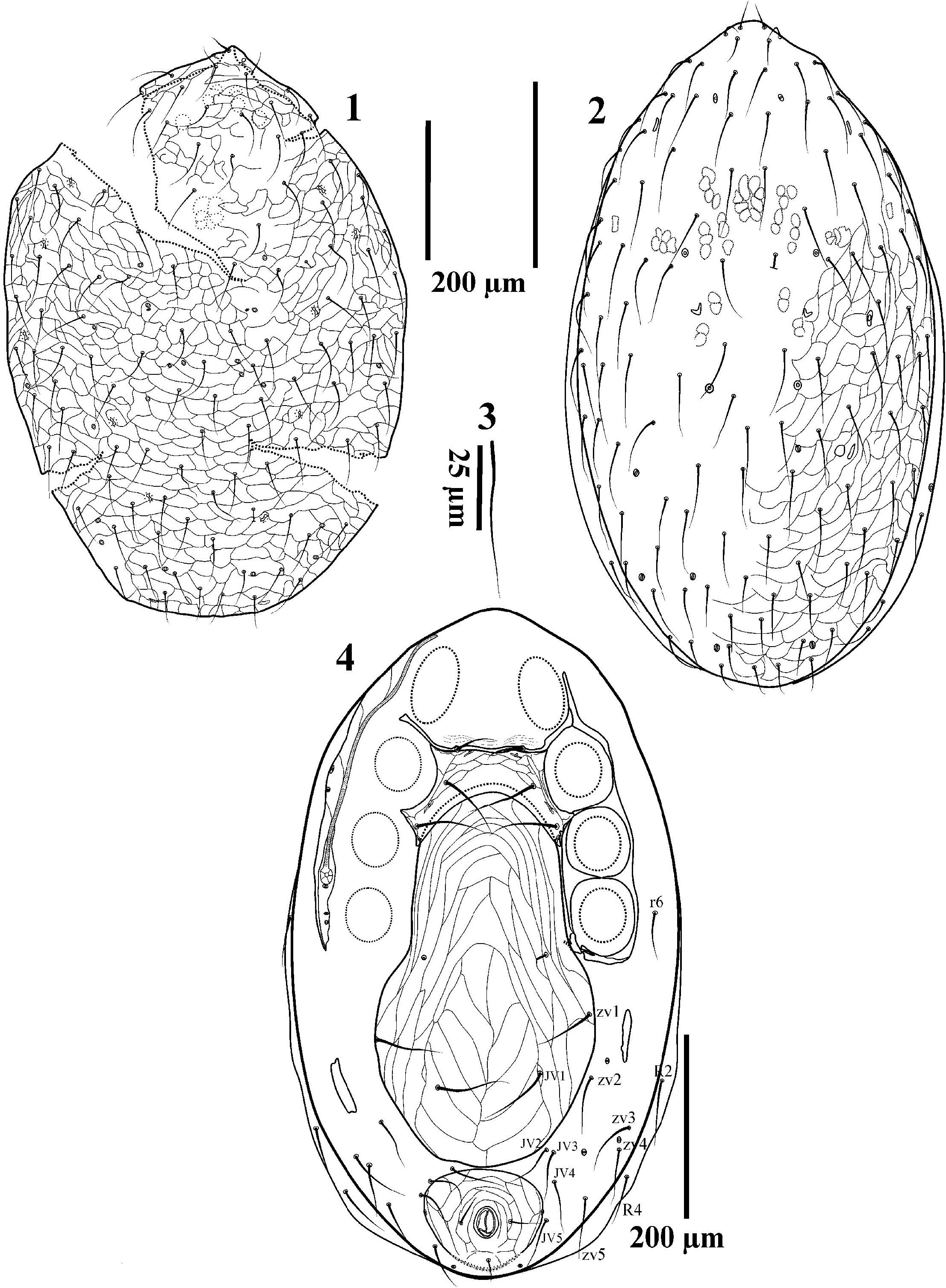
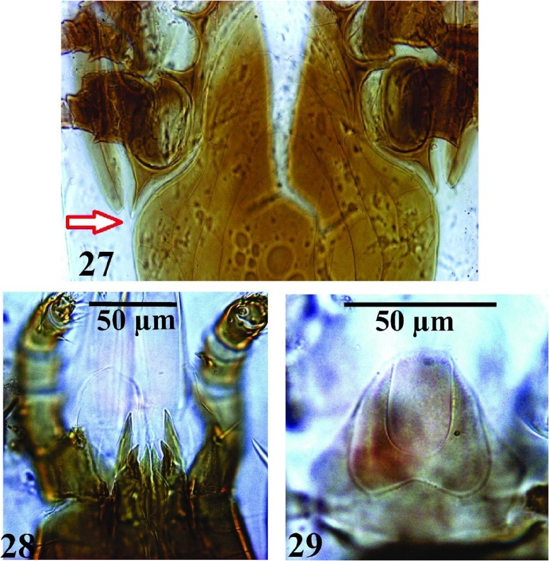
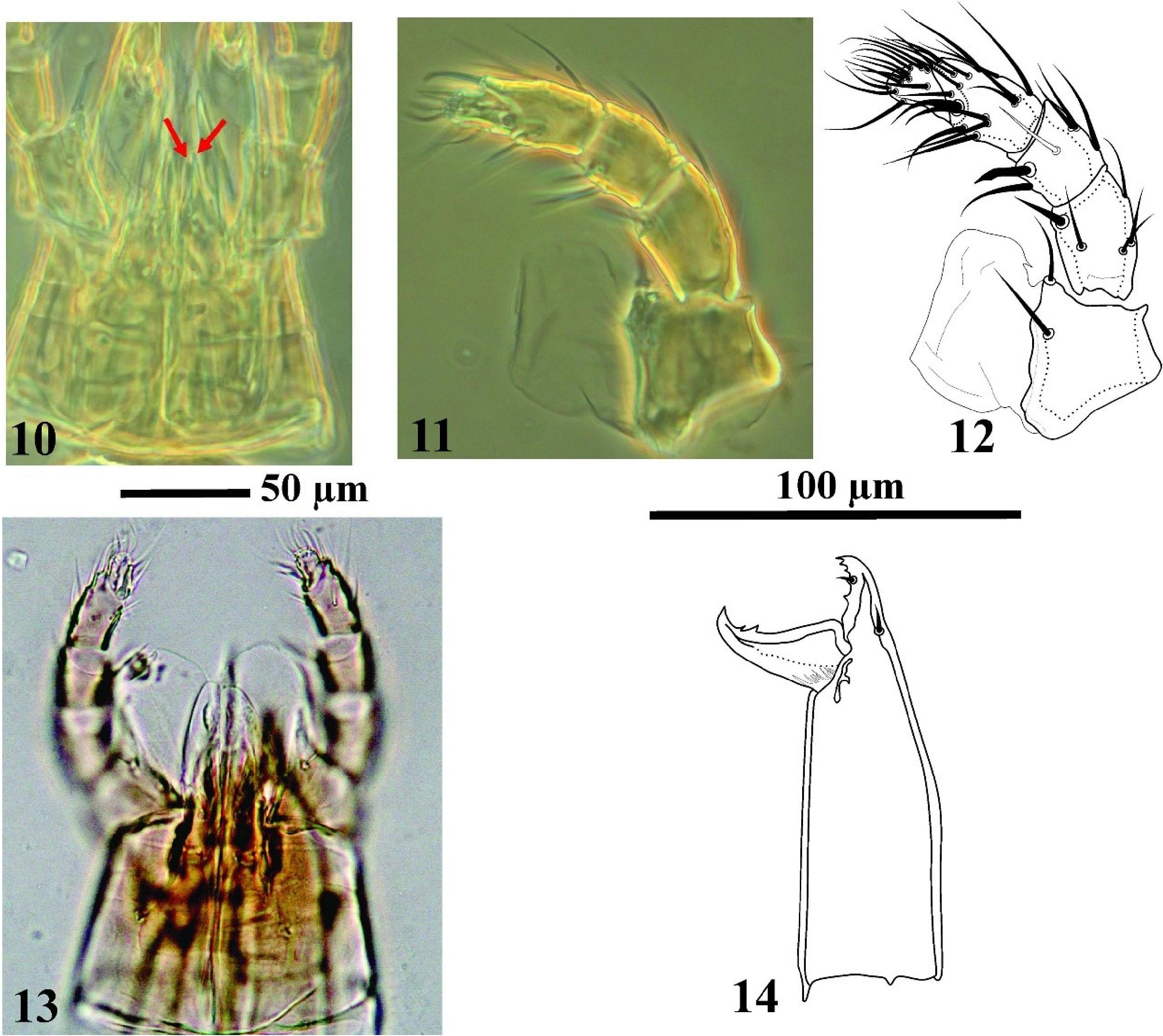
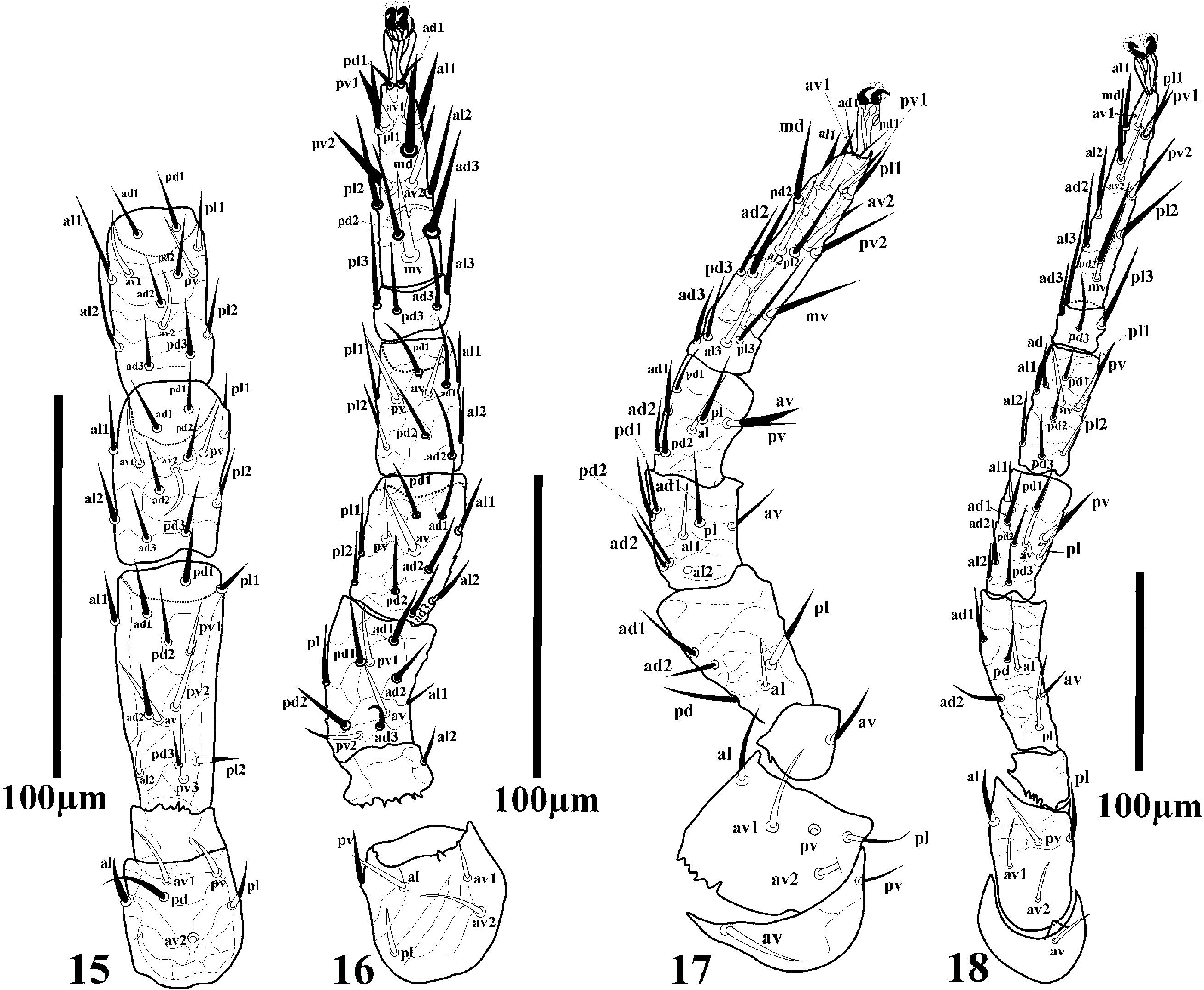
The genus is characterized by a well-sclerotized holodorsal shield with slender setae ( Figs. 1–3 View Figures 1–4 ). Tritosternum with small basal part and laciniae fused together for half their length ( Figs. 5–7 View Figures 5–8 ). Four character states are present in the presternal area of different Reticulolaelaps species – (1) presternal plates absent ( faini ); (2) presternal plates present, separate from sternal shield ( lativentris, jilinensis ); (3) presternal plates fused to sternal shield ( hallidayi , costai ); (4) presternal plates represented by lightly sclerotized transverse lines ( elsae ). Female sternal shield with three pairs of simple sternal setae, extending at least to the midlevel of coxa III, with iv1-3 on shield, poroids iv3 present on the posterolateral extensions ( Fig. 4 View Figures 1–4 ). Metasternal setae st4 absent ( Figs. 4 View Figures 1–4 , 25 View Figures 25–26 , 30 View Figure 30 ). Exopodal II-III, III- IV, parapodal and endopodal II-III plates fused, surrounding coxae; parapodal plate rounded or triangular, more or less contiguous with but separate from peritrematal and genitoventral shields; endopodals III-IV joined with podal plate posteriorly and with posterolateral extension of sternal shield anteriorly, endopodals II-III fused with lateral margins of sternal shield, and anterolateral corners acutely produced into narrow arms (endopodal extensions) flanking coxae II and joining exopodals extension between coxae I-II ( Figs. 4–9 View Figures 1–4 View Figures 5–8 View Figure 9 ). The left and right endopodals at the anterior level of coxae II are connected by a sclerotized rod-like bridge almost fused with anterior margin of sternal shield ( Figs. 4 View Figures 1–4 , 9 View Figure 9 , 30 View Figure 30 ). Genitoventral shield large, expanded posterior to coxae IV, fused with metapodal plates ( Figs. 25 View Figures 25–26 , 30 View Figure 30 ) or free rod like metapodal plates present ( Fig. 4 View Figures 1–4 ), shield bearing 3–6 pairs of smooth setae including: the genital setae (st5) and five (ZV1–2 and JV 1–3 in R. faini , R. hallidayi , and R. costai ), three (ZV1–2, JV 1 in R. lativentris ) or two (ZV1, JV 1 in R. elsae ) additional pairs of setae on its surface, extending near or abutting the anal shield, with strong reticulated ornamentation. Anal shield large with gv3 on marginal surface ( Figs. 4 View Figures 1–4 , 8 View Figures 5–8 ). Surface of pistome faintly reticulated, its anterior margin smooth ( Fig. 29 View Figures 27–29 ). Chelicera with small and robust digits with few teeth. Two large membranous flaps originate near the inner side of the palp trochanter ( Figs. 11–13 View Figures 10–14 , 28 View Figures 27–29 ) or the underside of the hypostome (based on Costa 1968; see note below). Corniculi robust and hornlike. Internal malae smaller but similar to corniculi and with two smooth hornlike lobes ( Fig. 28 View Figures 27–29 ). All the sclerotized parts of the body are well ornamented throughout, including the legs ( Figs. 15–18 View Figures 15–18 ). Legs significantly shorter than idiosoma, genu III (2 2/1 2/0 1) and IV (2 2/1 3/1 1) with eight and ten setae respectively. Male with sterno-genitiventral or holoventral shield with ten pairs of setae, with separate anal shield similar to that of the female.
Note
Our observations on dissected specimens of all our Reticulolaelaps species in APAS, including specimens identified as R. faini from Iran, revealed the attachment of membranous flaps on the inner side of the palp trochanter, but we have not had the opportunity to check the type material of R. faini from Israel. Costa (1968) stated that gnathosoma bears ventrally two large membranous flaps that originated in front of the anterior hypostomal setae. Study of dissected specimens of type materials or specimens collected from that area is needed to understand the exact location of these membranous flaps.
Diagnosis
Holodorsal shield reticulated. Tritosternum with small basal part and laciniae fused for half their length. Female sternal shield with concave posterior margin, bearing iv1–3. Metasternal setae st4 absent; left and right endopodals connected by a sclerotized rod-like bridge at the anterior level of coxae II, almost fused with anterior margin of sternal shield. Genitiventral shield large, expanded posterior to coxae IV, extending close to or abutting the anal shield. Surface of epistome faintly reticulated, its anterior margin smooth. With two large membranous flaps anterior to hypostome (see above note). Internal malae smaller than corniculi, with two smooth horn-like lobes. All the sclerotized parts of the body are well ornamented throughout. Male with sterno-genitiventral shield with 10 pairs of setae, with separate anal shield similar to that of the female.


No known copyright restrictions apply. See Agosti, D., Egloff, W., 2009. Taxonomic information exchange and copyright: the Plazi approach. BMC Research Notes 2009, 2:53 for further explanation.
|
Kingdom |
|
|
Phylum |
|
|
Class |
|
|
Order |
|
|
Family |
Reticulolaelaps Costa, 1968
| Nemati, Alireza, Khalili-Moghadam, Arsalan & Gwiazdowicz, Dariusz J. 2019 |
Reticulolaelaps
| Costa, M. 1968: 26 |