
Reticulolaelaps elsae ( Joharchi, Babaeian & Jalalizand, 2016 ) Nemati & Khalili-Moghadam & Gwiazdowicz, 2019
|
publication ID |
https://doi.org/ 10.22073/pja.v8i2.43017 |
|
publication LSID |
lsid:zoobank.org:pub:B1FB2D9B-9080-42EC-93ED-CA6EB2497BF4 |
|
persistent identifier |
https://treatment.plazi.org/id/4B1D9B72-FFEC-B84B-0619-1AD2FA50FBDA |
|
treatment provided by |
Felipe |
|
scientific name |
Reticulolaelaps elsae ( Joharchi, Babaeian & Jalalizand, 2016 ) |
| status |
comb. nov. |
Reticulolaelaps elsae ( Joharchi, Babaeian & Jalalizand, 2016) comb. nov.
Reticulolaelaps elsae ( Joharchi, Babaeian and Jalalizand, 2016)
Laelaspisella elsae Joharchi, Babaeian and Jalalizand, 2016 ; in Joharchi et al. (2016).
Note on materials examined
Nineteen females, Iran, Esfahan, March-April 2002, coll. A. Jalalizand, from bark of elm trees. Six of them used in the original description of Laelaspisella elsae by Joharchi et al. (2016), were precisely studied. The senior author of the present article (A. Nemati) described this species in 2009– 2010 based on 19 specimens mentioned above, but that work was not published because the genus assignment has been an enigma. In this paper, we will use some of the unpublished results mentioned above, including morphological traits and morphometric data of type materials of this species. Comparison with data presented in the original description of L. elsae by Joharchi et al. (2016) shows a number of discrepancies in the description of morphological traits and range of measurements, which could hardly be avoided. Below, we present the characters consistently different between Laelaspisella and the more closely related genus Reticulolaelaps .
Notes on genus assignment of Reticulolaelaps elsae ( Joharchi, Babaeian & Jalalizand, 2016) comb. nov.
Reticulolaelaps elsae was described in the genus Laelaspisella as mentioned above ( Joharchi et al. 2016). Laelaspisella was erected by Marais and Loots (1969) to accommodate two new species: L. macrodorsalis Marais & Loots and L. epigynialis Marais & Loots , from forest soil in South Africa, Lesotho and Congo. Subsequently, this genus was considered as a subgenus of Hypoaspis s. lat. by Karg (1989), who described two additional species, H. (Laelaspisella) foramenis Karg, 1989 and H. (Laelaspisella) cavitatis Karg, 1982 , and as a distinct genus by Joharchi and Halliday (2013), Joharchi et al. (2016) and Nemati and Gwiazdowicz (2016).
Joharchi and Halliday (2013) considered Laelaspisella canestrinii and L. kabitae as species of this genus but excluded the two species described by Karg. Subsequently, Joharchi et al. (2016) defined the scope of the genus definition more broadly, and introduced in it other species with more diverse attributes ( L. macrodorsalis , L. epigynialis , L. tonsilis , L. kabitae and L. canestrinii ).
Joharchi et al. (2016) with the description of L. elsae , along with various species mentioned above which they considered in this genus, expanded the genus concept; at the same time they had to make several changes in the genus attributes in order to accommodate various characteristics of species. On the other hand, in order to consider these heterogeneous species within a single genus ( Laelaspisella ), many characters must be defined as variable: (1) dorsal seta of chelicera present ( beaulieui , canestrinii, elsae , kabitae and tonsilis ) or absent ( epigynialis and macrodorsalis ); (2) podonotal shield hypertrichous [ epigynialis , macrodorsalis and tonsilis : many pairs (about 13 pairs) of setae on podonotal part of dorsal shield have not been drawn by Karg (1989) in the original description of Gymnolaelaps tonsilis (his Fig. 1a, p View Figures 1–4 . 336) based on observations that have been done on holotype and paratypes of Pseudoparasitus tonsilis Karg, 1989 in the Museum für Naturkunde Berlin (see materials and methods)], or with normal chaetotaxy ( beaulieui , canestrinii, elsae and kabitae ); (3) setae Jv5 and Zv5 expanded ( epigynialis and macrodorsalis ) or normal ( beaulieui , canestrinii, elsae , kabitae and tonsilis ); (4) seta pd3 on genu I present ( beaulieui , canestrinii, elsae , kabitae and tonsilis ) or absent ( epigynialis and macrodorsalis ); (5) epigynal shield with rounded posterior margin separate from anal shield ( epigynialis and macrodorsalis ), or with rounded posterior margin extending to and touching anal shield ( beaulieui , canestrinii, elsae , kabitae and tonsilis ); (6) anterior margin of epistome smooth rounded ( elsae ) or subtriangular ( beaulieui and canestrinii ), mucronate ( epigynialis and macrodorsalis ), with irregular minute denticulation ( kabitae ) or with deep denticles ( tonsilis ); (7) distinct sclerotized presternal plates present ( epigynialis , kabitae , macrodorsalis , tonsilis ), or presternal area with lineate reticulation ( beaulieui , canestrinii ) or nearly absent ( elsae ); (8) deutosternal groove with six multidentate rows ( beaulieui , canestrinii , epigynialis , kabitae and macrodorsalis ) or only with four rows possessing 2–5 denticles ( elsae ); (9) the palp apotele two-tined ( elsae , epigynialis , macrodorsalis and tonsilis ) or three-tined ( beaulieui , canestrinii and kabitae ); (10) dorsal shield with simple acicular setae ( elsae , epigynialis , kabitae , macrodorsalis and tonsilis ) or setae with a small knob at base ( beaulieui and canestrinii ); (11) internal malae free medially and densely fringed with very elongate hairs ( epigynialis and macrodorsalis ), in addition it possesses two groups of dense and very elongate hairs at basal part of each internal mala ( beaulieui and canestrinii ); or internal malae normal and lacking very elongate hairs ( elsae , kabitae and tonsilis ); (12) setae on epigynal shield: all setae located on margins ( beaulieui , canestrinii, elsae , epigynialis , kabitae and epigynialis and tonsilis ) or some of those well inside ( elsae ).
To resolve some of these problems, Nemati and Gwiazdowicz (2016) recently clarified the diagnosis of Laelaspisella , proposing a new definition considering the species which should be included in this genus. Based on this work (see Nemati and Gwiazdowicz, 2016) and in the mention of two important apomorphic attributes [spatulate and pilose setae JV5 and ZV5 and genu I with only two postero-dorsal setae (2 3/2 2/1 2)], only Laelaspisella macrodorsalis and L. epigynialis can be considered as members of this genus, excluding the other species mentioned above ( Nemati and Gwiazdowicz 2016). On the other hand, for the above species that would require a great variety of character states in Laelaspisella, Nemati and Gwiazdowicz (2016) erected the genus Pogonolaelaps to accommodate P. canestrinii (Berlese) and described P. beaulieui Nemati & Gwiazdowicz ; the appropriate genus for L. kabitae and L. tonsilis still requires further study.
Taking into account the morphological characters of L. elsae given by Joharchi et al. (2016), the variety of character states in Laelaspisella would become more complicated (some special characters of L. elsae were not mentioned above). Such changes in the definition of the genus resulted in several ambiguities in the diagnosis and scope of genus as cited above. In fact, due to the inappropriate genus assignment, its main traits are not consistent with the genus definition which are confirmed by our studies and observations on type series and other specimens of L. elsae . Here while we clarify the genus placement of this species, the morphological characters and the conflicts of the original description are reviewed and presented below. For this purpose, the definition of Reticulolaelaps is revised and presented.
Redescription (female, n = 19)
Material examined Nineteen females collected from Esfahan (as cited above).
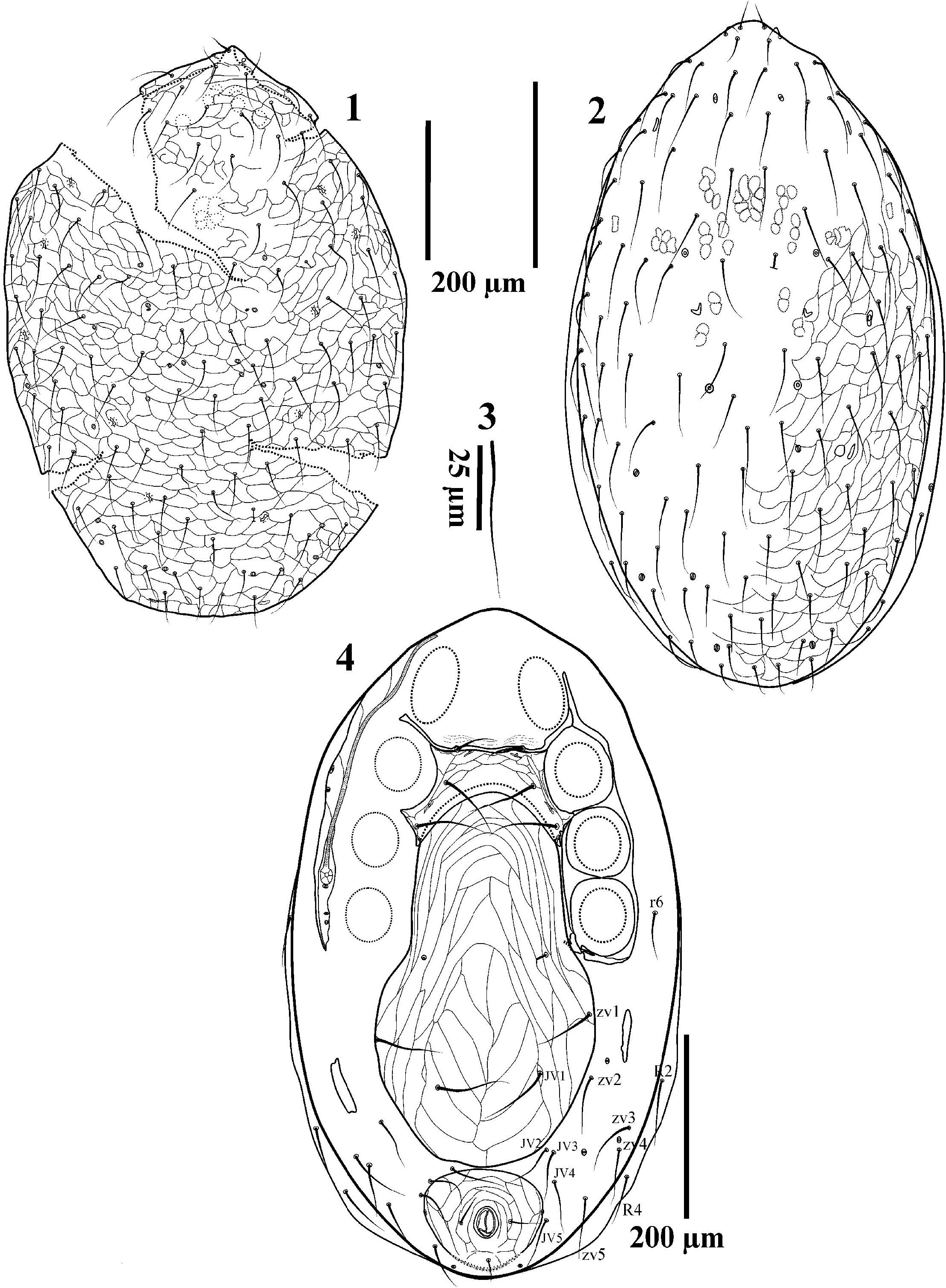
Dorsal idiosoma – Dorsal shield length 515–573 (400–449), width 300–330 (280–333) ( Figs. 1–2 View Figures 1–4 ). Shield oval shaped with convex dorsum and nearly flat venter; shield well-sclerotized and with clear reticulation; with about 111–114 long delicate simple setae ( Fig. 3 View Figures 1–4 ), with unpaired and asymmetrical setae especially in opisthonotal region. The length of j1, z1 17–22 (13–15), J5 22–25 and Z5 19–24 are among the shortest setae. Some other setae j2 38–42, j3–6 41–51, z2–6 39–42, r1– 6 40–51 and s1–6 50–56 shorter than the other nominated dorsal shield setae (48–58). Due to the hypertrichy of the dorsal shield (especially on opisthonotal part), recognition of individual setae based on the current standard dorsal setae system is not possible. Shield with ca. 18 pairs of pore-like structures; seven pairs with large slit-like appearance including one pair at the base of z1 setae, others smaller, circular or ovoid ( Fig. 1 View Figures 1–4 ).
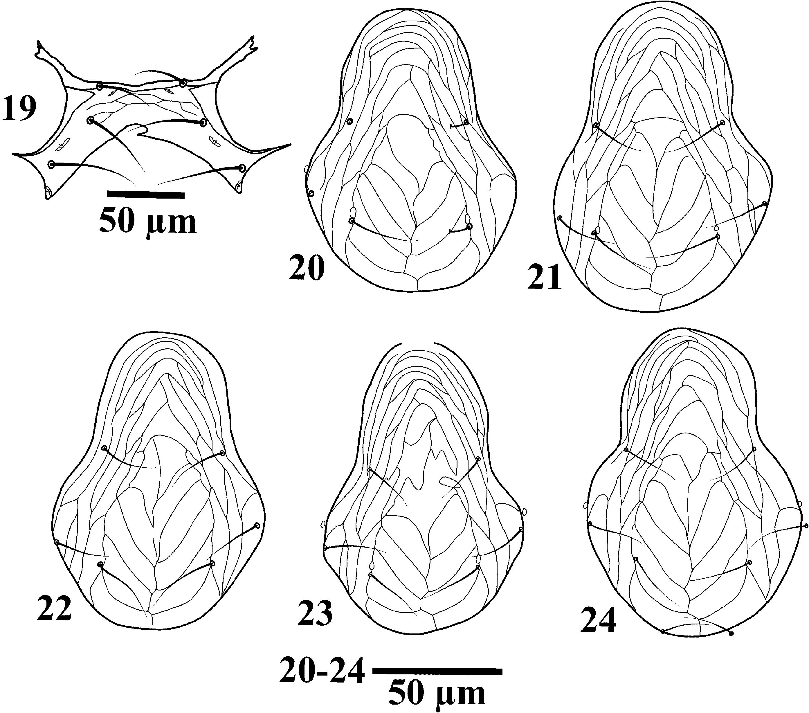
Ventral idiosoma ( Fig. 4 View Figures 1–4 ) – Tritosternum ( Figs. 5–7 View Figures 5–8 ) with 74–87 (base and lacinia) long, with a small arrow-like base ( Fig. 7 View Figures 5–8 ) 10–15 (12–13), paired pilose laciniae 64–72 (33–36): fused at their base for 30–35 (47–49% of their total length) and free for 34–37 (51–53% of their total length); presternal plates fused with sternal shield, this area consists of some lineate reticulation. Sternal shield medially 35–37 (35–43) narrowest between coxae II 97–114 (99–100) widest between coxae II and III 171–185 (120–128), with convex anterior margin, posterior margin concave, some specimens with variation in posterior margin ( Fig. 19 View Figures 19–24 ), posterolateral margins extending to mid-level of coxae III; shield bearing three pairs of smooth pointed setae: st1 32–34 (27–32), st2 46–56 (35–40), st3 58–61 (40–43), distances between st1-st1 54–59, st2-st2 73–76, st3-st3 122–127, st1-st2 27–29, st2-st3 37– 41 and three pairs of lyrifissures, the lyrifissures (iv1) between setae st1 and the second (iv2) between st2 and st3 and closer to st3; the third one (iv3) located at posterolateral corners of sternal shield extensions ( Figs. 4 View Figures 1–4 , 19 View Figures 19–24 ). Surface with reticulate ornamentation in anterior and lateral margins, extending to st2 level. Surface of sternal shield medially and posteriorly smooth. Metasternal setae st4 absent.
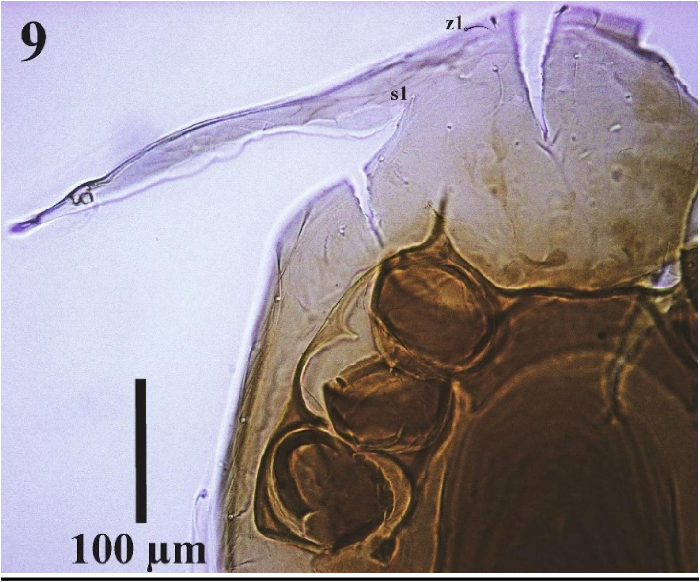
Endopodal plates II/III fused to lateral margins of sternal shield, endopodal plates III/IV angular, not elongate, narrow in anterior and posterior parts and wide in the middle, posterior tip not fused with suboval podal plate. Genitoventral shield broad, length 271–281 (258–267), length at level of st5 134–139, maximum width 193–198 (188–195), posterior edge rounded, extending near anterior margin of anal shield, surface with distinct polygonal ornamentation, bearing the genital setae st5 51– 61 (50–55) and two additional pairs of setae on its surface: ZV1 54–61 and JV1 61–64 (50–62), paragenital pores present on podal plates adjacent to the genitoventral shield at level of st5. The reticulation and the placement of setae on its surface varies in different specimens as in Figures 20– 24 View Figures 19–24 . Anal shield suboval and large 78–85 × 98–103 (64–73 × 82–88), wider than long, with distinct gv3 at lateral margins at level of anterior margin of anal opening ( Figs. 4 View Figures 1–4 , 8 View Figures 5–8 ); anterior and posterior margins slightly rounded, surface with polygonal ornamentation, para-anal setae 22–24, longer than unpaired post-anal seta 14–16 [in original description of Joharchi et al. (2016) para-anal setae length cited as 12 and post-anal as 20]; cribrum relatively narrow ( Fig. 4 View Figures 1–4 ). Opisthogastric integument with seven pairs (ZV2–4, JV2–5) and lateral membrane between dorsal and ventral sides of idiosoma with three pairs of smooth setae (r6, R2 and R4) with 41–51 long and four pairs of pores. Metapodal plates rode-like 36–44 × 7–9 (34–37 × 9–11). Podal plates posterior to coxae IV rod-like. Stigmata located at midlevel of coxae III-IV, peritremes extending anterior to coxae I. Peritrematal shields wide and reticulate, fused anteriorly with dorsal shield at distances between z1-s1 setae ( Fig. 9 View Figure 9 ) and extending behind stigmata to well behind coxa IV; with three small pore-like structures behind stigma and two on peritrematal shield, at level of coxae II-III.
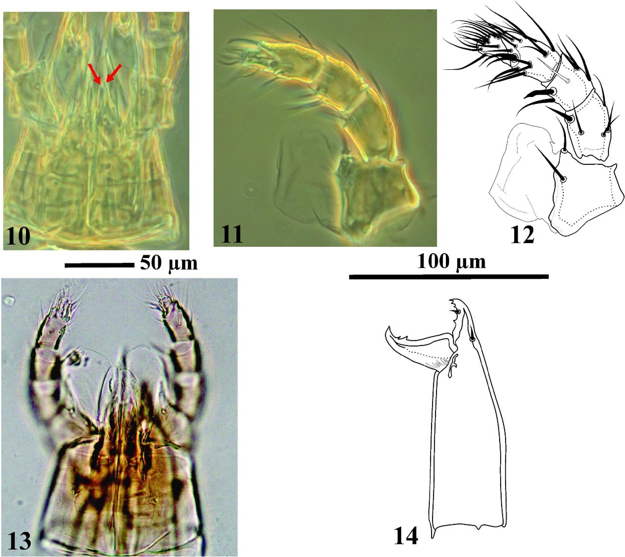
Gnathosoma ( Figs. 10–14 View Figures 10–14 ) – Hypostomal groove with four rows of denticles each with 2–4 small teeth. Corniculi sclerotized, moderately long, extending near midlevel of palp femur. Internal
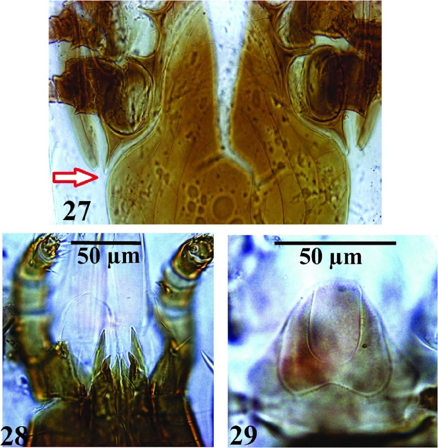
malae complex, with two pairs of lobes, inner lobes narrow, with smooth edges, outer lobes longer, narrow, pointed ( Fig. 10 View Figures 10–14 ), similar to those in R. faini ( Fig. 28 View Figures 27–29 ); labrum elongate, pubescent, extending beyond corniculi. Two large membranous flaps located at anterior part of hypostome attached to the inner surface of palp trochanter ( Figs. 11–12 View Figures 10–14 ), consists of two different parts: inner part smaller adjacent to the corniculi and more condensed, outer part larger balloon-like ( Fig. 13 View Figures 10–14 ), somewhat extending beyond the tip of palp genu. Several specimens were dissected to determine the place of connection of membranous flaps to the gnathosoma. For this purpose, the palps from the basal part (trochanter), were excised from the gnathosoma with special care. This was done to diagnose the connective tissue of this member to the hypostome or palp. Costa (1968) pointed out the origin of membranous flaps slightly in front of the anterior hypostomal setae (h1). According to our previous observations, it seemed that the flaps were not connected to the anterior part of hypostome. By separating the palp, it was observed that this membranous flap is connected to the inner surface of the palp trochanter ( Figs. 11, 12 View Figures 10–14 ). Usually in the hypostomal region, when microslides are prepared and due to pressure, the membranous flaps part is torn and only the remains of this flap are seen in the form of strings.
Hypostome with three hypostomal setae (h1–3): rostral seta h1 37–39 (42–45), h2 17–24 (28–
30), h3 56–59 (67–70), palp coxal seta 24–29 (32–35). Palp chaetotaxy: trochanter 2, femur 5, genu 6, tibia 14, tarsus 15, all setae smooth and needle-like except seta al on palp femur long and slightly thicken, al1 on palp genu short and arrow-like, al2 longer and pointed; palp tarsal claw with two pointed tines of unequal length ( Fig. 12 View Figures 10–14 ). The lengths of palp segments are as follows: palp- trochanter 29–34, palp-femur 30–32, palp-genu 24–34, palp-tibia 25–32 and palp-tarsus 17–20. Epistome subtriangular, anterior part membranous, posterior well sclerotized, posterior half with lineate ornamentation similar to R. faini ( Fig. 29 View Figures 27–29 ). Fixed digit 39–41 (40–41) of chelicera with four teeth including terminal hook ( Fig. 14 View Figures 10–14 ), second segment 76–85 pilus dentilis moderately robust, dorsal seta short, thick, prostrate, movable digit 35–37 (36–38) with two large teeth, arthrodial membrane with a rounded flap and a row of short filaments.
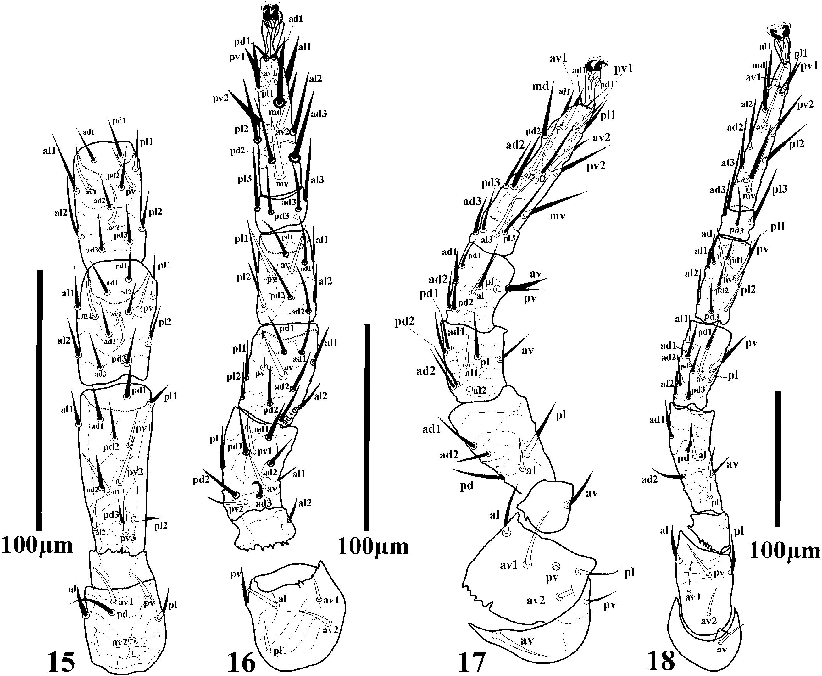
Legs ( Figs. 15–18 View Figures 15–18 ) – Legs short, well ornamented, legs II and III shorter 295–300, 283–295 (309–320, 302–310), I and IV longer 344–410, 449–464 (349–360, 431–447) (excluding pre-tarsus). Chaetotaxy normal for free-living Laelapidae : Leg I ( Fig. 15 View Figures 15–18 ): coxa 0 0/1 0/1 0, trochanter 1 0/2 1/1 1, femur 2 2/1 3/3 2, genu 2 3/2 3/1 2, tibia 2 3/2 3/1 2. Leg II ( Fig. 16 View Figures 15–18 ): coxa 0 0/1 0/1 0, trochanter 1 0/2 0/1 1, femur 2 3/1 2/2 1, genu 2 3/1 2/1 2, tibia 2 2/1 2/1 2. Leg III ( Fig. 17 View Figures 15–18 ): coxa 0 0/1 0/1 0, trochanter 1 0/2 0/1 1, femur 1 2/1 1/0 1, genu 2 2/1 2/0 1, tibia 1 2/1 2/1 1. Leg IV ( Fig. 18 View Figures 15–18 ): coxa 0 0/1 0/0 0, trochanter 1 0/2 0/1 1, femur 1 2/1 1/0 1, genu 2 2/1 3/1 1, tibia 2 1/1 3/1 2. All setae fine and needle-like. Tarsi I-IV with 18 setae 3 3/2 3/2 3 + mv, md. All pre-tarsi with a pair of claws and a long thin membranous ambulacrum.
Discrepancies of Reticulolaelaps elsae with specimens examined
The main discrepancies between the original description and the specimens that we checked (including the holotype and paratypes) are: (1) Dorsal shield with ca. 18 pairs of pore-like structures; seven pairs with large slit-like appearance including one pair at the base of z1 setae (shield with 12 pairs of pore-like structures, apparently including three pairs of gland pores and eight pairs of poroids in the original description and illustration); (2) Tritosternum 74–87 (base and lacinia) long, with a small arrow-like base 10–15 (12–13), paired pilose laciniae 64–72 (33–36): fused at their base for a length of 30–35 (47–49% of their total length) and free for 34–37 (51–53% of their total length) [tritosternum with paired pilose laciniae (33–36), columnar base (12–13 × 5–6 wide) in the original description and illustration]; (3) Sternal shield surface with reticulate ornamentation in anterior and lateral margins, extending to st2 level, medially and posteriorly smooth (surface with distinct reticulate ornamentation in the original description and illustration); (4) Metasternal setae st4 absent (Metasternal setae st4 apparently absent in the original description); (5) Endopodal plates III/IV angular, not elongate, narrow in anterior and posterior parts and wide in the middle (endopodal plates III/IV elongate, narrow, curved in the original description); (6) Podal plates not enlarged, suboval shaped (podal plate large and triangular in the original description but suboval in illustration); (7) With distinct gv3 at lateral margins at level of anterior margin of anal opening (anal pores indistinct in the original description and illustration); (8) Para-anal setae 22–24 longer than unpaired post-anal setae 14–16 [para-anal seta (12) shorter than post-anal seta (20) in original description, but para-anal setae as long as or slightly longer than postanal seta in illustration]; (9) Opisthogastric integument with ten pairs of setae, seven pairs: ZV2–4, JV2–5 on opisthogastric integument and lateral membrane between dorsal and ventral sides of idiosoma with three pairs of smooth setae (r6, R2 and R4 ) with 41–51 long [opisthogastric integument with eight pairs of smooth setae (55–65) in the original description and illustration]; (10) Peritrematal shields wide and reticulate, fused anteriorly with dorsal shield at distances between z1-s1 setae (not mentioned in original description); (11) Internal malae complex, with two pairs of lobes, inner lobes narrow, with smooth edges, outer lobes longer, narrow, pointed similar to situation which has been observed in R. faini (see Fig. 28 View Figures 27–29 ) [Internal malae complex, with two pairs of lobes, inner lobes narrow and long, with smooth edges, outer lobes very short, narrow, branched]; (12) All palp setae smooth and needle-like except seta al on palp femur long and slightly thicken, al1 on palp genu short and arrow-like, al2 longer and pointed (all setae smooth and needle-like in original description and without illustration); (13) Palp tarsal claw with two pointed tines of unequal length (palp tarsal claw with two pointed tines of equal length in original description and without illustration); (14) Fixed digit of chelicera with four teeth (fixed digit of chelicera with two small pointed teeth in original description but with four teeth including terminal hook in illustration); (15) Cheliceral dorsal seta short, thick, prostrate (dorsal seta not detected in the original description and illustration); (16) Two large membranous flaps located at anterior part of hypostome attached to the inner surface of palp trochanter (not mentioned in original description and illustration) .

No known copyright restrictions apply. See Agosti, D., Egloff, W., 2009. Taxonomic information exchange and copyright: the Plazi approach. BMC Research Notes 2009, 2:53 for further explanation.
|
Kingdom |
|
|
Phylum |
|
|
Class |
|
|
Order |
|
|
Family |
|
|
Genus |
Reticulolaelaps elsae ( Joharchi, Babaeian & Jalalizand, 2016 )
| Nemati, Alireza, Khalili-Moghadam, Arsalan & Gwiazdowicz, Dariusz J. 2019 |
Reticulolaelaps elsae
| Nemati & Khalili-Moghadam & Gwiazdowicz 2019 |
Laelaspisella elsae
| Joharchi, Babaeian and Jalalizand 2016 |
Laelaspisella
| , Nemati and Gwiazdowicz 2016 |
Laelaspisella
| , Nemati and Gwiazdowicz 2016 |
H. (Laelaspisella) foramenis
| Karg 1989 |
L. tonsilis
| Karg 1989 |
H. (Laelaspisella) cavitatis
| Karg 1982 |