
Fecenia protensa Thorell, 1891
|
publication ID |
https://doi.org/ 10.11646/zootaxa.3741.3.4 |
|
publication LSID |
lsid:zoobank.org:pub:F8DB922D-70D0-49EE-ACAD-934B18912247 |
|
DOI |
https://doi.org/10.5281/zenodo.6165528 |
|
persistent identifier |
https://treatment.plazi.org/id/43775172-972F-FFCD-FF3B-FED05856948D |
|
treatment provided by |
Plazi |
|
scientific name |
Fecenia protensa Thorell, 1891 |
| status |
|
Fecenia protensa Thorell, 1891 View in CoL
Fecenia protensa Thorell, 1891: 31 (Description of immature ♀). Levi 1982: 136 (Synonymy with F. macilenta ). Bayer 2011: 29 (Description and illustration of ♂ and ♀, removed from synonymy with F. macilenta ).
Fecenia travancoria Pocock, 1899: 750 (Description of ♀). Lehtinen 1967: 234 (Synonymy with F. macilenta ). Murphy 1986: 65 (Removed from synonymy with F. macilenta ). Sebastian and Peter 2009: 277 (Description of ♀). Bayer 2011: 36 (Description and illustration of ♀). New synonymy.
Fecenia sumatrana Kulczyński, 1908: 568 , pl. 23, fig. 20 (Description and illustration of ♀). Lehtinen 1967: 234 (Synonymy with F. macilenta ). Murphy 1986: 65 (Removed from synonymy with F. macilenta , Synonymy with F. travancoria ). Bayer 2011: 30 (Removed from synonymy with F. travancoria , Synonymy with F. protensa ).
Psechrus nicobarensis Tikader, 1977: 208 , fig. 27A–E (Description and illustration of ♂ and ♀). Bayer 2011: 31 (Synonymy with F. protensa ).
Fecenia nicobarensis —Levi 1982: 138 (Transfer from Psechrus ). Bayer 2011: 31 (Synonymy with F. protensa , see above).
Fecenia macilenta —Levi 1982: 136, figs 83–87, ad part, figs 83, 86–87 misidentified (figs 83, 86–87: Illustration of s.a. ♀ and ♀).
Material examined: (DASHC 821105) - 5 adult males: India, Kerala, Kottayam, Kumarakom bird sanctuary (9o37'35''– 9o37'60''N and 76o25'5''– 76o26'44''E), 1600 m alt., Jobi J. Malamel leg. 30. June. 2013, by hand; (DASHC 821205): 4 subadult males, same data as for male specimens; (DASHC 821305): 4 adult females, same data as for male specimens.
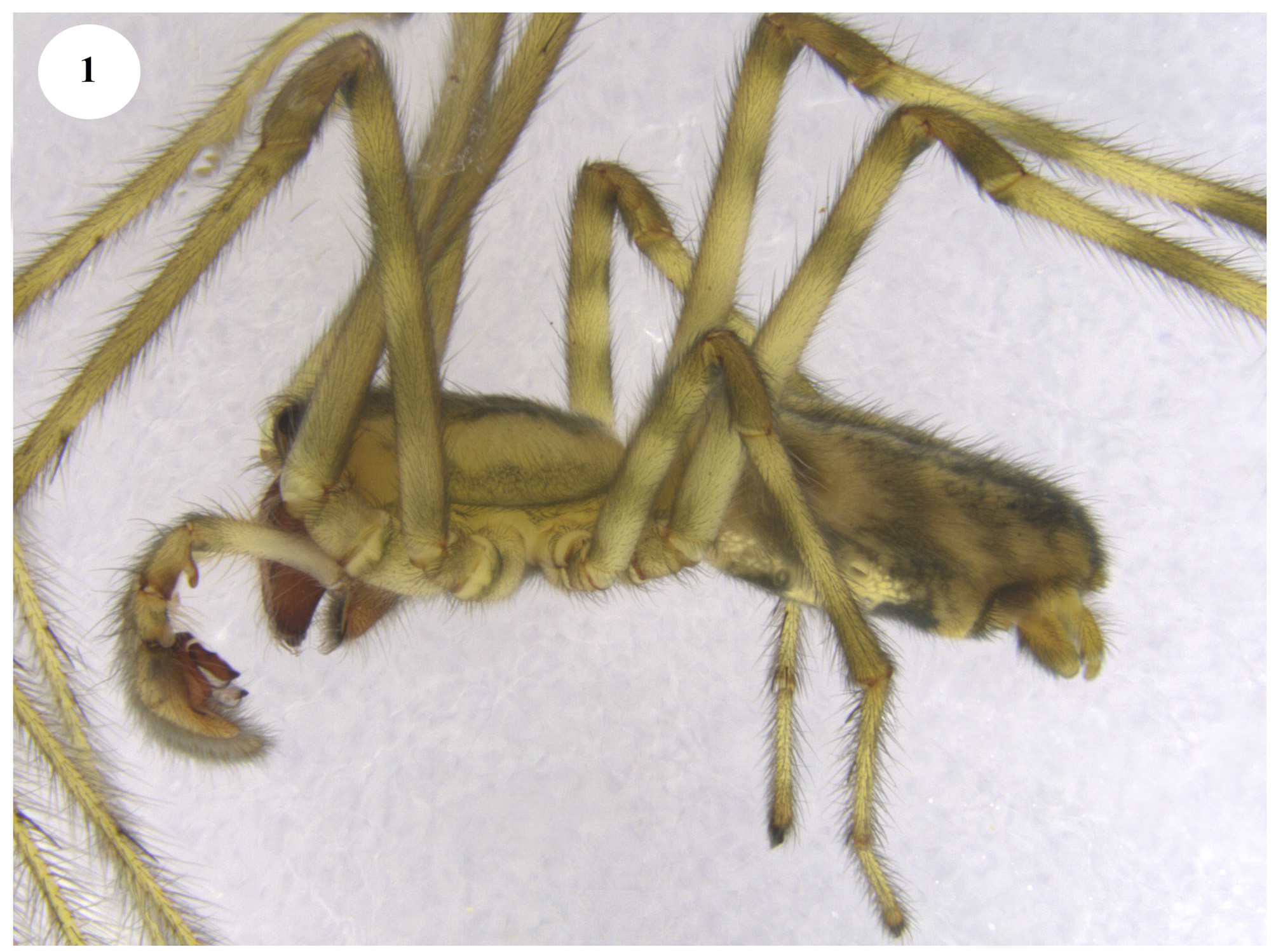
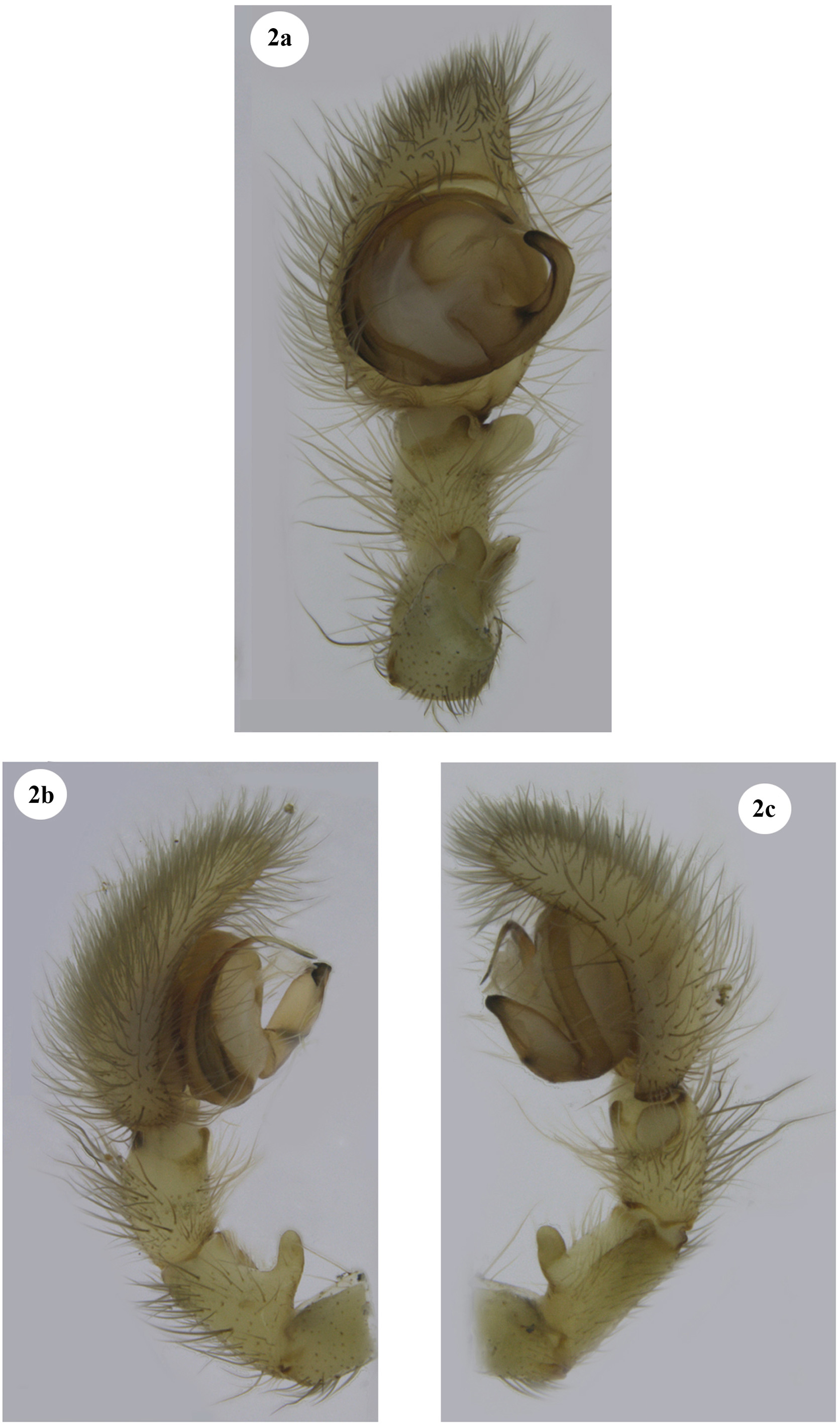
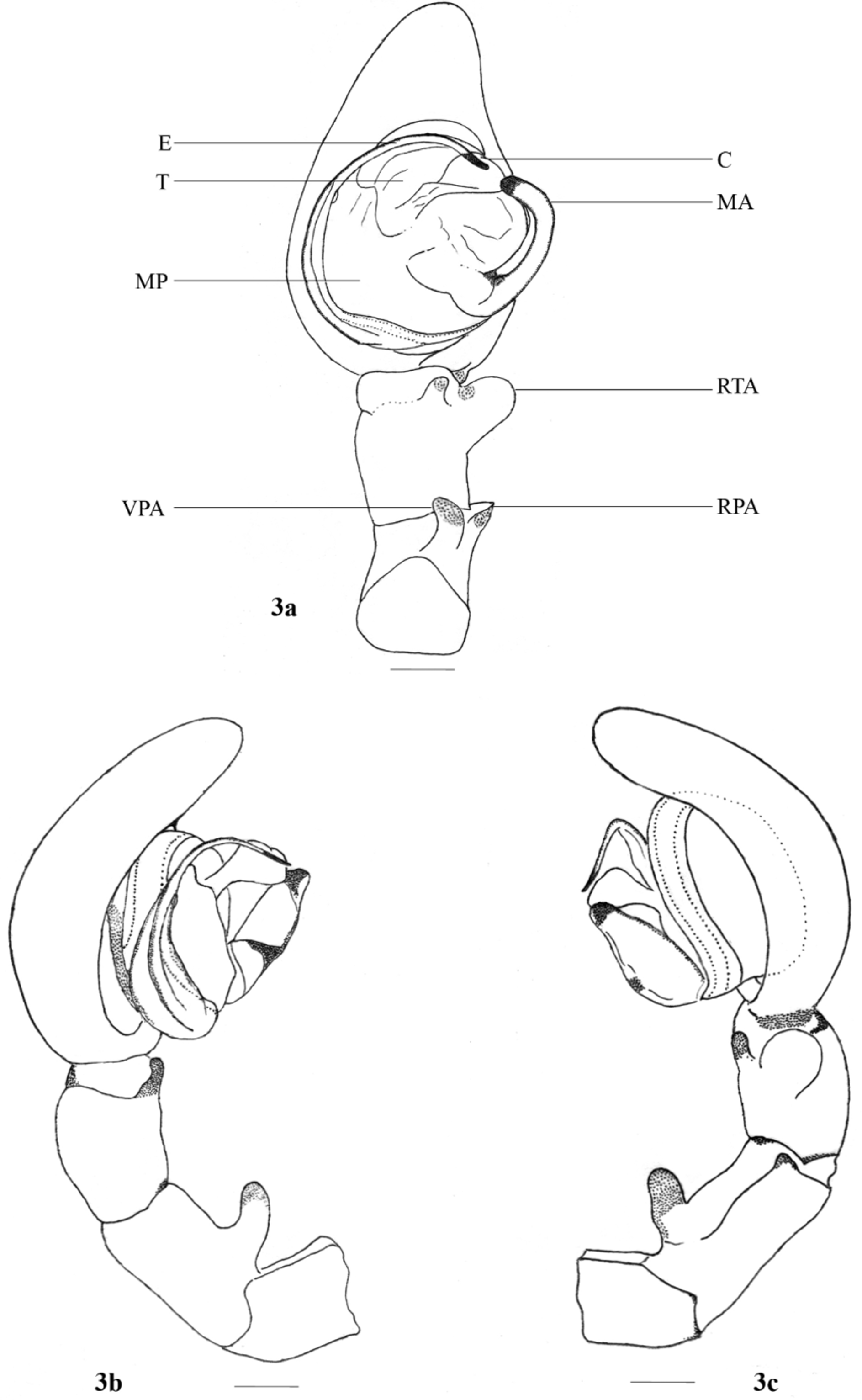
Description. MALE: ( Fig. 1 View FIGURE 1 ). Body length 9.12–10.14 mm. Prosoma length 3.11–3.71 mm, prosoma width 2.98–3.12. Opisthosoma length 6.01–6.42 mm, opisthosoma width 2.60–2.84. Eyes diameter: AME 0.24–0.28 mm. ALE 0.12–0.13 mm. PME 0.17–0.19 mm. PLE 0.11–0.13 mm. Eye interdistance: AME–AME 0.16–0.18 mm. PME–PME 0.22–0.24 mm. AME–ALE 0.97– 0.10 mm. ALE–PLE 0.01–0.03 mm. PME–PLE 0.19–0.21 mm. AME–PME 0.13–0.14 mm. Clypeus height at AMEs 0.25–0.28 mm. Measurements of palp and legs. Palp 4.97– 5.27 [1.99–2.11, 0.83–0.87, 0.54–0.59, 1.61–1.70], I 32.44–38.76 [11.82–13.65, 1.98–2.01, 11.70–13.84, 4.82– 6.03, 2.12–3.23], II 19.51–23.6 [5.58–6.65, 1.22–1. 70, 5.76–6.81, 4.98–5.53, 1.97–2.91], III 8.91–12.82 [2.19– 3.03, 1.02–1.36, 2.2–3.32, 2.36–3.30, 1.14–1.81], IV 16.89–20.72 [5.18–6.34, 1.19–1.51, 5.02–6.16, 3.28–3.98, 2.22–2. 73]. Leg formula: 1243. Spination. Palp: 0 0 0 0, 0 0 0 0, 0000; legs: femur I 0 0 0 0 (right: 1000), II 0 0 0 0, III 0 0 0 0, IV 0000; patellae I–IV 0000; tibia I 0 0 0 0 (right: 0200), II 0 0 0 0 (right: 0100), III–IV 0000; metatarsi I–II 0 601, III 0 300 (right: 1400), IV 0501; tarsi I–IV 0 0 0 0. Copulatory organ ( Figs 2–3 View FIGURES 2 a – c View FIGURES 3 a – 3 c ): conductor membranous and transparent with expanded distal portion and narrow proximal part; the arising point of conductor is at 12’o clock position, while its tip directed at 1’o clock position. Median apophysis large with a small distinct bulge at its proximal third; median apophysis arises more or less centrally, its distal portion curved prolaterally to touch the distal part of the conductor. Embolus long and narrow (almost filiform) and with semi-circular course; distal portions of embolus and MA lie confronting each other; membranous process of tegulum with wavy- inner margin and wide middle portion; retrolateral tibial apophysis stout and somewhat club-shaped, lies near the basal part of cymbium; ventral patellar apophysis finger shaped and originates from the proximal part of patella; retrolateral patellar apophysis is short.
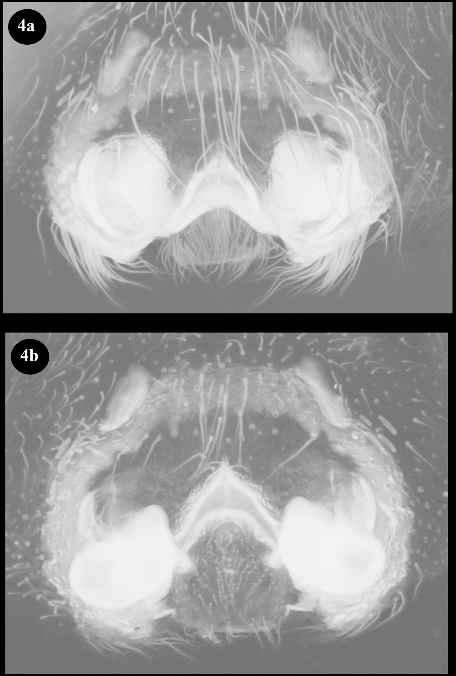
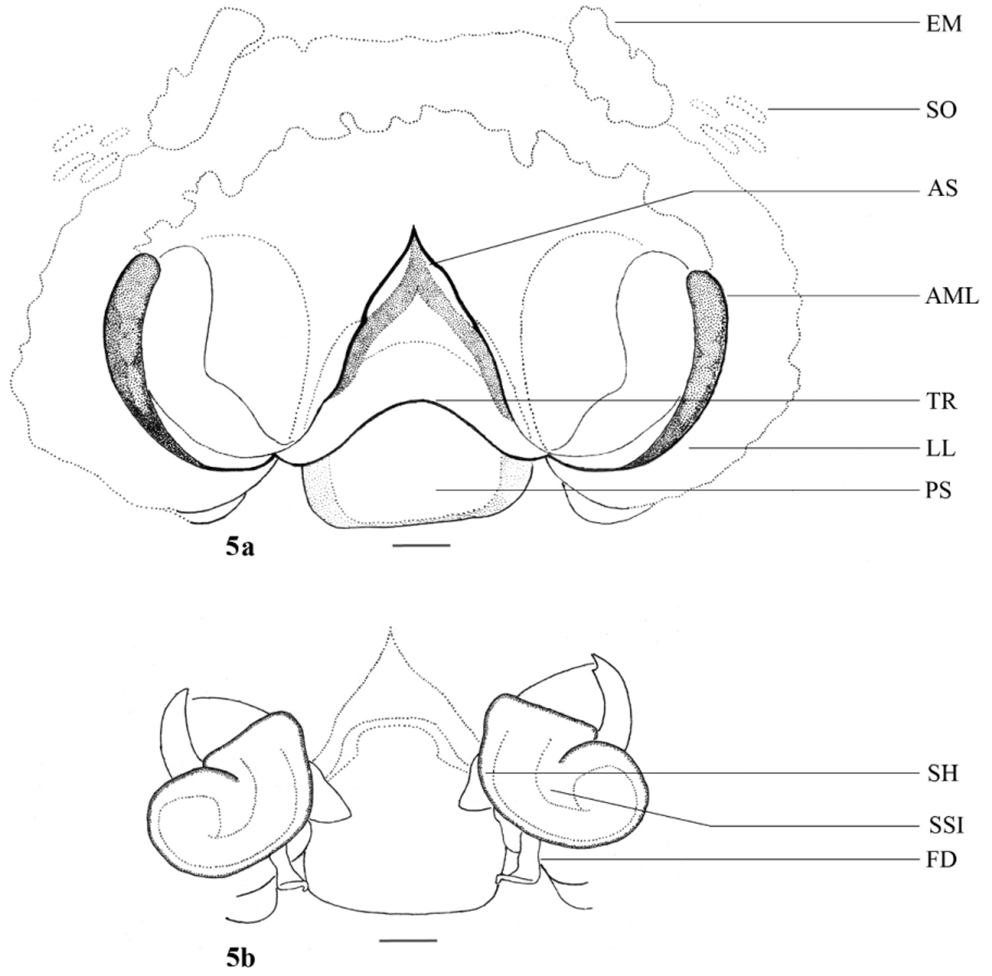
FEMALE: (For description of female, see Bayer 2011; Photos and illustrations of the female copulatory organ of specimens from Kumarakom, Kerala Province, are provided herein, see Figs 4–5 View FIGURES 4 a – 4 b View FIGURES 5 a – 5 b ).
Intraspecific variation. In the present study five males ( Figs 6–10 View FIGURES 6 – 10 ) and four females ( Figs 11–14 View FIGURES 11 – 14 ) of F. travancoria (= F. protensa , see above) collected from the Kumarakom bird sanctuary were examined. It was found that the copulatory organs of all the observed male and female specimens showed some morphological variations. The male copulatory organs showed minimal differences in the size and shape of cymbium, the degree of curvature and direction of median apophysis as well as its size and shape and the direction of embolus tip; size and direction of VPA varied slightly, size of RPA varied a little more. The epigynes and vulvae differed in size and shape of AS and PS, size of AML, size and direction of copulatory duct. However, all these fine morphological differences are recognised as intraspecific variation existing within this species.


No known copyright restrictions apply. See Agosti, D., Egloff, W., 2009. Taxonomic information exchange and copyright: the Plazi approach. BMC Research Notes 2009, 2:53 for further explanation.
|
Kingdom |
|
|
Phylum |
|
|
Class |
|
|
Order |
|
|
Family |
|
|
Genus |
|
Kingdom |
|
|
Phylum |
|
|
Class |
|
|
Order |
|
|
Family |
|
|
Genus |