
Sangabasis furcata ( Brauer, 1868 )
|
publication ID |
https://doi.org/ 10.11646/zootaxa.3815.1.1 |
|
publication LSID |
lsid:zoobank.org:pub:85DD7449-EBB5-4BDF-AB6A-465D926B21EA |
|
DOI |
https://doi.org/10.5281/zenodo.5671764 |
|
persistent identifier |
https://treatment.plazi.org/id/4370710E-D778-FFF9-FF2A-D35BFB39FD7D |
|
treatment provided by |
Plazi |
|
scientific name |
Sangabasis furcata ( Brauer, 1868 ) |
| status |
|
Sangabasis furcata ( Brauer, 1868) View in CoL
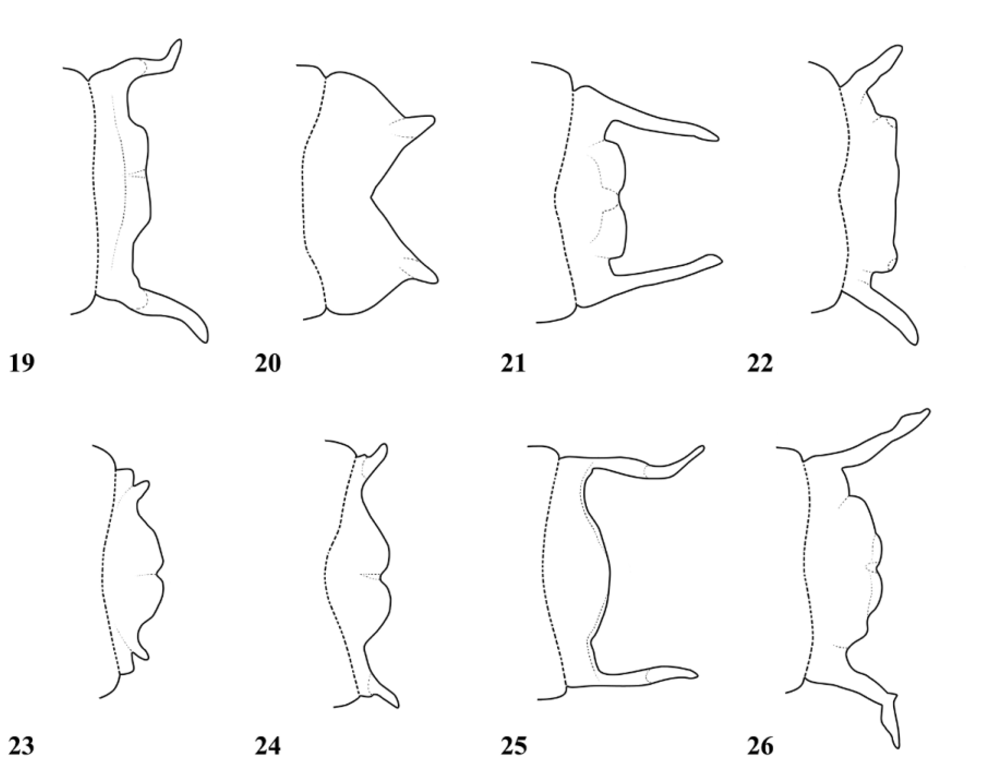
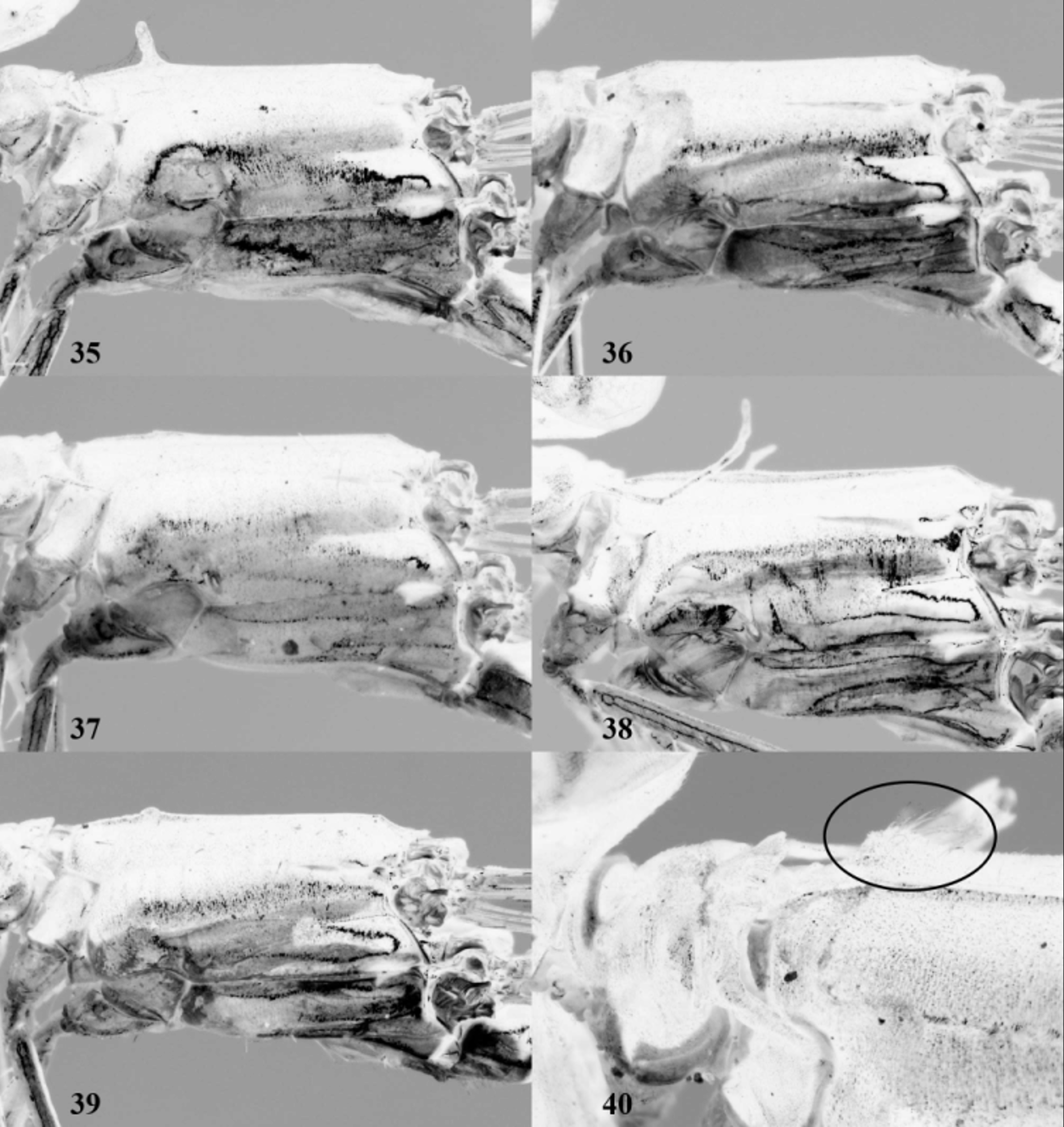
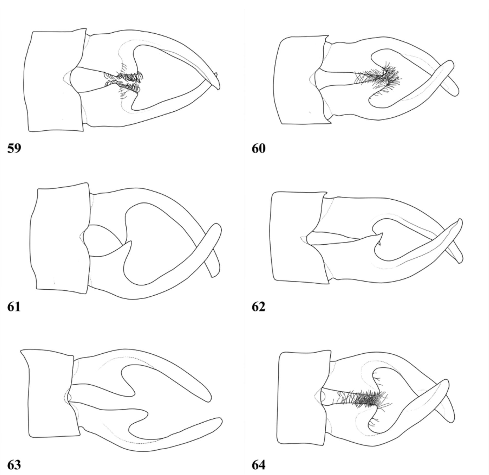
( Figs. 14 View FIGURES 8 – 15 , 26 View FIGURES 19 – 26 , 36, 40 View FIGURES 35 – 40 , 49 View FIGURES 47 – 52 , 61 View FIGURES 59 – 64 , 66 View FIGURE 66 )
Amphicnemis furcata Brauer 1868: 543 View in CoL –544 (original description ♂, Luzon);—Selys (1877: 127–128);— Needham & Gyger (1939: 298–299);— Lieftinck (1940: 362);— Lieftinck (1957: 165–168, Figs.5–6 View FIGURES 1 – 5 View FIGURES 6 – 7 );— Hämäläinen & Müller (1997: 259);— Villanueva (2005: 80, comparison with S. braulitae View in CoL );—Villanueva et al. (2012: 28, Fig. 35 View FIGURES 35 – 40 ).
Sangabasis furcata (Brauer) View in CoL ;—Villanueva (2012: 581, 582, 595, transferred to Sangabasis).
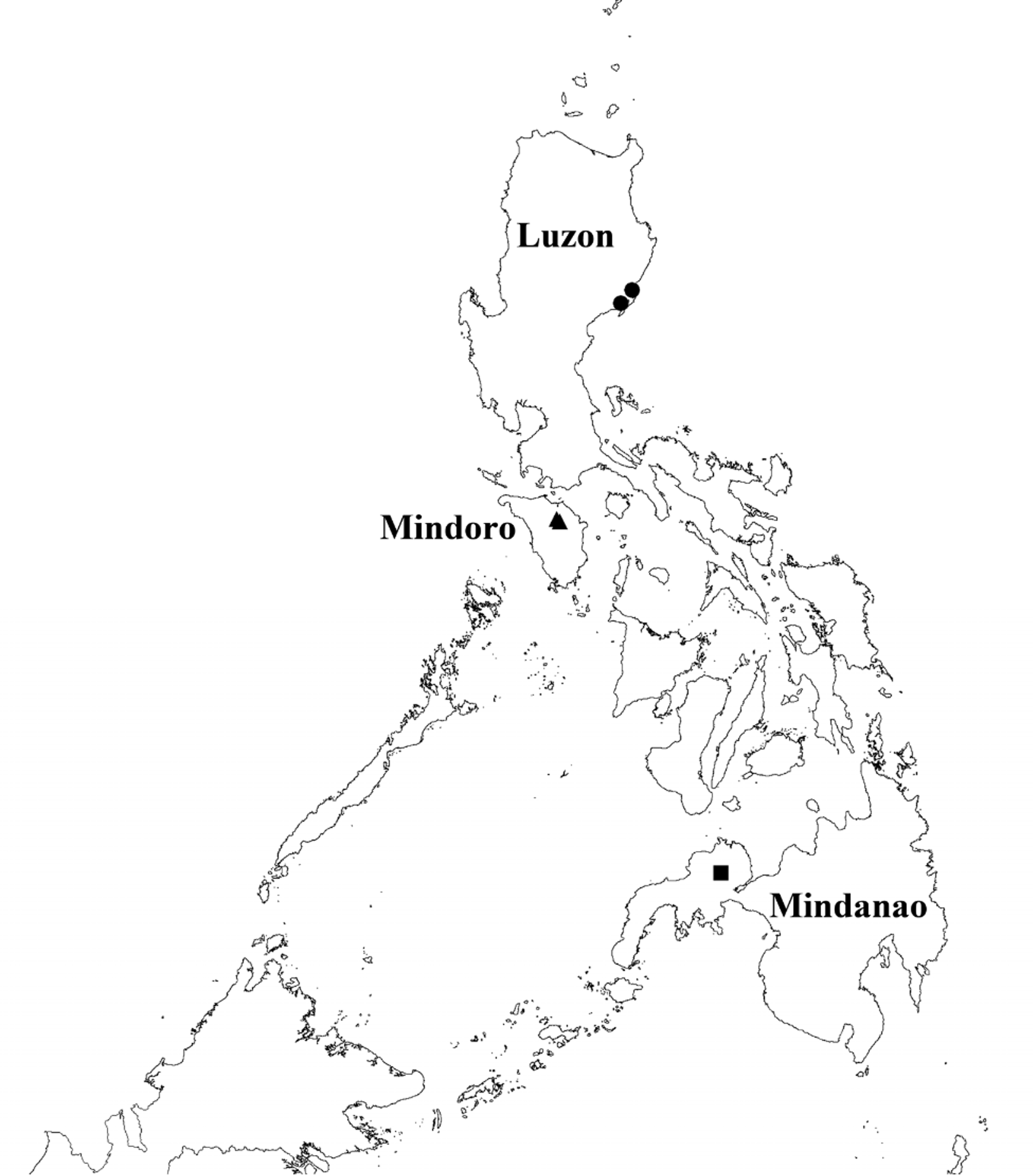
Material. ♂, Philippines, Luzon, Kasiguran, 30 viii 1915, in SMF, leg. G. Boettcher; 3 ♂, ♀, Luzon, Aurora, Dilasag swamp, 5–20 xi 2012, in RMNH, leg. HC.
Description of male (based on specimens from Dilasag swamp): Head: Labium and mandible bases pale, latter with dark central mark. Labrum shiny black except for yellow anterior quarter. Genae pale with dark central mark, pale colour continued to eye margin and narrowly along eye margin from ridge of frons to level of antennal sockets. Anteclypeus pale blue with broad dark central band beneath postclypeus. Postclypeus black. Frons black, with distinct ridge, anterior face with paired transverse yellow streaks separated centrally. Vertex black with metallic reflection. Antennae brown. Remainder of head dark metallic green. Distinct but shallow, ridge-like tubercle situated anteriorly beside eye margin in postocular area.
Thorax: Prothorax dark metallic green except for pale transverse streak on crest of anterior lobe, and lower part of propleuron. Shelf of posterior pronotal lobe ( Fig. 14 View FIGURES 8 – 15 ) short, simple. Horns of posterior pronotal lobe long, rearward and slightly outward directed, running approximately parallel to top of mesinfraepisternum, bent more strongly outward in terminal third. Synthorax ( Fig. 36 View FIGURES 35 – 40 ) with dorsal carina almost smooth. Mesinfraepisternum metallic green with irregular pale borders. Mesepisternum metallic green, with low tubercle covered in setae on either side of middorsal carina immediately behind level of mesinfraepisternum ( Fig. 40 View FIGURES 35 – 40 ). Mesepimeron mostly metallic green with pale streak along interpleural suture in its distal half and along lower margin ( Fig. 36 View FIGURES 35 – 40 ). Metepisternum pale, large metallic green marking adjacent to antealar carina. Metepimeron pale except for black marking at metapleural suture near antealar carina, contiguous with marking on metepisternum. Legs with coxa and trochanter yellowish, femur and tibia pale with dark spines, blackish streaks on extensor surfaces of femur and tibia and dark markings around joint of femur and tibia. Tarsi brown, without denticle. Wings hyaline with dark brown veins. Arc just off Ax2; Ac near Ax2; petiolation ceases before level of Arc. R4 arising at subnodus; IR3 distal to subnodus. 13 Px in Fw, 12 Px in Hw. Pt rectangular with pale margin, costal 2/3 length of subcostal side.
Abdomen: S1–2 dorsum black, becoming pale lower on sides; S3–7 dark brown, paler lower laterally with pale basal ring; S8–10 black with pale lateral marks on S10 and apically on S9. Cerci whitish, more than twice length of S10. Fork of upper branch at ca 1/4 length of upper branch ( Fig. 61 View FIGURES 59 – 64 ). In lateral view upper branch shaped as in Fig. 49 View FIGURES 47 – 52 , with small black tooth at tip. In dorsal view spur triangular, directed strongly inwards, visible in lateral view, almost as long as distance from S10 to fork. Lower branch very short, not reaching base of spur, hidden in normal lateral view. Paraprocts typical for genus.
Measurements (mm): abdomen including cerci 38–41; Hw: 19–21.
Female. Very similar to male except for more pronounced tubercle on mesepisternum. Mesinfraepsternum with broader pale borders. S8–10 with more extensive pale markings: S8 and S10 almost entirely yellow laterally, S9 yellow except for dark greenish dorsal part.
Measurements (mm): abdomen including cerci 36; Hw: 21–23.
Diagnosis. Distinguished from all species except S. bulba , S. cahilogi , S. carmelae , S. hamis and S. janvantoli by the lack of a distinct tubercle on the dorsal carina of the synthorax. Distinguished from S. bulba and S. cahilogi by the structure of the posterior pronotal lobe, from S. hamis by having a lower branch of the cercus and from S. janvantoli by the length of the horns of the posterior pronotal lobe. It differs from S. carmelae in having the tips of the horns of the posterior pronotal lobe somewhat swollen, in lacking a prominent tubercle on the mesinfraepisternum and in the more basal position of the fork of the upper branch of the cercus.


No known copyright restrictions apply. See Agosti, D., Egloff, W., 2009. Taxonomic information exchange and copyright: the Plazi approach. BMC Research Notes 2009, 2:53 for further explanation.
|
Kingdom |
|
|
Phylum |
|
|
Class |
|
|
Order |
|
|
Family |
|
|
Genus |
Sangabasis furcata ( Brauer, 1868 )
| Villanueva, R. J. T. & Dow, R. A. 2014 |
Amphicnemis furcata
| Hamalainen 1997: 259 |
| Lieftinck 1940: 362 |
| Brauer 1868: 543 |