
Hersilia sundaica Baehr & Baehr, 1993
|
publication ID |
https://doi.org/10.5281/zenodo.154560 |
|
DOI |
https://doi.org/10.5281/zenodo.5823118 |
|
persistent identifier |
https://treatment.plazi.org/id/423C87CF-E259-FFCF-FF51-704AFEA0EF80 |
|
treatment provided by |
Carolina |
|
scientific name |
Hersilia sundaica Baehr & Baehr, 1993 |
| status |
|
Hersilia sundaica Baehr & Baehr, 1993 Figs 6-10, 28, 31
Hersilia sundaica Baehr & Baehr, 1993: 58 , figs 38c-f.
NEW MATERIAL: MHNG-PDC-1488756462151465454; Thailand, Phetchabun Province, Khao Kho NP, forest behind park headquarters, 650 m; 1 male ; 10.- 15.11.2006; collected by Malaise trap; leg. P, Dankittipakul. – MHNG-PDC-545464654545787; Thailand, Sakon Nakhon Province, Phu Phan NP, 800 m; 1 female ; 12.- 15.9.2007; collected by Malaise trap; leg. P. Dankittipakul. – TNHM-PDC-8782511532551454514; Thailand, Loei Province, Phu Kradueng NP, 1200 m; 1 female ; 18.- 20.9.2007; collected by Malaise trap; leg. P. Dankittipakul.
REMARKS: Hersilia sundaica belongs to the impressifrons -group which can be easily recognized by the peculiar structure of the male palp: the TA is complicated, provided with: 1) a membranous apical flange with serrated margin (Fig. 8, AF), and a bifurcated apical prong directed postero-retrolaterad (Fig. 8, AP); 2) lateral process of TA (lP) a large sclerotized, C-shaped plate, partially membranous, retrolaterally with a spoon-shaped projection (Figs 6-8, lPr), anteriorly with a median tubercle clearly visible in retrolateral view (Fig. 8, lPt), a membranous flange (Figs 7-8, lPf), and an elongated prong directed posteriad, its apex bifurcated (Fig. 7, lPp); 3) median process of TA (mP) with two large prongs, a basal prong abruptly bent, obliquely directed anteriad (Fig. 7, mPb), apical prong elongated, its apex membranous, fan-like (Fig. 7, mPa). Females are recognized by the protruded epigyne extending posteriorly (Fig. 9), copulatory orifices situated close to excavated posterior margin; vulva (Figs 10, 28) provided with parallel insemination ducts running mid-longitudinally, ascending anteriorly then curving laterally to form large glandular apparatus (Fig. 28); two pilose, spherical receptacula (Fig. 31) with short stalks situated anteriorly. The females examined lack a glandular patch which is present in the female paratype from Indonesia .
NATURAL HISTORY: All specimens examined were collected by means of a Malaise trap suggesting that this species is rather active and does not stay on the same tree as previous observations made us believe.
Hersilia sundaica . (6) Left male palp, prolateral view. (7) Ditto, ventral view. (8) Ditto, retrolateral view. (9) Epigyne, ventral view. (10) Vulva, dorsal view. Scale lines = 1.0 mm.
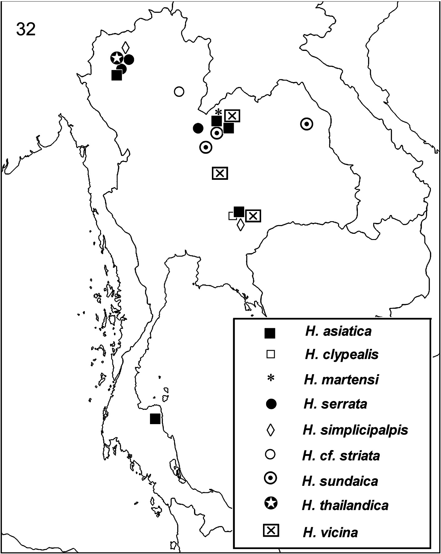
DISTRIBUTION: Indonesia (Lombok, Sumbawa) and Thailand (new record, Fig. 32 View FIG ). Although strong resemblance in genital morphology leave no doubt that the specimens examined belong to this species, it is important to note that the new specimens were collected very far away from the type localities on the Lesser Sunda Islands. Hersilia sundaica seems to have a broad distribution range. Additional material will hopefully become available from SE Asian countries in the future to confirm this .
| NEW |
University of Newcastle |
| TA |
Timescale Adventures Research and Interpretive Center |
No known copyright restrictions apply. See Agosti, D., Egloff, W., 2009. Taxonomic information exchange and copyright: the Plazi approach. BMC Research Notes 2009, 2:53 for further explanation.
|
Kingdom |
|
|
Phylum |
|
|
Class |
|
|
Order |
|
|
Family |
|
|
Genus |
Hersilia sundaica Baehr & Baehr, 1993
| Dankittipakul, Pakawin & Singtripop, Tippawan 2011 |
Hersilia sundaica
| BAEHR, M. & BAEHR, B. 1993: 58 |