
Mannheimsia stylodactyla ( Liu, 1995 ), 2005
|
publication ID |
https://doi.org/10.5281/zenodo.7666888 |
|
persistent identifier |
https://treatment.plazi.org/id/3E7AF841-323A-EB59-5615-FABF3244A84F |
|
treatment provided by |
Felipe |
|
scientific name |
Mannheimsia stylodactyla ( Liu, 1995 ) |
| status |
comb. nov. |
Mannheimsia stylodactyla ( Liu, 1995) View in CoL , comb. n.
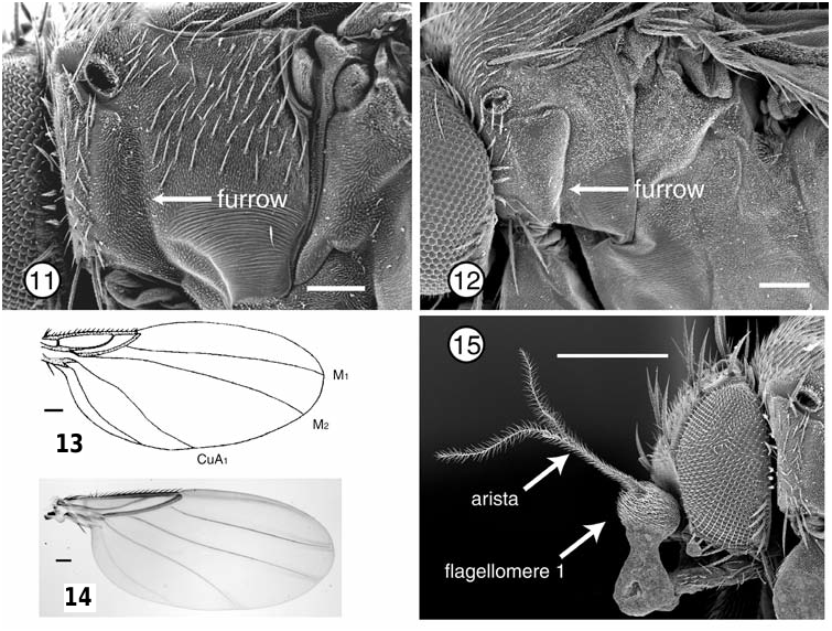
Figs 7–9 View Figs 7–9 , 11 View Figs 11, 12 , 14
Chouomyia stylodactyla Liu, 1995: 186–188 View in CoL , figs 11–20.
Chouomyia ramai Mostovski, 1997: 876 View in CoL , figs 1–8. Syn. n.
Chouomyia dactyloformis Liu, 2001: 129 View in CoL , 231. Lapsus View in CoL for M. stylodactyla View in CoL . Syn. n.
Female abdomen: Tergites 1–6 present; tergite 6 divided medially, but otherwise normal. Venter of abdomen with small setae; posterior margin of segment 6 with longer setae. Segment 7 lacking tergite, but with elongate, narrow, slightly apically clavate sternite. Segments 8–10 without tergites or sternites; cerci present, but relatively short.
Type material: C. stylodactyla Liu , Holotype ^, CHINA: Yunnan: Menglun , 22.v.1991, G. Liu, Y. Wang (Shenyang Agricultural University Collection; examined).
Figs 13, 14. Wings: (13) drawing of Mannheimsia tianzena (Liu) (modified from Liu, 1995); (14) photograph of Mannheimsia stylodactyla (Liu) .
Fig. 15. Scanning electron micrograph of head, Mannheimsia stricta Beyer , right lateral (material adhering to lower margin of flagellomere 1 and palpus is conducting paint).
Scale bar = 0.5 mm, except Fig. 15 = 0.25 mm.
Material examined: THAILAND: Chiang Mai: Doi Inthanon National Park , 1260 m, 1^ 31.i–7.ii.1989, Malaise trap, T . Thormin ( LACM); Maerim (18.29°N: 98.98°E), 1^ 3–26.i.1995, 1^ viii–ix.1995, 4ơ 1^ xi–xii.1995, 1ơ 1^ i–ii.1996, Malaise trap, R GoogleMaps . Beaver ( LACM); Nakhon Ratchasima: Khao Yai National Park , semi-evergreen forest, 780 m, 3^ 11–16.iv.1990, B. Brown, 2^ 11–18.iv.1990, Malaise trap, E. Fuller ( LACM) .
Remarks: Mostovski described C. ramai from Thailand from a specimen extremely similar in appearance to illustrations of C. stylodactyla . In particular, illustrations of the terminalia of the two were almost identical, although the illustrations by both authors, especially of the male terminalia, were not greatly detailed.Mostovski stated that his new species differed from both of Liu’s previously described species by having fewer setae on the palpus, lacking a large seta above the thoracic spiracle, by the different costal index and costal sector measurements, the distribution of setae on the midtibia, and by shape of the epandrium and hypandrium. Furthermore, he stated that C. ramai differed from C. stylodactyla by the dark club of the halter and longer setae on the cercus.
I have several specimens of a species I consider conspecific with C. ramai , as it agrees well with all of Mostovski’s illustrations.After reviewing the supposed differences between this species and the illustrations of C. stylodactyla , however, I consider them to be conspecific. The number of setae on the palpus in my specimens is the same as in C. stylodactyla . The large seta above the spiracle—basal postpronotal seta in the terminology of McAlpine (1981) —is present in my specimens. The differences in wing measurements and small differences in location in midtibial setae are errors in the illustrations of Liu. Part of the difference in male terminalia noted by Mostovski is due to an error in his illustration. The long digitiform structure he illustrates in his fig. 4 as the right surstylus, continuous with the right side of the epandrium, is in fact the medial process of the subepandrial process ( Figs 7, 8 View Figs 7–9 ). The differences in the ventral view of the hypandrium appear to be small variations in angle of observation in the two species. Finally, the halter of both species is dark-coloured, and the illustrations of the terminalia by Liu depict the length of the cercal setae too short.
It is clear from the illustrations of the male terminalia that the subepandrial process has exactly the same structure in both species. Given that there are small errors in the illustrations of both authors, and without other substantive characters that might separate the two, I herein synonymize the names. Additionally, I synonymize the new name C. dactyloformis that Liu (2001) introduced in error in his book on Chinese phorids.
Neither author illustrated the full structure of the hypandrium, probably confusing part of this structure with the aedeagus. Specifically, the right side of the hypandrium has an unusual bilobed secondary process dorsal to the large, more obvious process ( Fig. 9 View Figs 7–9 ).

| T |
Tavera, Department of Geology and Geophysics |
| LACM |
Natural History Museum of Los Angeles County |
| R |
Departamento de Geologia, Universidad de Chile |
No known copyright restrictions apply. See Agosti, D., Egloff, W., 2009. Taxonomic information exchange and copyright: the Plazi approach. BMC Research Notes 2009, 2:53 for further explanation.
|
Kingdom |
|
|
Phylum |
|
|
Class |
|
|
Order |
|
|
Family |
|
|
Genus |
Mannheimsia stylodactyla ( Liu, 1995 )
| Brown, Brian V. 2005 |
Chouomyia ramai
| MOSTOVSKI, M. B. 1997: 876 |
Chouomyia stylodactyla
| LIU, G. 1995: 188 |