
Eumetriochroa araliella Kobayashi, Huang & Hirowatari
|
publication ID |
https://doi.org/10.11646/zootaxa.3619.2.1 |
|
publication LSID |
lsid:zoobank.org:pub:CD8D33DF-DF33-437F-B0C9-5AC26EC36C11 |
|
DOI |
https://doi.org/10.5281/zenodo.6159532 |
|
persistent identifier |
https://treatment.plazi.org/id/3C3BCD7F-A31C-FFEB-FF54-DF26589E84C9 |
|
treatment provided by |
Plazi (2016-04-13 19:54:37, last updated 2024-11-27 02:46:53) |
|
scientific name |
Eumetriochroa araliella Kobayashi, Huang & Hirowatari |
| status |
sp. nov. |
Eumetriochroa araliella Kobayashi, Huang & Hirowatari View in CoL sp. nov.
Figs. 2 View FIGURE 2 E–H, 5, 9, 10, 17.
Diagnosis. All Eumetriochroa species possess a forewing with vein R1 ( Fig. 5 View FIGURE 5 A). The forewing pattern of this species is easily distingished from other species by the three dark greyish-brown oblique streaks ( Fig. 2 View FIGURE 2 E–H). The genital structure of this species is similar to E. hederae Kumata and E. miyatai Kumata , but it is distinguished from them by the bowl-shaped vinculum with saccus long virgulate in the male genitalia ( Fig. 5 View FIGURE 5 D, E) and very small signum in the female genitalia ( Fig. 5 View FIGURE 5 G).
Adult. ( Fig. 2 View FIGURE 2 E–H). Wing expanse 6.0 mm in holotype, 5.0– 8.1 mm (6.9 mm in average of eleven paratype specimens) in paratypes. Vertex and frons lustrous white; vertex with lustrous white scales appressed on occiput. Labial palpus whitish, porrect, slightly upcurved, with pale blackish brown scales in the base. Maxillary palpus absent. Antennae as long as forewing, lustrous white annulated with whitish brown. Thorax white to pale brown. Abdomen dark grey. Anal tuft grey. Forewing. White with dark greyish-brown oblique streaks; first triangular patch from base to 1/5, second broad, at costal 1/3, third linear at costal 1/2, obscure and narrow from middle to dorsum, apical patches at 9/10 of wing. Cilia white and dark grey at costal area with one apical dark grey transverse strigula; sometimes a blackish apical spot at apex; terminal cilia white with fuscous fringe line near termen. Hindwing whitish grey or grey; cilia white. Wing venation ( Fig. 5 View FIGURE 5 A, B).
Male genitalia ( Fig. 5 View FIGURE 5 C–F). Tegumen as long as valva. Vinculum bowl-shaped, with saccus long virgulate. Valva slender, acute at apex, with plumose setae occuring on interior part of apex. Aedeagus tubular, as long as valva; vesica without spines.
Female genitalia ( Fig. 5 View FIGURE 5 G). Apophysis anterioris and apophysis posterioris slender. Ostium bursae membranous; ductus bursae long, tubular. Corpus bursae small, with very small signum on central part.
Pupa. ( Fig. 17 View FIGURE 17 ). Pale yellow to ochreous, 2.8–3.0 mm in length, 0.3–0.4 mm in diameter. Vertex with a short, triangular frontal process ( Fig. 17 View FIGURE 17 B, C, E). Clypeus with a pair of short setae ( Fig. 17 View FIGURE 17 A, B, F). Dorsum of A2–A10 with a concentration of small spines in anterior portion ( Fig. 17 View FIGURE 17 G, H). A10 furcated with a pair of beak-shaped processes from caudal apex, rolled dorsally ( Fig. 17 View FIGURE 17 I, J–L).
Host plant. Dendropanax trifidus (Thunb.) Makino ex Hara , Evodiopanax innovans (Siebold & Zucc.) Nakai , Eleutherococcus sciadophylloides (Franch. & Sav.) H. Ohashi and Fatsia japonica (Thunb.) Decne. & Planch. (Araliaceae) .
Distribution. Japan (Mie, Nara, Fukuoka, Kagoshima (Amami Is.) Prefectures).
Specimens examined
Type material. 15 (53 4Ƥ 6 exs).
Adults: Holotype 3, Japan: Kumawata, Soni, Uda, Nara, 12.x.2011 em., S. Kobayashi, Host: Evodiopanax innovans , 9.x.2011 (ex pupa) (genitalia slide no. OPU-SK366) in OPU. Paratypes 233Ƥ5exs. Same host plants as holotype, [Nishi–rokuban–cho, Yuri–gaoka, Nabari, Mie]: 1Ƥ, 23.xi.2009 em., S. Kobayashi & S. Teramura, 8.xi.2009 (ex larva); 13 1Ƥ 1 ex, 23.x.2010 em., S. Kobayashi, 16.x.2010 (ex larva). [Hikosan, Fukuoka, H. Kuroko leg.]: 2 exs, 10 & 14.xi.1954; 13 1Ƥ, 4 & 22.x.1955. 1 ex, 11.ix.1959, Host: Eleutherococcus sciadophylloides . 1 ex, 3.v. 1957 in OPU. [Kuninao, Yamato, Amami, Kagoshima, S. Kobayashi leg., 6.iii.2012 (ex pupa)]: 13 1Ƥ 1ex, 9–14.iii.2012 em., Host: Dendropanax trifidus ,; 13, 20–22.iii.2012 em., Host: Fatsia japonica Pupae : 5 exs.
[Host: Evodiopanax innovans, S. Kobayashi leg.]: [Nishi–rokuban–cho, Yuri–gaoka, Nabari]: 1 ex, 23.x.2010, 16.x.2010 (larva); 4 exs, 12 & 26.ix.2011, 10.ix.2011 (ex larva).
Etymology. The specific epithet, araliella , is derived from the family name of the host plant, Araliaceae .
Biology. This species has 2–3 generations per year. The larvae emerged from July to November in Nara and Mie Prefectures. We observed larvae on Evodiopanax innovans forming a narrow, long serpentine mine; about 30~ cm in length, clear and colorless. The mines ( Fig. 9 View FIGURE 9 A–D) were only found on the abaxial epidermis of leaves, usually 1–3 mines per leaf. The late instar larva is 3.0–4.0 mm long and pale greenish yellow in coloration ( Fig. 9 View FIGURE 9 E–G). A pupal cocoon fold (white to creamy white, 4.5–5.0 mm in length, 0.8–1.0 mm in width) situated at the end of the mine, usually found along leaf margins ( Fig. 9 View FIGURE 9 H). We also observed the mined leaf of Fatsia japonica to be a narrow, long linear mine (whitish, about 20~ cm in length; 0.6–5 mm in width; brownish frass line: ~1.0 mm in width) ( Fig. 10 View FIGURE 10 C, E, F). A pupal cocoon fold (white, 9.0 mm in length, 2.0 mm in width) situated along leaf margins.
Biotope. The Kumawata valley (type locality of E. araliella ) is part of Tokai Nature Trail connecting Soni Vilage and Uda City (Murō Vil.), Nara Prefecture with a planted forest of Japanese cedar and cypress mixed with fagaceous trees and with few host plants ( Fig. 1 View FIGURE 1 E).
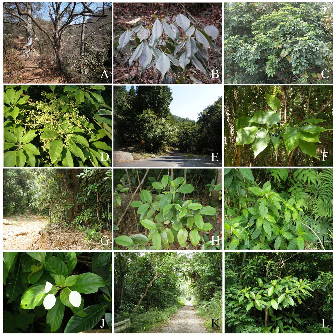
FIGURE 1. Habitats and hostplants of Oecophyllembiinae species. A. Type locality of Metriochroa symplocosella sp. nov., Tianpingshan, Badagongshan NNR, Hunan, China, asl 400 m. B. The shoot of Symplocos sumuntia at the type locality. C. Type locality of Guttigera schefflerella sp. nov., Kuninao, Amami – Oshima, Kagoshima Pref., Japan, asl 100 m. D. The leaves and flowers of Schefflera octophylla at the type locality. E. Type locality of Eumetriochroa araliella sp. nov., Kumawata, Soni, Nara Pref., Japan, asl 700 m. F. The leaves of Evodiopanax innovans at the type locality. G. Type locality of Corythoxestis tricalysiella sp. nov., Maesato, Ishigaki Is., Okinawa Pref., Japan, asl 100 m. H. The shoot of Tricalysia dubia at the type locality. I. Habitat of Corythoxestis sunosei, Takeda, Ishigaki Is., Okinawa, Japan, asl 150 m. J. The leaves and flower of Mussaenda parviflora in the habitat. K. Habitat of Cryphiomystis yaeyamensis, Maesato, Ishigaki Is., Okinawa, Japan, asl 100 m. L. The leaves of Saurauia tristyla in the habitat.

FIGURE 2. Adult of Oecophyllembiinae species from China and Japan. A. Metriochroa symplocosella sp. nov., holotype 3. B. Paratype ♀. C. Guttigera schefflerella sp. nov., holotype 3. D. Paratype ♀. E. Eumetriochroa araliella sp. nov., holotype 3. F. Paratype ♀ mining Evodiopanax innovans. G. Paratype 3 mining Dendropanax trifidus. H. Paratype 3 mining Fatsia japonica. I. Corythoxestis tricalysiella sp. nov., holotype 3. J. Paratype ♀. K. Corythoxestis sunosei (Kumata, 1998) 3 in Japan. L. Corythoxestis yaeyamensis (Kumata, 1998).

FIGURE 5. The wing venation and genitalia of Eumetriochroa araliella sp. nov .. A. Forewing ♀. B. Hindwing ♀. C. Male genitalia, lateral view. D. Left valva, lateral view. E. Male genitalia, ventral view .. F. Aedeagus, lateral view. G. Female genitalia, ventral view.
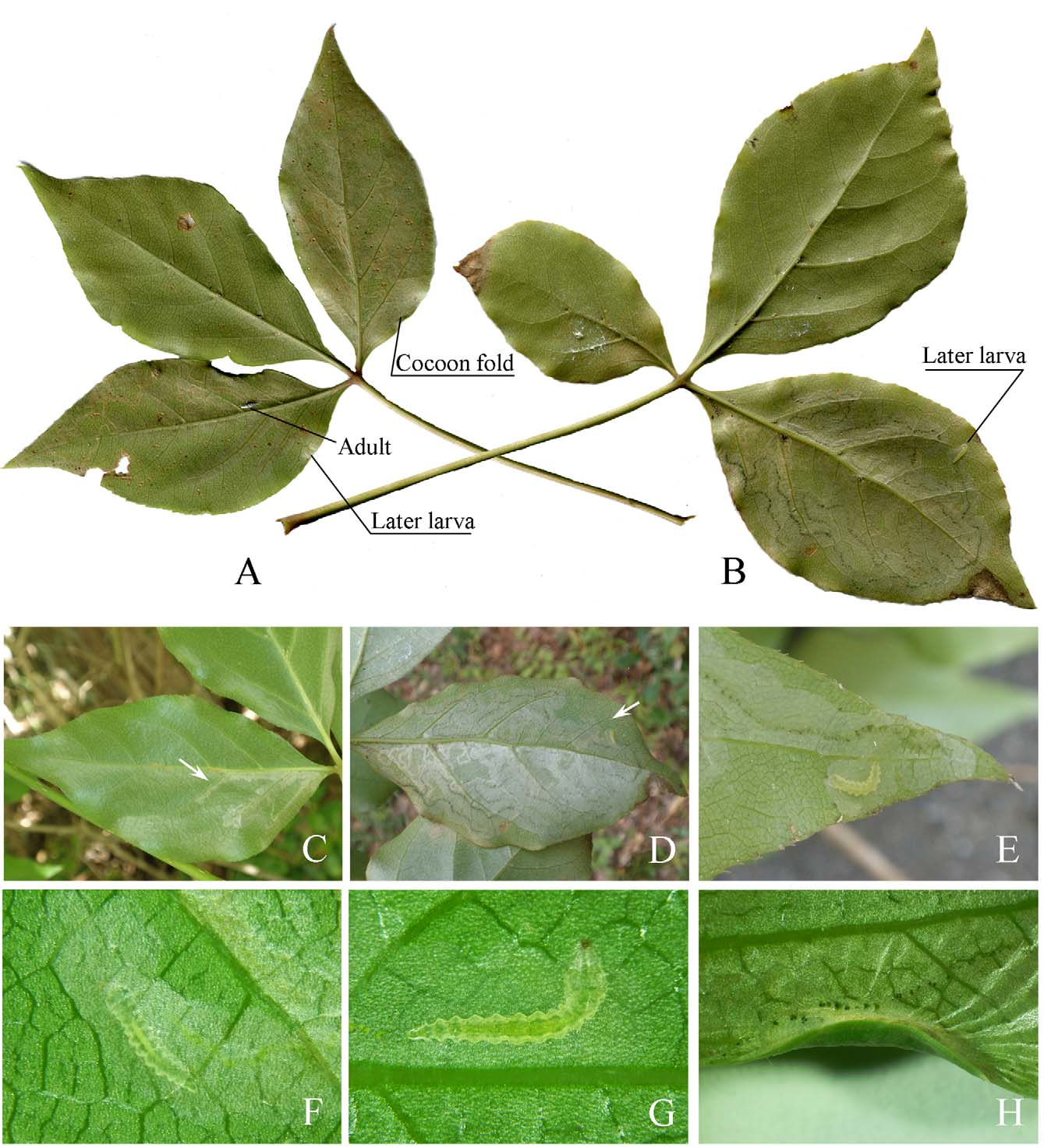
FIGURE 9. Biology of Eumetriochroa araliella sp. nov. on the abaxial epidermis of the leaves of hostplant, Evodiopanax innovans. A, B. The immature stages oh the leaves. C. Mine by young larva. D. Mine by later instar larva. E. Last instar larva. F. Young larva. G. Later instar larva. H. Cocoon fold.
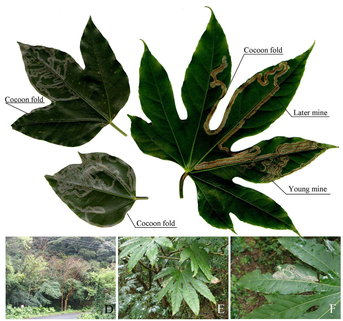
FIGURE 10. Biology of Eumetriochroa araliella sp. nov. on the abaxial epidermis of the leaves of hostplants. A, B. Mines and cocoon folds on Dendropanax trifidus. C. Mines and cocoon folds on Fatsia japonica. D. Habitat of Fatsia japonica, Yu wan, Amami – Oshima, Kagoshima Pref. asl 490 m. E, F. Mines and leaves of Fatsia japonica.

FIGURE 17. Pupa of Eumetriochroa araliella sp. nov .. A. Head, ventral view. B. Cocoon cutter, ventral view. C. Cocoon cutter, dorsal view. D. Head, lateral view. E. Cocoon cutter, lateral view. F. Seta of clypeus. G. Spines on 6 th abdominal tergum. H. Lateral view. I. A 8 – A 10, ventral view. J. lateral view. K. A 6 – A 10, lateral view. L. A 8 – A 10, dorsal view. ds: dorsal seta; ls: lateral seta.
No known copyright restrictions apply. See Agosti, D., Egloff, W., 2009. Taxonomic information exchange and copyright: the Plazi approach. BMC Research Notes 2009, 2:53 for further explanation.
|
Kingdom |
|
|
Phylum |
|
|
Class |
|
|
Order |
|
|
Family |
|
|
Genus |
1 (by plazi, 2016-04-13 19:54:37)
2 (by ImsDioSync, 2016-12-22 17:03:17)
3 (by ImsDioSync, 2016-12-22 17:05:14)
4 (by ImsDioSync, 2017-06-25 14:37:39)
5 (by ExternalLinkService, 2019-09-26 17:35:40)
6 (by ExternalLinkService, 2022-01-30 10:00:40)
7 (by ExternalLinkService, 2022-02-19 09:23:04)
8 (by plazi, 2023-10-26 12:31:49)