
Sticholotis dohrni Weise, 1885
|
publication ID |
https://doi.org/10.11646/zootaxa.2658.1.3 |
|
DOI |
https://doi.org/10.5281/zenodo.5309598 |
|
persistent identifier |
https://treatment.plazi.org/id/3A1BFE7B-FFD0-DC33-2BEF-B2FCFF44FECF |
|
treatment provided by |
Felipe |
|
scientific name |
Sticholotis dohrni Weise |
| status |
|
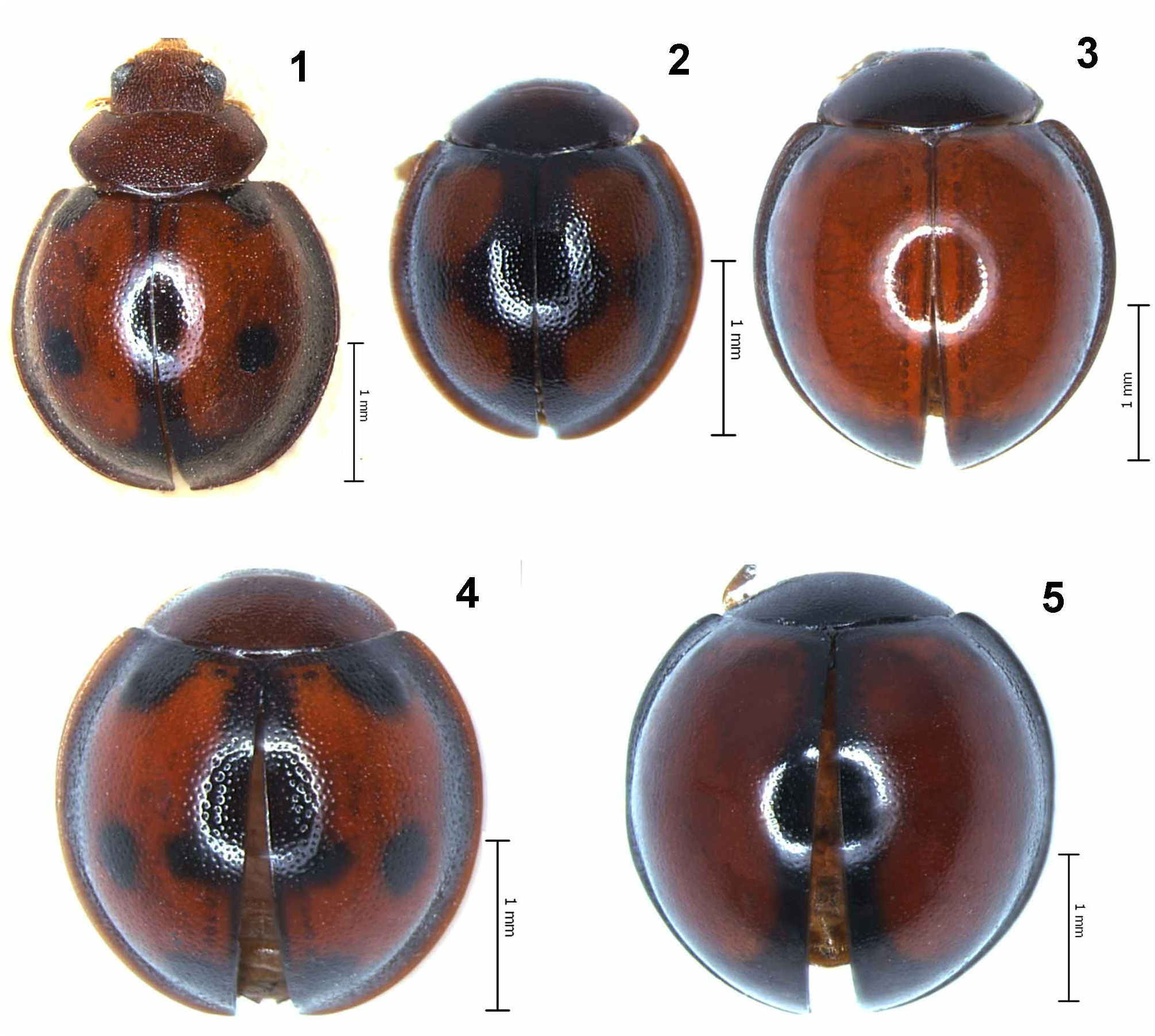
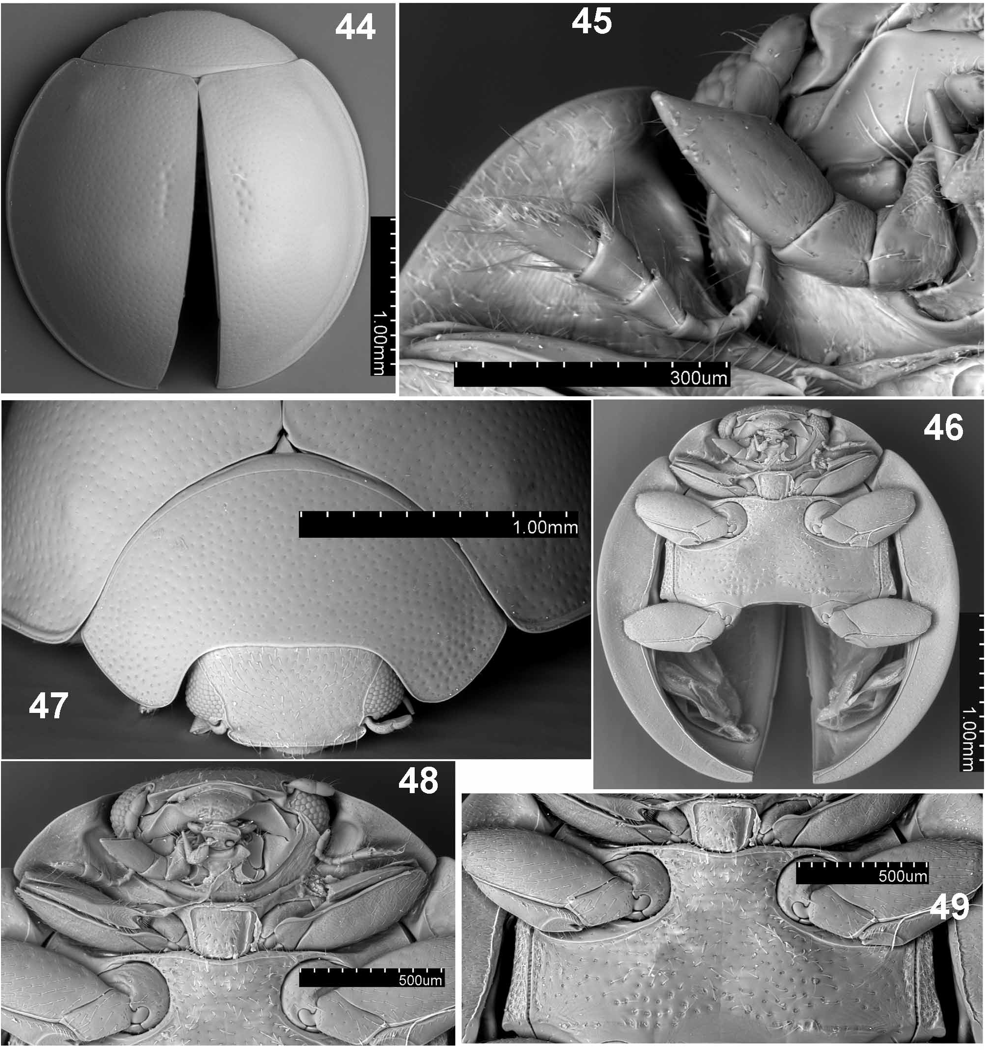
( Figs. 4 View FIGURES 1–5 , 44–55 View FIGURES 44–49 View FIGURES 50–55 )
Sticholotis dohrni Weise, 1885: 240 .
Diagnosis. S. dohrni resembles S. bipunctata , but can be separated by having four, black spots on each elytron, the elytra irregularly punctate with additional short, somewhat regular rows of coarse punctures in mid length near suture, and the venter of prothorax with deep antennal grooves.
Redescription. Male. Length 2.65 mm; TL/EW = 1.1; PL/PW = 0.47; EL/EW = 0.85; GD/TL = 0.6.
Body ( Figs. 4 View FIGURES 1–5 , 44 View FIGURES 44–49 ) rounded, strongly convex; pronotal margins very narrow, hardly visible from above; elytral margins moderately explanate, entirely visible from above. Head and pronotum dark reddish brown; labrum, ventral mouthparts and antennae dark yellowish brown. Scutellum blackish. Elytra predominantly reddish brown with black lateral margins (except for lateral edges reddish brown), and black stripe along suture running from scutellum to about mid length of elytra; each elytron additionally with four, moderately large, black round spots – first one near mid length of basal margin (touching margin), second one on disk slightly posteriad of half length of elytron, third one (smallest one) at posterior end of sutural stripe, appearing as a lateral expansion of this stripe, fourth one just before elytral apex. Punctures on pronotum 1.0–1.5 diameters apart, moderately coarse and dense; punctures on elytra slightly finer and shallower than those on pronotum, 1.5–2.5 diameters apart; additional irregular, short rows of coarse punctures along suture in mid length of elytra; surfaces between elytral and pronotal punctures feebly microreticulate and shiny; dorsum apparently glabrous. Ventral surface dark reddish brown with prosternal process, meso-, metaventrite and intercoxal process of abdominal ventrite I infuscate.
Head ( Fig. 47, 48 View FIGURES 44–49 ) flat medially, punctate, covered with dense and moderately long setae. Clypeus weakly arcuate anteriorly, scarcely reflexed at anterior edge. Eyes moderately large, rather coarsely faceted, dorsally separated by about 4 times eye width; interocular distance nearly 0.65 times head width; inner margins of eyes slightly rounded, convergent anteriorly. Maxilla ( Fig. 45 View FIGURES 44–49 ) with terminal palpomere about 2 times longer than wide, subparallel along basal 2/3 of its length, strongly and obliquely truncate apically; labial terminal palpomere narrowed and acuminate, distinctly narrower than penultimate palpomere. Antenna ( Fig. 45 View FIGURES 44–49 ) 11- segmented with narrow 3-segmented club.
Prothorax ( Fig. 47 View FIGURES 44–49 ) about 0.92 times basal width of elytra; pronotum with groove extending along most of basal margin, disappearing laterally before reaching hind angles; pronotal hypomeron anteriorly and lateral prosternum with distinct antennal groove ( Fig. 45 View FIGURES 44–49 ); anterior lobe of prosternum distinctly bordered with anterior edge straight; prosternal process ( Fig. 48 View FIGURES 44–49 ) subtruncate at apex, with distinct lateral carinae, moderately coarsely and densely punctate, punctures with long setae. Mesoventral intercoxal process ( Figs. 46, 49 View FIGURES 44–49 ) about 1.4 times mesocoxal diameter. Metaventrite ( Fig. 49 View FIGURES 44–49 ) with complete discrimen, densely and moderately coarsely punctate medially and sparsely punctate laterally; postcoxal lines curved and complete. Elytral epipleuron ( Fig. 46 View FIGURES 44–49 ) broad with maximum width at metaventrite, narrowing posteriorly but complete to apex, without distinct foveae. Wings well-developed.
Abdomen ( Fig. 50 View FIGURES 50–55 ) with 5 ventrites; ventrite I along mid line about 3.5 times longer than ventrite II; postcoxal line of first ventrite curved posteriorly and laterally, closely paralleling posterior margin, incomplete laterally; postcoxal disk microreticulate and sparsely punctate ( Fig. 51 View FIGURES 50–55 ); ventrite V arcuate. Abdominal segment VIII with sternite divided in two parts ( Fig. 52 View FIGURES 50–55 ). Male genital segment ( Fig. 53 View FIGURES 50–55 ) with sternite round-oval, apophysis absent.
Male genitalia as in Figs. 54, 55 View FIGURES 50–55 . Tegminal basal piece with distinct strut and additional, dorsal strut-like projection of nearly the same length; parameres long and thin with long setae at their apices; penis with large capsule at base, and with two small teeth near apex.
Female not known.
Material examined. Types. Neotype (here designated), male: “Tenasserim Meetan, Fea. Apr. 1887 / Gorham type/ Orcus bipunctatus Gorham / Syntypus, Orcus bipunctatus var. Gorham, 1895 / Museo Civico di Genova ” ( MCSN).
Note. After an extensive and unsuccessful search for the type of S. dohrni in many European museums where the Dohrn and Weise collections are deposited, we consider it lost. Therefore, the incorrectly placed syntype of O. bipunctatus , which is actually a member of S. dohrni , is designated here as the neotype of this species. The designation of the neotype for S. dohrni Weise, 1885 , is made to fix the taxonomic status of this species.

| MCSN |
Museo Civico di Storia Naturale, Verona |
No known copyright restrictions apply. See Agosti, D., Egloff, W., 2009. Taxonomic information exchange and copyright: the Plazi approach. BMC Research Notes 2009, 2:53 for further explanation.
|
Kingdom |
|
|
Phylum |
|
|
Class |
|
|
Order |
|
|
Family |
|
|
Genus |
Sticholotis dohrni Weise
| Tomaszewska, Wioletta & Łączyński, Piotr 2010 |
Sticholotis dohrni Weise, 1885: 240
| Weise, J. 1885: 240 |