
Russula purpureozonata K.Das, A.Ghosh & Buyck, 2021
|
publication ID |
https://doi.org/ 10.5852/ejt.2021.782.1595 |
|
DOI |
https://doi.org/10.5281/zenodo.5788665 |
|
persistent identifier |
https://treatment.plazi.org/id/370987F0-FFAE-FFAF-A28E-FAA4FD5FFE02 |
|
treatment provided by |
Felipe |
|
scientific name |
Russula purpureozonata K.Das, A.Ghosh & Buyck |
| status |
sp. nov. |
Russula purpureozonata K.Das, A.Ghosh & Buyck View in CoL sp. nov.
Diagnosis
Russula purpureozonata sp. nov. is a blackening species with a dark purplish concentric zonation on the pileus surface and otherwise typical features of subsect. Decolorantes; it differs from the microscopically similar, North American R. burkei Burl. in its mild taste and ectomycorrhizal association with Abies densa , and from North American R. californiensis and R. magna in its distinctly smaller spores.
Etymology
Refers to the dark purplish concentric zones on the pileus surface.
Material examined
Holotype INDIA • Sikkim, East district, Memeinchu ; 27°21.108′ N, 88°49.660′ E; alt. 3539 m a.s.l.; on the soil under Abies densa ; 2 Aug. 2018; K. Das, KD 18-003; GenBank: MN267570 View Materials (ITS); CAL[1817] . GoogleMaps
Additional material
INDIA • Sikkim, East district, opposite to Gnathang firing range forest; 27°18.605′ N, 88°48.794′ E; alt. 3885 m a.s.l.; on the soil under Abies densa ; 5 Aug. 2018; K. Das, KD 18-15 (CAL 1818); GenBank: MN269951 View Materials (ITS); CAL[1818] GoogleMaps .
MycoBank: MB 838572; Index Fungorum number: IF558127; Facesoffungi number: FoF 09580
Description
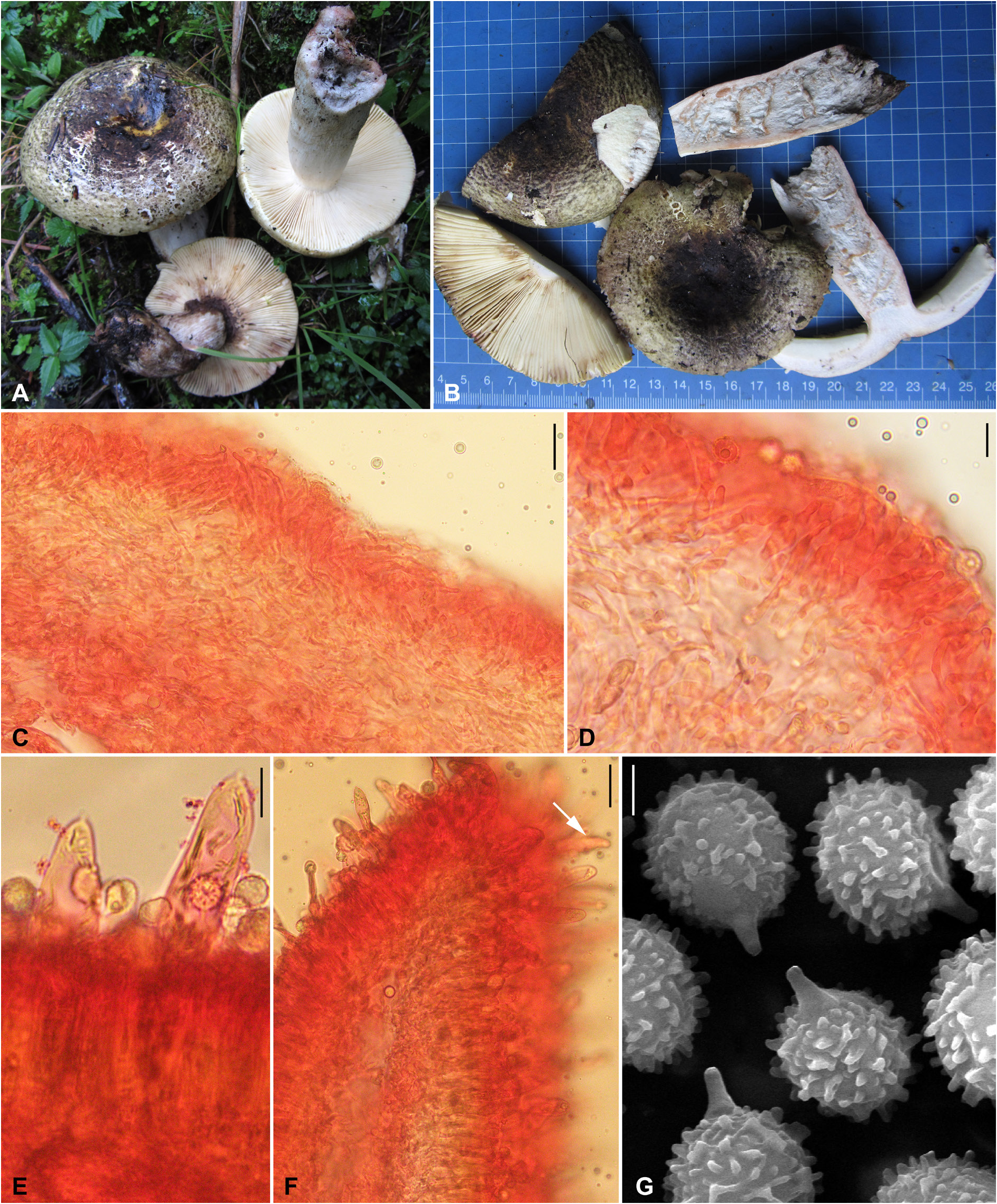
Pileus large-sized, 85–105 mm diam., hemispherical when young, then convex, plano-convex to applanate, broadly but shallowly depressed in the centre when mature; margin decurved to plane with maturity, entire; surface viscid when moist, then dry, finely areolate, peeling to ½ of the radius, greyish yellow (2C3–4) or olive to linden green (2D5–6), centrally dark brown (7F4–5) to dark purple or purple-black with pastel yellow to light yellow patches (3A4–5), fading towards mid in distinctive concentric zones. Pileus context up to 8 mm thick, thinning towards margin, firm, brittle, chalky white (1–2A2), changing first orange-red, then black when cut or bruised; turning greyish red to reddish brown (8C–D5) and dull green (26E3–4) with guaiacol and FeSO 4, respectively. Lamellae adnexed to free, subdistant (6–7/cm at pileus margin), yellowish white (2–3A2), forked near the stipe apex; edge even and concolorous. Stipe 70–100 × 20–30 mm, subclavate to clavate, tapered at the apex, central, solid; surface dry, finely longitudinally venose, chalky white (1–2A1) with yellowish white to pale yellow (4A2–3) flush at one side of the centre, changing first orange-red, then black when cut or bruised. Stipe context solid, chalky white (1–2A1), changing first orange-red, then black when cut or bruised; turning greyish red to reddish brown (8C–D5) and dull green (26E3–4) with guaiacol and FeSO 4, respectively. Odour indistinctive. Taste mild. Spore print yellowish white (3A2).
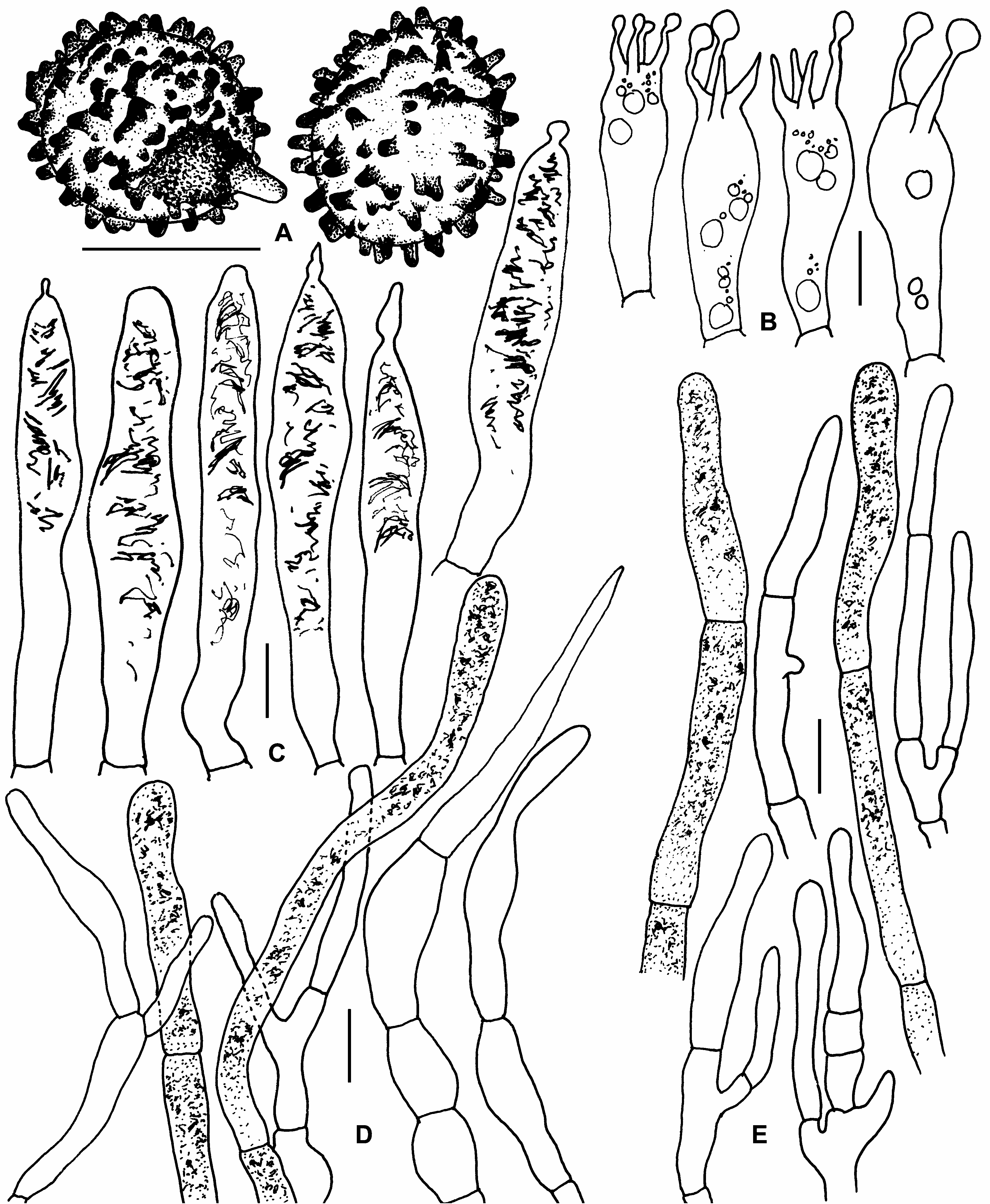
Basidiospores globose, subglobose to broadly ellipsoid, (7.4–)7.6–8.02–8.4(–8.9) × (6.5–)6.8–7.2–7.7 (–8) μm, Q = (1.04–)1.07–1.11–1.15(–1.23), ornamentation amyloid, composed of somewhat cylindric (mostly with rounded or obtuse apices) isolated very small (0.3 μm) to high (up to 1.2 μm) spines and most of spines fused laterally and connected by thin to thick ridges (like small crests) forming partial reticulum; suprahilar plage amyloid; apiculi up to 2 μm high. Basidia (35–)41–49–57(–68) × 10–11–12(–14) μm, 4-spored, subclavate to clavate. Subhymenium layer 20–25 μm thick, made up of pseudoparenchymatous cells. Hymenial cystidia on lamellar sides (63–)71.5–86–101(–134) × (9–) 9.5–11–12.5(–14) μm, subcylindrical, cylindrical to ventricose with capitate, mucronate, moniliform or appendiculate (up to 7 μm long appendage) apex, emergent up to 55 μm beyond the basidiole tips; contents dense, heteromorphous and partly or completely filled with fibrous to somewhat crystalloid components, hardly staining in sulfovanillin. Lamellae edges fertile with frequent basidia. Hymenial cystidia on lamellar edges (72–)75–84–92.5(–98) × (9–)9.6–10.5–11.5(–12) μm, subcylindrical, cylindrical to ventricose with obtuse-rounded, capitate, mucronate or appendiculate apex; contents dense, heteromorphous and partly or completely filled with fibrous to somewhat crystalloid components, hardly staining in sulfovanillin. Hymenophoral trama composed of numerous sphaerocytes and connecting hyphae; sphaerocytes globose to elliptical. Pileipellis orthochromatic in Cresyl Blue, sharply delimited from the underlying sphaerocytes of the context, 120–180 μm thick, two-layered, distinctly divided in 50–80 μm deep suprapellis composed of erect or ascending hyphal terminations, arranged in a densely turf of trichodermal structure and dispersed pileocystidia, and subpellis 70–100 μm deep, composed of more or less horizontally irregularly oriented, moderately dense, 2.5–3.5 μm wide pilear hyphae. Acid-resistant incrustations absent. Hyphal terminations near the pileus margin usually branched at the subterminal cells or the cells just below, occasionally slightly flexuous, thin-walled; terminal cells (13–)15.5–24.5–34(–53) × 3–3.5–4.5(–5) μm, mainly subulate to tapering towards tips or cylindrical to subcylindrical, apically obtuse or slightly narrowed towards tips and wider near base; subterminal cells usually equal in size, rarely with lateral branches or nodulose, equally wide or more or slightly wider. Hyphal terminations near the pileus centre with shorter but slightly wider terminal cells measuring (9–)17–22.5–28.5(–35) × (2.5–)3–4–4.5(–6) μm, mainly tapering to subulate towards tips or cylindrical or ventricose or occasionally lageniform, apically obtuse or slightly narrowed towards tips and wider near base; subterminal cells equally wide, sometimes slightly wider or ventricose, rarely with nodulose or with lateral branches. Pileocystidia near the pileus margin 1–4-celled, numerous, cylindrical, usually originating deep in subpellis and often originating from branched subterminal cells, thin-walled; terminal cells (30–)32.5–52–71(–108) × (4–)4.5–5–6(–6.5) μm, cylindrical or sometimes slightly tapered towards tips, rounded-obtuse, without any incrustations; contents heteromorphous-crystalline, without reaction in sulfovanillin. Pileocystidia near the pileus centre with often more septa (1–7); terminal cells (14–) 22.5–38–54(–73) × (4–)4.5–5–6(–7) μm, cylindrical or slightly tapered towards tips, rounded-obtuse apex. Clamp connections absent from all tissues.
| MB |
Universidade de Lisboa, Museu Bocage |
No known copyright restrictions apply. See Agosti, D., Egloff, W., 2009. Taxonomic information exchange and copyright: the Plazi approach. BMC Research Notes 2009, 2:53 for further explanation.
|
Kingdom |
|
|
Phylum |
|
|
Class |
|
|
Order |
|
|
Family |
|
|
Genus |