
Russula adwanitekae A.Ghosh, K.Das & Buyck, 2021
|
publication ID |
https://doi.org/10.5852/ejt.2021.782.1595 |
|
DOI |
https://doi.org/10.5281/zenodo.5788663 |
|
persistent identifier |
https://treatment.plazi.org/id/370987F0-FFAD-FFA3-A2F5-FD85FDF8FB6B |
|
treatment provided by |
Felipe |
|
scientific name |
Russula adwanitekae A.Ghosh, K.Das & Buyck |
| status |
sp. nov. |
Russula adwanitekae A.Ghosh, K.Das & Buyck View in CoL sp. nov.
Diagnosis
Russula adwanitekae sp. nov. is mainly separated from similarly looking species with reddish lilac, orchid purple or greyish to deep magenta colored pileus in subsect. Laricinae by its sequence data (nrITS) and geographic distribution.
Etymology
After the name of the forest locality, Adwani-Teka.
Material examined
Holotype INDIA • Uttarakhand, Pauri Garhwal, Adwani-Teka forest ; 30°05.681′ N, 78°43.890′ E; alt. 1989 m a.s.l.; in temperate mixed forest; 3 Oct. 2016; A. Ghosh, AG 16-1430; GenBank: MN263242 View Materials (ITS); CAL[1821] . GoogleMaps
Paratype INDIA • Uttarakhand, Pauri Garhwal, Adwani-Teka forest ; 30°05.675′ N, 78°43.880′ E; alt. 1953 m a.s.l.; in temperate mixed forest; 5 Oct. 2016, A. Ghosh, AG 16-1435; GenBank: MN263243 View Materials (ITS); CAL[1822] GoogleMaps .
MycoBank: MB 838571; Index Fungorum number: IF558126; Facesoffungi number: FoF 09581
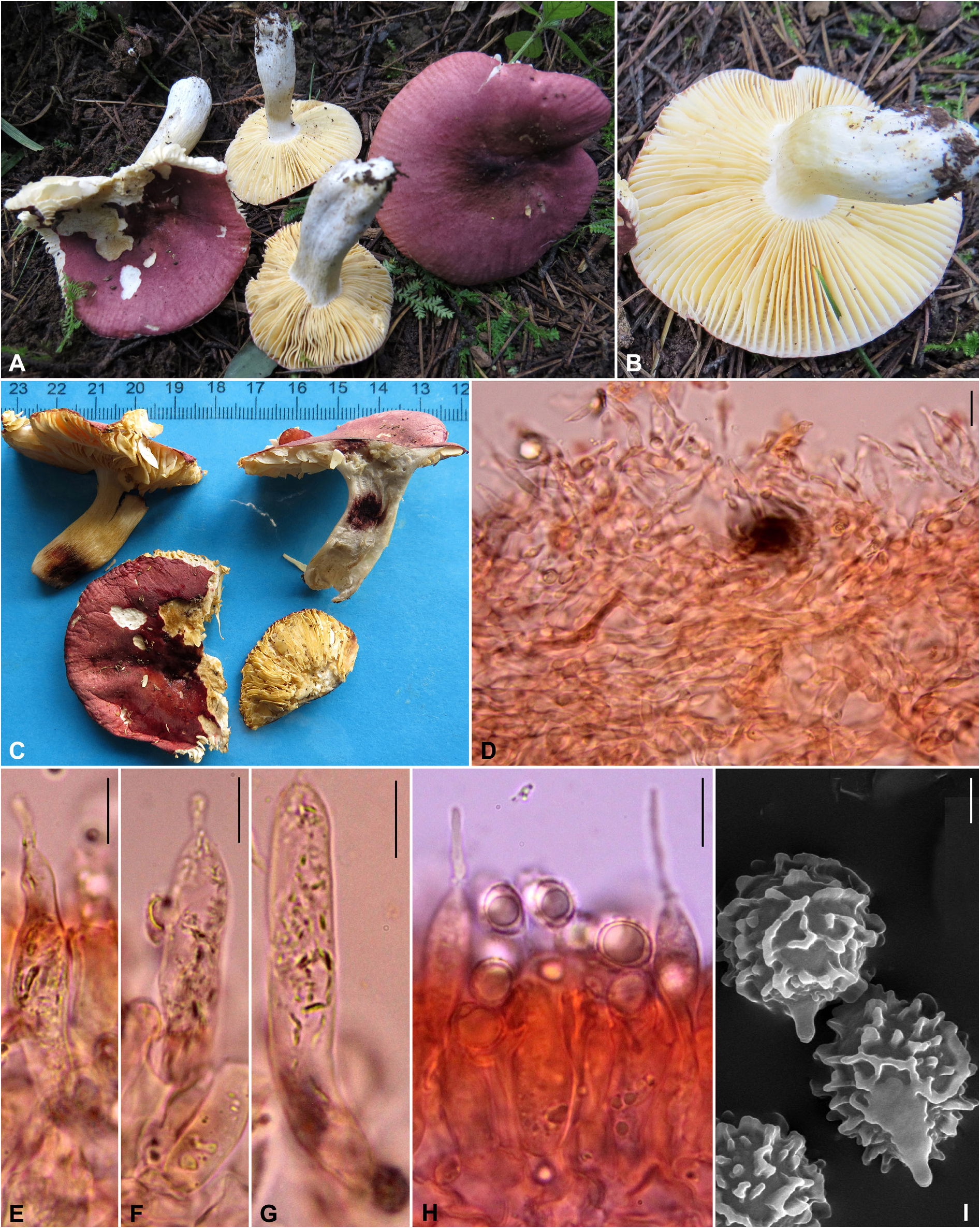
Description
Pileus small to medium-sized, 36–53 mm diam., convex when young, becoming plano-convex, applanate to uplifted with maturity, broadly depressed centre, near the margin becoming tuberculately striate and more or less straight with age, entire; pileipellis peeling to mid-radius; surface dry, shiny and viscid when moist, glabrous, reddish lilac (14C3–5), orchid purple (14C8) or greyish magenta (14D5–7) to deep magenta (14D8) and centrally dark magenta (13–14F4–8). Pileus context firm, brittle, yellowish white (1–2A2), unchanging after bruising, on exposure or with age. Lamellae adnexed to subdecurrent, equal but mixed with some dispersed lamellulae of different length, close (10–12/cm at pileus margin), cream to pale yellow or light yellow (4A2–4), up to 6 mm broad, occasionally forked near the stipe apex; lamella edge even and concolorous. Stipe 35–51 × 7–14 mm, cylindrical to subclavate, tapered at the apex, central, solid but not firm; surface dry, finely longitudinally venose, chalky white (1–2A1) but with yellowish white to pale yellow (4A2–3) flush near its middle portion, changing greyish white (1–2B1) after bruising. Stipe context solid, chalky white (1–2A1), changing dirty white or greyish white (1–2B1) after bruising or on exposure; turning reddish brown (8E7–8) to dark brown (8F7–8) with application of guaiacol. Odour indistinctive. Taste mild. Spore print not obtained.
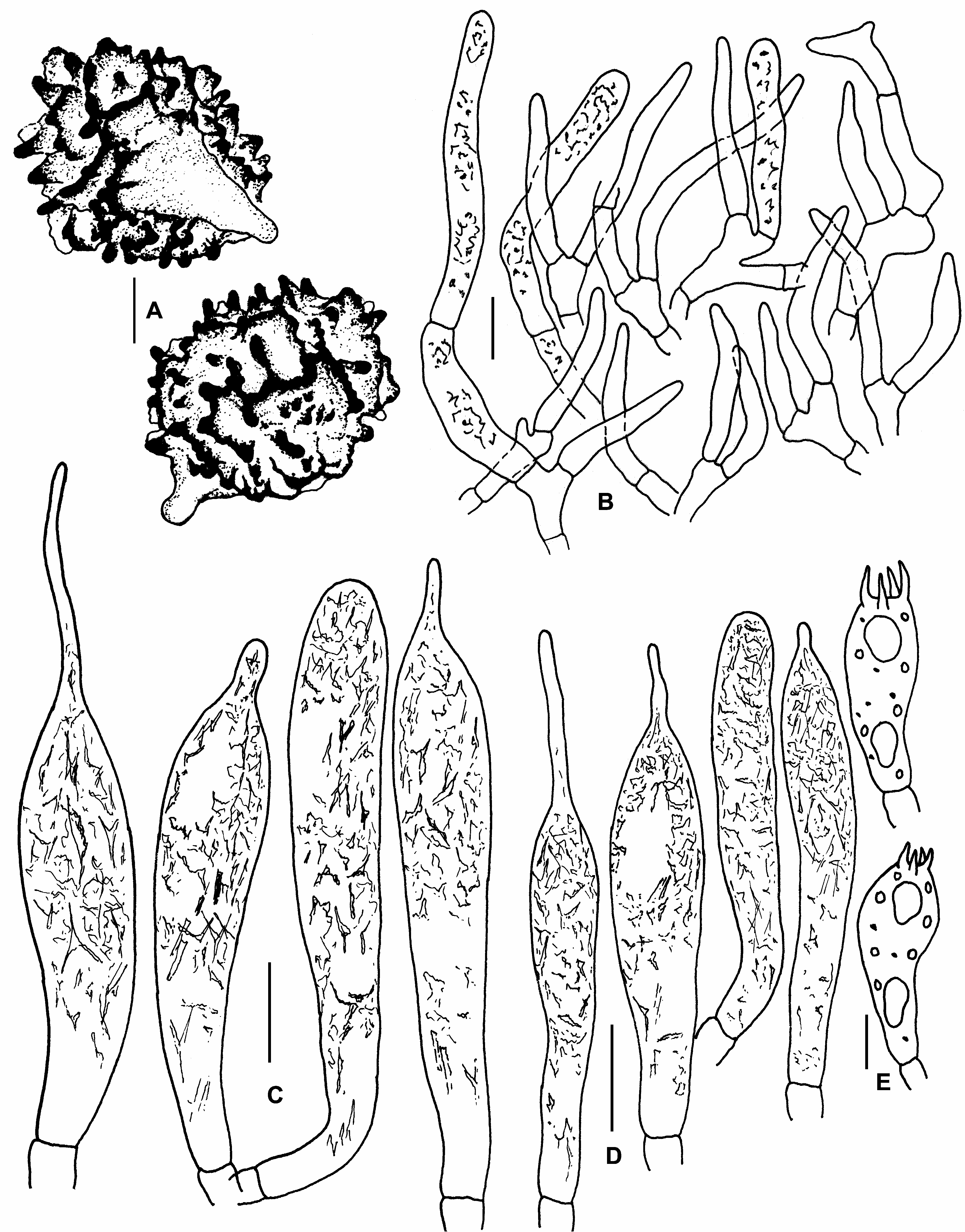
Basidiospores subglobose to broadly ellipsoid, rarely ellipsoid, (7–)7.73–8.23–8.72(–9.5) × (6–)6.6– 6.93–7.3(–8) μm, Q = (1.07–)1.13–1.19–1.25(–1.33), ornamentation amyloid (up to 1.7 µm high), consisting of thick ridges and warts forming incomplete reticulum, with some isolated intermediate warts; suprahilar plage amyloid; apiculi up to 2 µm high. Basidia (29–)32–35–38(–40) × (10–)10–11–13 (–15) µm, 4-spored, subclavate to clavate; sterigmata up to 8 µm long. Subhymenium layer up to 37 µm thick, pseudoparenchymatous. Hymenial cystidia on lamellar sides (55–)61.5–71.5–81(–92) × (7–)8– 9.5–10.5(–11) µm, cylindrical, subclavate to fusiform with frequent lageniform (up to 23 µm long) or appendiculate or few rounded-obtuse apices, emergent up to 32 µm beyond the basidiole tips; contents crystalline, without reaction in sulfovanillin. Lamellae edges fertile with basidia and cystidia. Hymenial cystidia on lamellar edges (37–)42.5–52.5–62.5(–66) × (6–)6–7–8(–9) µm, cylindrical to subclavate with lageniform (up to 20 µm long) or appendiculate or rounded apex; contents crystalline, without reaction in sulfovanillin. Hymenophoral trama mainly composed of large nests of sphaerocytes and few hyphal elements. Pileipellis orthochromatic in Cresyl Blue, sharply delimited from the underlying sphaerocytes of the context, 100–120 μm thick, two-layered, distinctly divided in 40–50 μm deep suprapellis composed of erect or ascending hyphal terminations, arranged in a trichodermal structure and dispersed pileocystidia, and subpellis 60–70 μm deep, composed of more or less dense, horizontally oriented hyphae.Acid-resistant incrustations absent. Hyphal terminations near the pileus margin usually branched at the subterminal cells or the cells just below, thin-walled; terminal cells (11–)19–28.5–37.5(–50) × 3–4–4.5(–6) μm, mainly subulate, sometimes cylindrical to subcylindrical, apically obtuse or slightly narrowed towards tips and wider near base or sometimes attenuated; subterminal cells usually equal in size, sometimes with lateral branches; near the pileus centre with slightly shorter and less wide terminal cells, measuring (15–)18.5–25–32(–44) × (2–)2.5–3–3.5(–4) μm, but equally wide subterminal cells. Pileocystidia near the pileus margin 1–4-celled, cylindrical, usually originating deep in subpellis and often originating from branched subterminal cells, thin-walled; terminal cells (23–)40.5–62–83(–110) × (4–)4.5–5–6(–7) μm, cylindrical or sometimes slightly tapered towards tips, rounded-obtuse apex, without any incrustations; contents crystalline, without reaction in sulfovanillin; those near the pileus centre with 0–3 septa and shorter terminal cells (15–)25.5–38–51(–70) × (3.5–)4–5–5.5(–7) μm. Clamp connections absent from all tissues.
| MB |
Universidade de Lisboa, Museu Bocage |
No known copyright restrictions apply. See Agosti, D., Egloff, W., 2009. Taxonomic information exchange and copyright: the Plazi approach. BMC Research Notes 2009, 2:53 for further explanation.
|
Kingdom |
|
|
Phylum |
|
|
Class |
|
|
Order |
|
|
Family |
|
|
Genus |