
Omolicna mariajosae Bahder & Bartlett, 2021
|
publication ID |
https://doi.org/ 10.11646/zootaxa.4975.2.6 |
|
publication LSID |
lsid:zoobank.org:pub:67697F85-7E5A-4CCC-AB1C-0343FE1E86C6 |
|
DOI |
https://doi.org/10.5281/zenodo.4925352 |
|
persistent identifier |
https://treatment.plazi.org/id/3535E628-B827-FF83-719E-A427FD012D6D |
|
treatment provided by |
Plazi |
|
scientific name |
Omolicna mariajosae Bahder & Bartlett |
| status |
sp. nov. |
Omolicna mariajosae Bahder & Bartlett View in CoL sp. n.
( Figures 2–6 View FIGURE 2 View FIGURE 3 View FIGURE 4 View FIGURE 5 View FIGURE 6 )
Type locality. Costa Rica, Alajuela, Reserva Privada el Silencio de Los Angeles , Hotel Villa Blanca .
Diagnosis. Distinguished from congeners by the dark fuscous coloration, presence of tubercles on forewings along all major longitudinal veins and most branches, presence of a spurious vein (lacking tubercles) arising near fork of Sc and RA, the Pcu is fused with the CuP well in advance of the fusion of A1 with the composite vein; the medioventral process of pygofer with reduced lateral teeth, and endosoma with two strongly downcurved processes.
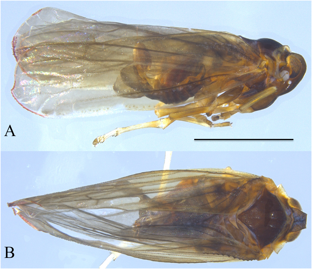
Description. Color. General body color dark fuscous, legs lighter brown, pronotum testaceous (yellowish in dried specimens). Wings uniformly light fuscous with reddish vein near wing apex ( Fig. 2 View FIGURE 2 ). Vertex lighter in color than frons.
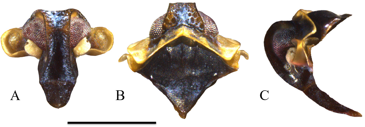
Structure. Body length (all measurements averages, males n = 2): 2.91 mm with wings; 1.90 mm without wings. Head. In lateral view, rounded on anterior margin with dorsal margin planar ( Fig. 3C View FIGURE 3 ). Frons relatively broad, widest at frontoclypeal suture, constricting just below ventral margin of eye, expanding slightly between eyes until reaching vertex, two rows of wax-producing pits on lateral margins running entire length of frons ( Fig. 3A View FIGURE 3 ). Vertex relatively broad, approximately twice as wide at posterior margin as long at midline, posterior margin concave, anterior margin straight, two rows of wax-producing pits along lateral margins, median carina obsolete ( Fig. 3B View FIGURE 3 ), transverse carina present at fastigium ( Fig. 3B View FIGURE 3 ). Vertex length: 0.12 mm; width at hind margin: 0.23 mm; width at distal margin: 0.14 mm. Frons length: 0.69 mm; dorsal width: 0.30 mm; width frontoclypeal margin: 0.38 mm; width at narrowest point: 0.24 mm. Clypeus length: 0.26 mm.
Thorax. Pronotum with anterior margin convex, appearing angular, posterior margin smoothly concave, pronotal disk tricarinate, median carina distinct, lateral carinae closely following posterior margin of head ( Fig. 3B View FIGURE 3 ). Paranota strongly foliately keeled and laterally projecting, lateral margin rounded from frontal view ( Fig. 3A View FIGURE 3 ), nearly covering antennae in lateral view ( Fig. 3C View FIGURE 3 ). Mesonotum tricarinate, lateral carinae sinuate, anteriorly distinct, converging and becoming obscure posteriorly; length at midline and width at tegulae approximately equal ( Fig. 3B View FIGURE 3 ). Pronotum length at midline: 0.79 mm. Mesonotum length at midline: 0.49 mm, width: 0.61 mm. Spinulation of hind tibia, basitarsus, and second tarsomere 6-6-6.
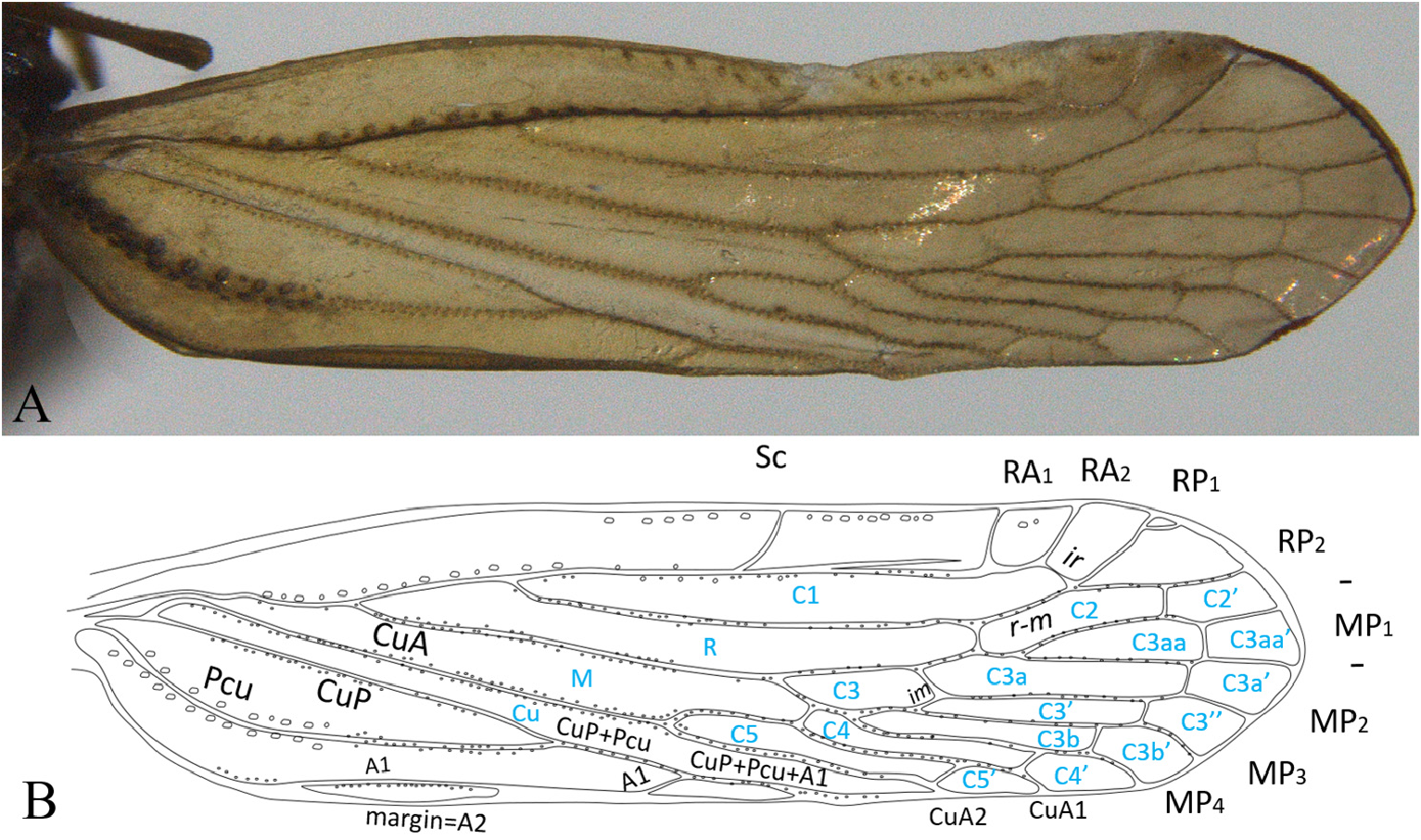
Forewing ( Fig. 4 View FIGURE 4 ) with large tubercles along costal, Sc+R(+M) and A1 veins. Smaller tubercles along all major longitudinal veins and branches. ‘Spurious’ vein present between Sc and RA. Vein branching pattern ( Fig. 4 View FIGURE 4 ): RA two-branched, RP three-branched (RP 1 apically forked), MP five-branched, CuA two-branched. Unusually, CuP fused with Pcu in distal quarter, well prior to fusion with A1, which closely tracks wing trailing margin Fork of R (Sc+RA from RP) well proximad to fork of CuA, about at level of fusion of CuP with Pcu. Forewing length: 2.53 mm.
Terminalia. Pygofer in lateral view narrow, with irregularly sinuate margins; narrowest dorsally, abruptly enlarged ventrally ( Fig. 5A View FIGURE 5 ); median process in ventral view ornate with apex rounded, subapical teeth and small lateral basal teeth ( Fig. 5B View FIGURE 5 ). Parameres in lateral view becoming wider distally, rounded ventrally, distal margin truncate, distal half of dorsal margin straight to dorsal process then strongly sloped ventrad; dorsal process with rounded apex, weakly sclerotized, angled anteriodorsad with smaller sclerotized process on lateral margin ( Fig. 5A View FIGURE 5 ). Parameres in ventral view spatulate, narrow proximally, with pair of medially directed teeth, proximal tooth small bifurcated, distal very large and cultrate, curved basad; distal margin straight ( Fig. 5B View FIGURE 5 ).
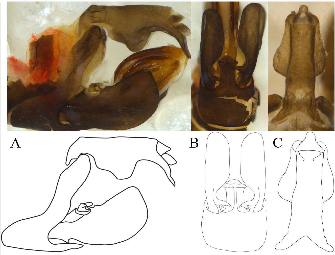
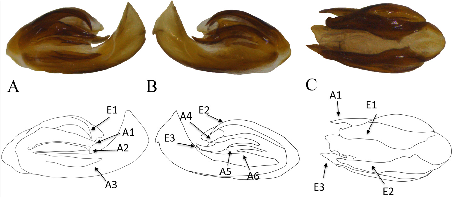
Aedeagus asymmetrical, all processes associated with aedeagal apex (A1–A3 on right side and A4–6 on left side) and endosoma (E1 on right side and E2 and E3 on left side); right side of aedeagus with three robust processes, approximately equal in length (A1–A3), right side of endosoma with single dorsal process (E1) strongly downcurved at apex ( Fig. 6A View FIGURE 6 ), aedeagus left lateral side with three processes ( Fig. 6B, A View FIGURE 6 4–A View FIGURE 4 6 View FIGURE 6 ), all more slender than processes on right lateral side, ventral two processes (A5, A6) significantly more slender and shorter, ventral-most process smallest ( Fig. 6B View FIGURE 6 ). Endosoma left lateral with dorsal process (E2) moderately downcurved, equal in length to the first endosomal process, third process on left side longer than all other processes, curved dorsad (E3) ( Fig. 6C View FIGURE 6 ). Anal tube in lateral view robust, with large, irregularly sinuate ventral lobe setting off strong distal concavity; apex elongate, and projecting to pointed apices ( Fig. 5A View FIGURE 5 ); dorsal margin straight. In dorsal view, lateral margins subparallel with ventral lobes visible past midlength, anal column short, nipple-like ( Fig. 5C View FIGURE 5 ).
Plant associations. Unknown. Collected at light trap in primary growth cloud forest at an elevation of approximately 1,100 meters.
Distribution. Costa Rica (Alajuela)
Etymology. The specific name is given in honor of the lead author’s daughter, Maria Jose.
Material examined. Holotype male “ Costa Rica, Alajuela / Los Angeles Cloud Forest / 15.V.2018 / Coll.: B.W. Bahder, light trap / Holotype Omolicna mariajosae ♂ ” ( FLREC) ; Paratype: 1 male, same data as holotype ( FLREC) .
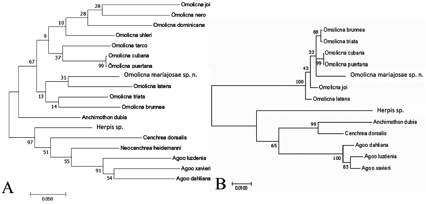
Sequence Data and Analysis. For Omolicna mariajosae sp. n., a 761 bp product was generated for the COI locus (GenBank Accession No. MT 422534 View Materials ). Based on the pairwise comparison of the COI loci for all taxa sampled, Omolicna mariajosae sp. n. is on average 15.4% (±0.005) different from other members of Omolicna , 17% different from Anchimothon dubia , 21% (±0.004) different from the genus Agoo, 19% different from Neocenchrea heidemanni , 20% different from Herpis metcalfi , and 23% different from Cenchrea dorsalis ( Table 2 View TABLE 2 ). Based on the phylogenetic analysis using the COI locus, Omolicna mariajosae sp. n. resolved well within the genus Omolicna ( Fig. 7A View FIGURE 7 ).
For the 18S locus, a 1,395 bp product was generated for Omolicna mariajosae sp. n. (GenBank Accession No. MT 424915 View Materials ). Based on the pairwise comparison of 18S, Omolicna mariajosae sp. n. differed, on average, by 3.3% (±0.0008) from other species of Omolicna , whereas excluding the novel taxon, the average variability among species with Omolicna was on average 0.7% (±0.001) ( Table 3 View TABLE 3 ). Omolicna mariajosae sp. n. differed from the genus Agoo by an average of 11.7% (±0.0009), 12.2% from Anchimothon dubia , 11.8% from Cenchrea dorsalis , and 13.5% from Herpis metcalfi ( Table 3 View TABLE 3 ). The phylogenetic analysis demonstrated Omolicna mariajosae sp. n. resolves within the genus Omolicna and that there is relatively strong support at the genus level for the cenchreine genera sampled in this study ( Fig. 7B View FIGURE 7 ).
Remarks. While the novel taxon generally exhibits the morphological features observed in many species of Omolicna , there are significant deviations that make O. mariajosae sp. n. unique among Omolicna . A striking feature, relative to other Omolicna , is the asymmetry of the aedeagus. Bahder et al. (2020) found that in dorsal view, the longest process was on the right side of all taxa studied whereas the longest aedeagal process in O. mariajosae sp. n. is on the left side. Furthermore, the forewing of O. mariajosae sp. n. appears unusual among Omolicna by the presence small tubercles along most veins, a spurious vein in the subcostal cell, and the fusion of the Pcu with CuP before the A 1 in the forewing. The combination of molecular data and morphological characters confirm the placement of this new species in the genus Omolicna .
Interestingly, available sequence data for both 18S and COI show O. cubana (from Jamaica) and O. puertana (from Puerto Rico) to be the same—100% identical for 18S and 99.8% for COI. Similarities in the aedeagus and terminalia (viz. Caldwell & Martorell 1951: 202, plate 24), Fennah 1952:135, figs. 13G, L,M,N) alongside the genetic evidence, seem to support that the taxa may be the same; however, we have not yet examined type material which is needed to confirm or refute the synonymy.
| MT |
Mus. Tinro, Vladyvostok |
No known copyright restrictions apply. See Agosti, D., Egloff, W., 2009. Taxonomic information exchange and copyright: the Plazi approach. BMC Research Notes 2009, 2:53 for further explanation.