
Glossogobius muscorum, Hoese, Douglass F. & Allen, Gerald R., 2009
|
publication ID |
https://doi.org/ 10.5281/zenodo.185296 |
|
DOI |
https://doi.org/10.5281/zenodo.5612014 |
|
persistent identifier |
https://treatment.plazi.org/id/34157120-FFB0-FFAD-27AB-F61FFA4FF880 |
|
treatment provided by |
Plazi |
|
scientific name |
Glossogobius muscorum |
| status |
sp. nov. |
Glossogobius muscorum View in CoL , sp. nov.
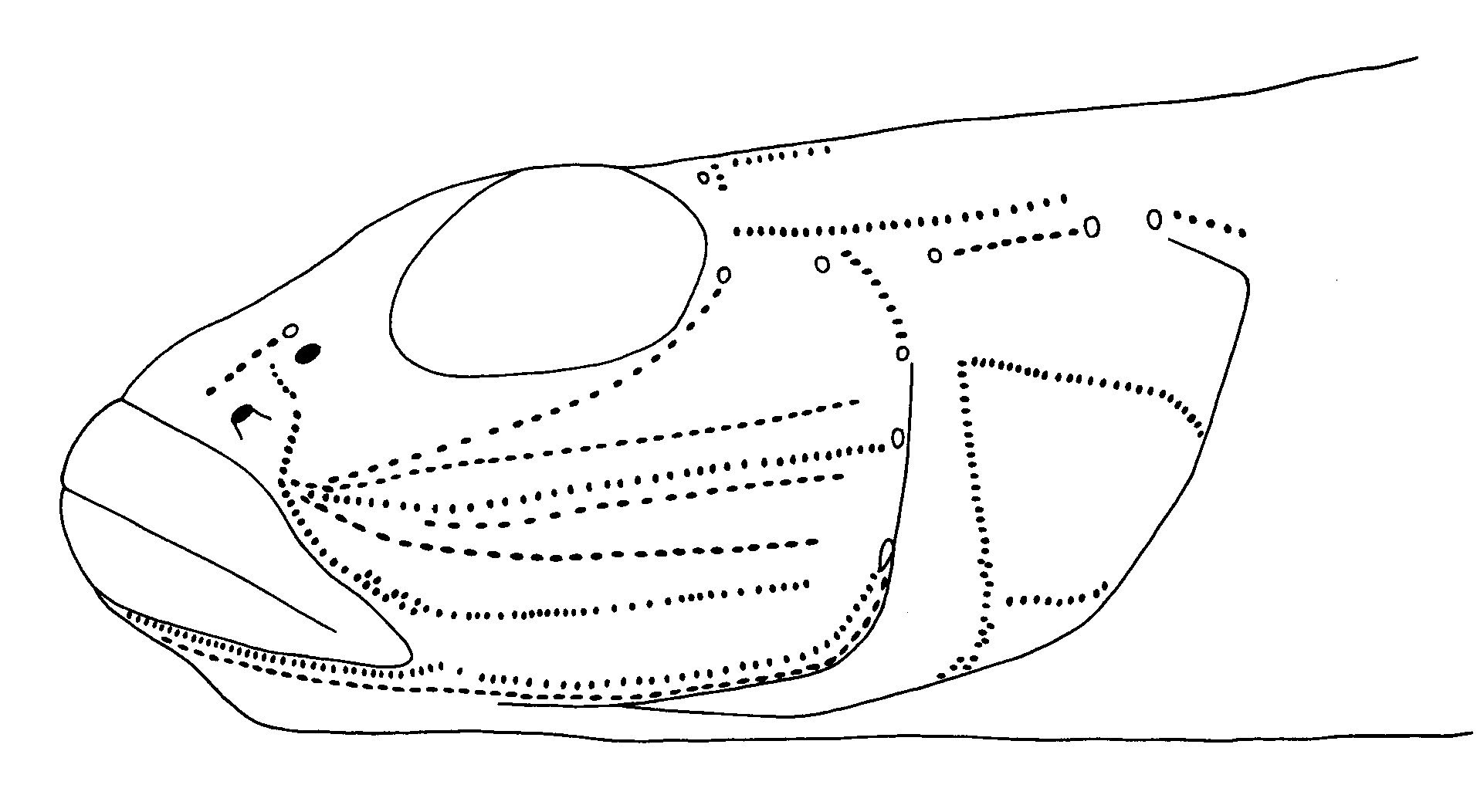
( Figs. 8 View FIGURE 8. a b–10, Tables 1–3 View TABLE 1 View TABLE 2 View TABLE 3 )
Glossogobius View in CoL sp. 11.— Allen, 1991: 186, pl. 16, fig. 13 ( Papua New Guinea).
Holotype. WAM P.27807–010, 45 mm SL female, creek 7 km N of Kiunga on Tabubil Road, 6°03'S, 141°18' E, 20 September, 1982, G. Allen & J. Paska.
Paratypes. Fly River System, Papua New Guinea: AMS I.44119-001 (formerly WAM P.27796-003), 2(26–27), small creek 32 km N of Kiunga on Tabubil Rd., 5°55'S, 141°17'E, 16 September, 1982, G. Allen & J. Paska; WAM P.27793–007, 1(39), small creek near Port at Kiunga, 6°07' S, 141°18'E, 16 September, 1982, G. Allen & J. Paska; WAM P.27795–003, 3(23–37), small creek 16 km N of Kiunga on Tabubil Rd., 5°58'S, 141°17'E, 16 September, 1982, G. Allen & J. Paska; WAM P.27807–004, 3(33–36), taken with holotype.
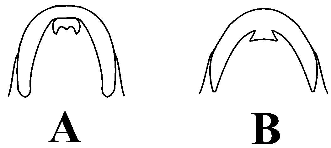
Diagnosis: A species of Glossogobius with bilobed mental frenum; predorsal area with scales extending forward to just before posterior preopercular margin; cheek, operculum, pectoral base, prepelvic area and anterior midline of belly naked; first dorsal fin with 3 dark spots, centred on fourth, fifth and sixth dorsal spine, respectively; second dorsal-fin rays usually I,10; anal-fin rays I,8; pectoral-fin rays 16–17; predorsal scale count 11-13; cheek papilla lines composed of single row of papillae; papilla line 6 absent and vertebrae 11+17.
Description: Based on 8 specimens 24–44 mm SL. First dorsal spines 6(8*); gill rakers on outer face of first arch 0+6(1), 0+1+6(1), 0+1+7(1), 1+1+6(2), 1+1+7(1); segmented caudal-fin rays 17(8*); branched caudal rays 7/6(1), 7/7(6*); vertebrae 11+17 (10*); predorsal scale count 11(2), 12(4*), 13(2); longitudinal scale count 27(2), 28(1), 29(5*); transverse scale count (TRB) 7.5(3), 8.5(5*). Second dorsal-fin rays usually I,10; anal-fin rays I,8; pectoral-fin rays 16–17 (see Tables 1–3 View TABLE 1 View TABLE 2 View TABLE 3 ).
Head depressed, 30.9–34.6% SL. Snout short, broadly rounded in dorsal view; convex (with notch before eye) in side view; 8.5–11.5% SL. Eye slightly shorter than snout, 9.5–12.3% SL. Cheeks tapering. Interorbital narrow, less than eye length. Upper jaw 9.7–12.3% SL. Small bump below anterior nostril present. Anterior nostril at end of short tube, just above upper lip. Posterior nostril 1 nostril diameter from anterior nostril and midway between eye and upper lip. Posterior preopercular margin without spine. Preoperculum short, distance from end of eye to upper posterior preopercular margin much less than eye (one-half to three-quarters of eye). Postorbital short, subequal to distance from tip of snout to mideye. Mental fraenum bilobed, with lateral lobes broad and attached to chin and not free distal margins. Mouth small, reaching to below anterior quarter of eye; jaws forming an angle of 35–40° with body axis; upper margin of upper jaw in line with middle of eye. Gill opening reaching to below or just behind posterior preopercular margin. Teeth in upper jaw: outer row of teeth conical, slightly enlarged and wideset, 2–3 inner rows of smaller depressible, inwardly directed teeth, innermost row larger than middle row. Teeth in lower jaw: teeth in outer row conical, slightly enlarged and wideset anteriorly, 2–3 inner rows of smaller depressible teeth. Tongue tip bilobed. Gill rakers on outer face of first arch slender and short; longest about one-fifth filament length. Rakers on inner face of first arch and other arches short and denticulate. Predorsal area with scales extending forward to just before posterior preopercular margin. Cheek naked. Operculum naked. Pectoral base naked. Prepelvic area naked. Belly with a large naked area behind pelvic insertion. Body covered mostly with large ctenoid scales, cycloid on midline of belly. First dorsal fin low, with rounded margin, spines 3–5 extending beyond other spines when fin depressed, origin well behind pelvic insertion. Second dorsal fin subequal in height to first dorsal fin. Anal fin slightly lower than dorsal fins. Pectoral fin with pointed margin, reaching to between anus and anal origin. Pelvic disc moderately thick, slightly longer than wide, reaching to below anus; fifth ray with 7–9 terminal tips.
Head pores: nasal pore above posterior nostril; anterior interorbital pore present; posterior interorbital pore present; postorbital pore behind eye present; infraorbital pore below postorbital present; lateral canal pore above preoperculum present; lateral canal pore above posterior preopercular margin absent; terminal lateral canal pore above anterior operculum present; short tube above operculum, with pore at each end present, represented by open trough or often absent in small specimens (below 20 mm SL); 3 preopercular pores, upper in line with lower margin of eye; widely separated from lower 2 pores.
Sensory Papillae: ( Figure 10 View FIGURE 10 ; lines listed below all composed of single row of papillae, except where noted). Line 1 (before nasal pore) present. Line 2 (between nasal pores) curved, with gap at midline of snout. Line 5 (suborbital) reaching to line 7 before eye. Line 6 (suborbital branch) absent. Lines 7, 9, 10 (VL cheek rows) each reaching near posterior preopercular margin. Lines 8 and 11 (VT row) ending short of posterior preopercular margin. Line 12 (Outer POP-mandibular) with a gap at end of jaws. Line 13 (Inner POP-mandibular) curved near end of jaws (line composed of two rows of papillae on chin only). Line 20 (OP VT) branched ventrally. Line 21 (Upper OT) strongly curved, becoming vertical posteriorly in some paratypes, often almost reaching line 22. Line 22 (Lower OT) without branches. Several vertical papillae rows on belly. A single curved line anteriorly on most body scales (often obscure dorsally and posteriorly).
Coloration: Head and body brown. Head with an elongate brown spot behind posterior margin of eye, an elongate bar above operculum, a small brown spot dorsoposteriorly on operculum, a dark brown bar from anteroventral margin of eye to middle of upper lip, a horizontal brown stripe on cheek below eye and an oval brown spot on anteromedian margin of operculum; posterior end of lips whitish. Pectoral base with a slightly oblique brown bar on upper base. Two rows of small brown spots on back extending from above pectoral base to below middle of second dorsal fin; midside with one large blotch on side of belly and a series of brown spots along midside (number of scales spots cover indicated in parentheses), first spot below second dorsal origin (2), 3 (1–2, 2, 1) below second dorsal fin, one (3) on middle of caudal peduncle and last (2) on caudal peduncle, confluent with triangular spot on base of caudal. Fins dark, dorsal and caudal fins spotted, caudal fin grey dorsally and ventrally, spots confined to middle rays; first dorsal fin with a small oval spot along fourth dorsal spine and a darker oval spot centred on fifth dorsal spine and similar spot centred on sixth dorsal spine; pectoral fin grey (lighter than other fins); pelvic disc dark brown; anal fin without spots.
Distribution: Glossogobius muscorum is known only from the upper Fly River system in the vicinity of Kiunga and Ningerum at distances between about 840–900 km upstream from the sea. It generally occurs in rainforest creeks at elevations below 50 m.
Similarity to other species: Glossogobius muscorum is most similar to G. bellendenensis from Queensland. All are characterised by reduced predorsal scale coverage, no prepelvic or pectoral base scales and small body size (see discussion of G. bellendenensis for comparison of these species).
The species can also be confused with the sympatric Glossogobius concavifrons , which has scales on the pectoral base and prepelvic area, predorsal scales reaching to near the eye, more numerous precaudal vertebrae (13–15) and a more compressed head. Separation of juveniles below 20 mm SL is difficult because the scales are not well developed in Glossogobius concavifrons at that size.
Etymology: From the genitive of the Latin muscus (fly) referring to the type locality, ‘of the Fly River’
| WAM |
Western Australian Museum |
No known copyright restrictions apply. See Agosti, D., Egloff, W., 2009. Taxonomic information exchange and copyright: the Plazi approach. BMC Research Notes 2009, 2:53 for further explanation.
|
Kingdom |
|
|
Phylum |
|
|
Class |
|
|
Order |
|
|
Family |
|
|
Genus |
Glossogobius muscorum
| Hoese, Douglass F. & Allen, Gerald R. 2009 |
Glossogobius
| Allen 1991: 186 |