
Wow Jałoszyński, Maruyama & Klimaszewski, 2023
|
publication ID |
https://doi.org/ 10.11646/zootaxa.5357.4.5 |
|
publication LSID |
lsid:zoobank.org:pub:122F5432-1468-4BE6-9566-73083903A050 |
|
DOI |
https://doi.org/10.5281/zenodo.10116336 |
|
persistent identifier |
https://treatment.plazi.org/id/336587DD-B938-0035-F9E7-FC3F51BE04BF |
|
treatment provided by |
Plazi |
|
scientific name |
Wow Jałoszyński, Maruyama & Klimaszewski |
| status |
gen. nov. |
Genus Wow Jałoszyński, Maruyama & Klimaszewski , gen. n.
Type species: Wow assingi Jałoszyński, Maruyama & Klimaszewski , sp. n. (here designated).
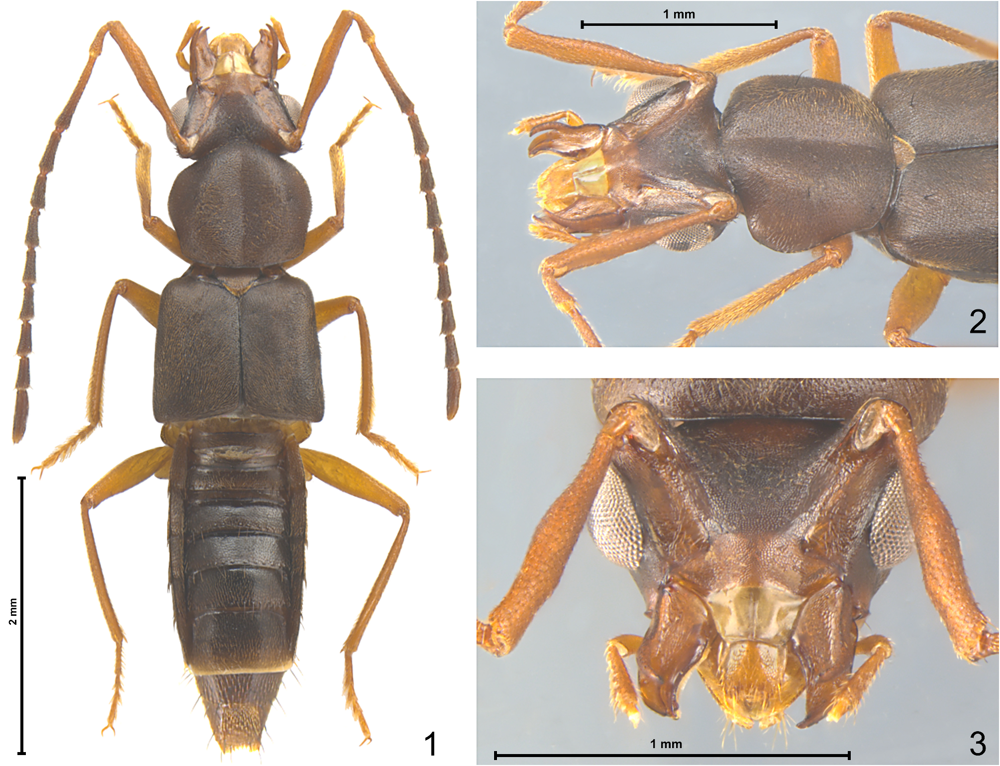
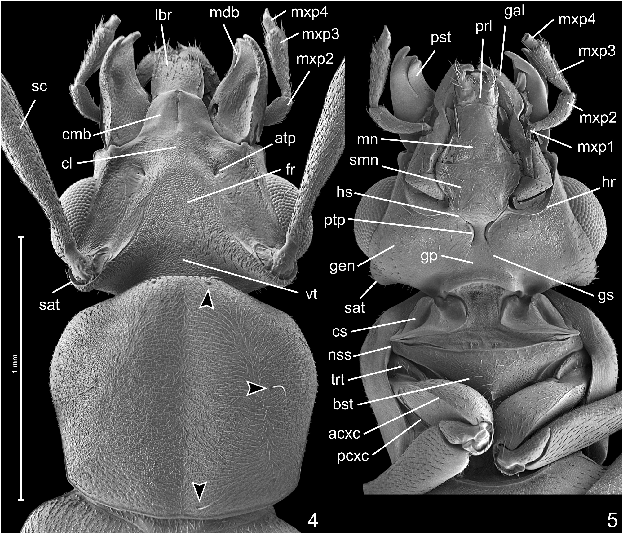
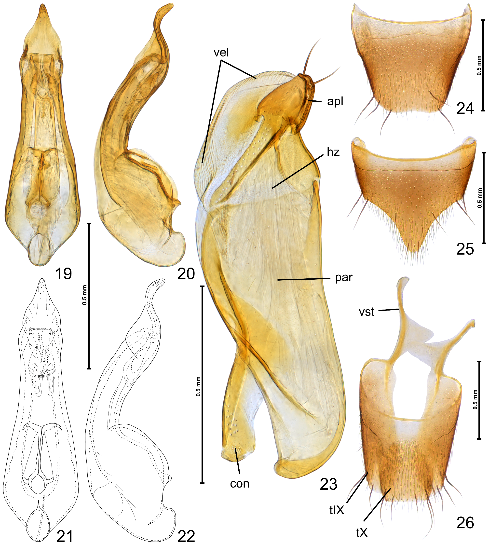
Diagnosis (based on male characters). Body ( Fig. 1 View FIGURES 1–3 ) strongly flattened; antennae inserted on posterodorsal margin of head capsule, behind line drawn between posterior margins of eyes ( Fig. 4 View FIGURES 4–5 ); supraantennal tubercles projecting posteriorly on posterior margin of head ( Fig. 4 View FIGURES 4–5 ); longitudinal subocular ridge (= occipital ‘suture’) absent; labrum ( Fig. 4 View FIGURES 4–5 ) subrectangular, weakly transverse, weakly emarginate anteriorly; mandibles ( Fig. 4 View FIGURES 4–5 ) nearly symmetrical, lacking preapical mesal teeth, each with outer dorsal preapical tooth; galea shorter than lacinia ( Figs 7‒8 View FIGURES 6–11 ); anteromedian region of prelabium posterad ligula with inversely drop-shaped sclerotization ( Figs 10‒11 View FIGURES 6–11 ); maxillary palpomeres 1 and 2 each bearing several long setae ( Figs 10‒11 View FIGURES 6–11 ); lateral lobes of labial apodeme short, curved at apices, median projection of labial apodeme short, rounded at apex ( Figs 10‒11 View FIGURES 6–11 ); scape ( Figs 1‒3 View FIGURES 1–3 ) conspicuously elongate, longer than pronotum; pronotum ( Fig. 4 View FIGURES 4–5 ) impressed along midline, with vestiture of setae on central region directed largely laterad, and on lateral regions directed largely mesad; mesoventral process ( Fig. 13 View FIGURES 12–18 ) subtriangular, barely separating mesocoxae and posteriorly separated from anterior metaventral process; abdomen ( Figs 1 View FIGURES 1–3 , 16 View FIGURES 12–18 ) clearly narrower than elytra; tergites III‒V each with flat basal carina laterally curving posterolaterad, on tergite VI only postspiracular portions of carina developed; sternite VIII ( Figs 18 View FIGURES 12–18 , 25 View FIGURES 19–26 ) in posterior half triangular and strongly projecting posterad; tarsomeres of all tarsi with conspicuously long ventral setae ( Figs 14‒15 View FIGURES 12–18 ); ostium of aedeagus ( Figs 19‒20 View FIGURES 19–26 ) distant from apex of parameral wall; parameres ( Fig. 23 View FIGURES 19–26 ) with narrow velar region of paramerite and broad, subtriangular apical lobe.
Description (male). Body ( Fig. 1 View FIGURES 1–3 ) strongly flattened, slender.
Head ( Figs 1‒5 View FIGURES 1–3 View FIGURES 4–5 ) with vestigial, barely discernible postocciput, transverse; eyes clearly separated from mandibular bases; clypeolabral connecting membrane ( Fig. 4 View FIGURES 4–5 ; cmb) nearly as long as broad and with median longitudinal groove; clypeus ( Fig. 4 View FIGURES 4–5 ; cl) strongly transverse and about 3 times as broad as long, its posterior margin marked by distinct invagination sites of anterior tentorial arms (i.e., anterior tentorial pits; Fig. 4 View FIGURES 4–5 ; atp); frons and vertex together subtriangular; antennal insertions and supraantennal tubercles ( Fig. 4 View FIGURES 4–5 ; sat) situated behind eyes on posterodorsal margin of head capsule and strongly elevated, ‘neck ’ absent; antennal foramen within supraantennal tubercle oval and much larger than articulating basal region of scape; longitudinal subocular ridge (= occipital ‘suture’) absent on entire length; gular plate ( Fig. 5 View FIGURES 4–5 ; gp) subtriangular and slightly narrower than 1/3 of head width (excl. eyes); gular sutures ( Fig. 5 View FIGURES 4–5 ; gs) complete; posterior tentorial pits ( Fig. 5 View FIGURES 4–5 ; ptp) distinct, each C-shaped (convex mesally) and strongly elongate, separated by distance shorter than length of pits; hypostomal sutures ( Fig. 5 View FIGURES 4–5 ; hs) complete and much shorter than submentum; hypostomal ridges ( Fig. 5 View FIGURES 4–5 ; hr) sharply marked, complete, mesally connected with hypostomal sutures; submentum inversely subtrapezoidal, strongly broadening anteriorly, with anterolateral corners not projecting anterad.
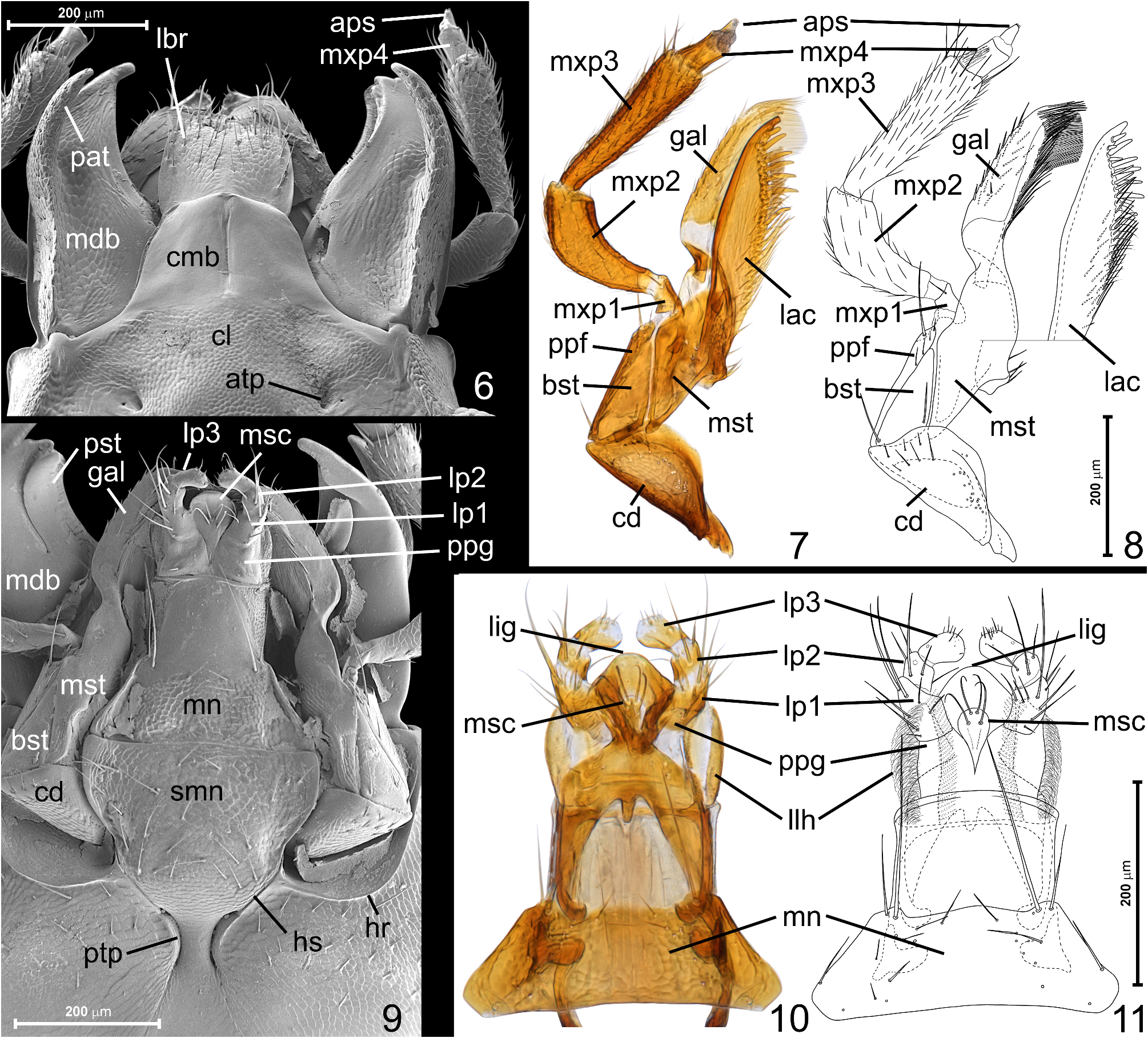
Labrum ( Fig. 6 View FIGURES 6–11 ; lbr) subrectangular, weakly transverse, weakly emarginate anteriorly, with weakly arcuate sides, subhorizontal, with sparsely and only partly symmetrically distributed numerous dorsal setae, basal labral region asetose; mandibles ( Fig. 6 View FIGURES 6–11 ; mdb) nearly symmetrical, elongate subtriangular, weakly curved mesally, lacking preapical mesal teeth, each with outer dorsal preapical tooth ( Fig. 6 View FIGURES 6–11 ; pat); lateral (outer) mandibular margin with distinct longitudinal ventral carina; prostheca ( Fig. 9 View FIGURES 6–11 ; pst) developed as elongate ventral process with dense and short mesal marginal microtrichia, mola absent. Maxilla ( Figs 7, 8 View FIGURES 6–11 ) with subtriangular and transverse cardo ( Figs 7, 8 View FIGURES 6–11 ; cd), subtriangular and elongate basistipes ( Figs 7, 8 View FIGURES 6–11 ; bst) and elongate mediostipes ( Figs 7, 8 View FIGURES 6–11 ; mst) distinctly longer than basistipes; palpifer ( Figs 7, 8 View FIGURES 6–11 ; ppf) strongly elongate; galea ( Figs 7, 8 View FIGURES 6–11 ; gal) slender, shorter than lacinia (but its anterior margin projecting anteriorly beyond lacinia), with row of short robust setae along outer margin and with long, dense, thin setae on mesal margin; lacinia ( Figs 7, 8 View FIGURES 6–11 ; lac) slender, with robust apical tooth, subapical mesal row of several thick and nearly rod-like setae and mesal proximal row of dense and thin setae. Maxillary palp tetramerous and slender; palpomere 1 ( Figs 7, 8 View FIGURES 6–11 ; mxp1) minute and slightly elongate; palpomere 2 ( Figs 7, 8 View FIGURES 6–11 ; mxp2) pipe-shaped, elongate, curved, sparsely setose; palpomere 3 ( Figs 7, 8 View FIGURES 6–11 ; mxp3) subequal in width to palpomere 2, strongly elongate and slender, weakly and gradually broadening from base to truncate apex, sparsely setose; palpomere 4 ( Figs 7, 8 View FIGURES 6–11 ; mxp4) elongate, with truncate apex, weakly and gradually narrowing distally, with subconical apical ‘pseudosegment’ ( Figs 7, 8 View FIGURES 6–11 ; aps). Labium with broadly subtrapezoidal mentum ( Figs 9‒11 View FIGURES 6–11 ; mn) and elongate prelabium with ventral inversely drop-shaped median premental sclerotization ( Figs 9‒11 View FIGURES 6–11 ; msc) bearing two pairs of setae with their insertion sites arranged in nearly a square, proximal (premental) prelabial region transverse, with one setal pore and two real pores, without pseudopores, ligula ( Figs 10, 11 View FIGURES 6–11 ; lig) in ventral view broadly rounded subtriangular and strongly sclerotized, as broad as distance between labial palps and twice as broad as median premental sclerotization; labial palps trimerous and shorter than prelabium, with palpomere 1 ( Figs 9‒11 View FIGURES 6–11 ; lp1) subcylindrical and distinctly elongate, bearing several long setae mainly on outer margin; palpomere 2 ( Figs 9‒11 View FIGURES 6–11 ; lp2) nearly annular, much shorter than 1, with several long setae; palpomere 3 ( Figs 9‒11 View FIGURES 6–11 ; lp3) shorter than 1 and longer than 2, distinctly broadening toward broadly rounded apex bearing several minute setiform sensilla. Lateral lobes of hypopharynx ( Figs 10, 11 View FIGURES 6–11 ; llh) narrow and elongate, with dense and thin marginal and ventral microtrichia.
Antenna ( Fig. 1 View FIGURES 1–3 ) shorter than body but much longer than head and thorax combined, geniculate. Scape conspicuously elongate and clavate, much longer than head, broadest distally to middle. Pedicel much shorter and narrower than scape, weakly clavate. Flagellum nearly filiform and loosely assembled, with antennomeres 3‒10 each broadening distally, antennomere 11 asymmetrical.
Pronotum ( Fig. 4 View FIGURES 4–5 ) broadest clearly in front of middle, impressed along midline, with conspicuous setal pattern: elongate area at both sides of midline with setae directed posterolaterad near anterior margin, laterad on large median region and anterolaterad near posterior margin; setae on elongate lateral areas of pronotum directed posteromesad near anterior margin, mesad on large median region, and anteromesad near posterior margin. Three pairs of macrosetae ( Fig. 4 View FIGURES 4–5 ; indicated by arrowheads) present submedially near anterior pronotal margin, sublaterally on disc in front of middle, and submedially near posterior margin. Lateral pronotal carinae largely shifted ventrally and in dorsal view visible only near posterior pronotal corners; hypomera exposed in lateral view, each as wide as about 1/5 width of basisternal region of prosternum (i.e., basisternum fused with preepisternum). Prosternum weakly convex, subtrapezoidal, laterally delimited by short notosternal sutures ( Fig. 5 View FIGURES 4–5 ; nss), with weakly arcuate anterior margin and broadly subtriangular posterior margin; furcasternum shorter than basisternal region.
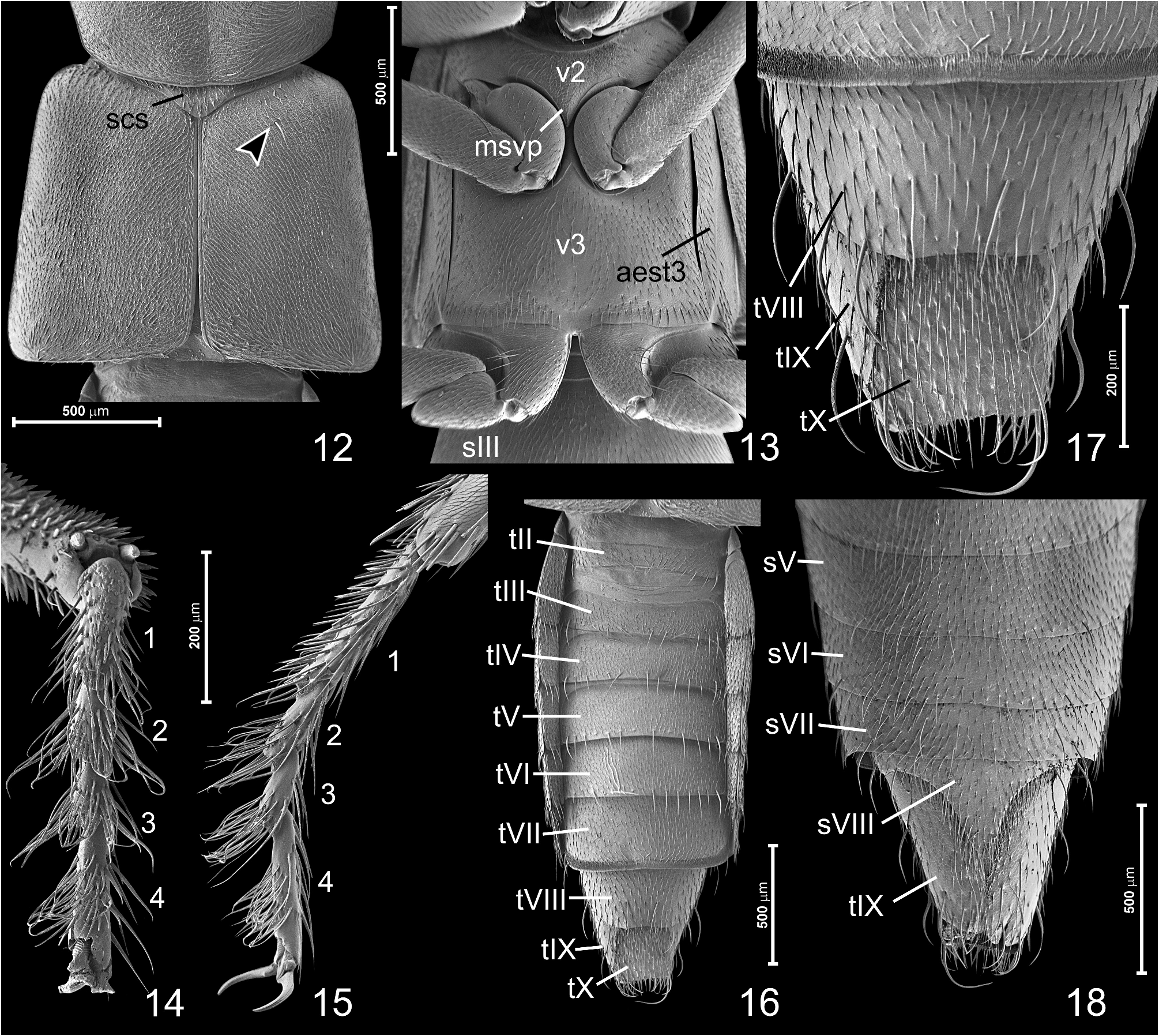
Mesoscutellar shield ( Fig. 12 View FIGURES 12–18 : scs) sub-pentagonal and transverse (exposed portion subtriangular), densely setose.
Mesoventrite ( Fig. 13 View FIGURES 12–18 ; v 2 View FIGURES 1–3 ) strongly transverse, with arcuate anterior carina demarcating vestigial prepectus, mesoventral process ( Fig. 13 View FIGURES 12–18 ; msvp) subtriangular, barely separating mesocoxae, posteriorly separated from anterior metaventral process.
Metanotum not studied.
Elytra ( Fig. 12 View FIGURES 12–18 ) broadening posteriorly, with weakly concave posterior margins, covered with setae largely directed posterolaterad and each with one macroseta inserted sub-basally on median region of disc ( Fig. 12 View FIGURES 12–18 ; indicated by arrowhead); epipleural carina absent.
Metaventrite ( Fig. 13 View FIGURES 12–18 ; v 3 View FIGURES 1–3 ) subquadrate, mesocoxal rests posteriorly and laterally with fine marginal carinae connected medially on flat and poorly differentiated anterior metaventral process; posterior metaventral margin weakly sinuate on sides and weakly projecting posteriorly at middle, so that metacoxae are subcontiguous. Metaventral discrimen indiscernible, katepisternal sutures marked by faint impression accentuated by transverse row of setae.
Abdomen ( Figs 1 View FIGURES 1–3 , 16‒18 View FIGURES 12–18 ) clearly narrower than elytra, nearly parallel-sided in slightly more than anterior half, in posterior half gradually narrowing and truncate at apex; broad bipartite paratergites present on segments II‒VII; tergites III‒V each with flat basal carina laterally curving posterolaterad, on tergite VI only postspiracular portions of carina developed; tergite VII with broad microtrichial field along posterior margin; sternite VIII triangularly projecting posterad; lateral portions of tergite IX ( Fig. 26 View FIGURES 19–26 ) strongly elongate and with broadly rounded distal regions bearing paired long setae; tergite X ( Fig. 26 View FIGURES 19–26 ) sub-pentagonal with truncate distal region bearing paired long setae.
Legs ( Figs 1 View FIGURES 1–3 , 5 View FIGURES 4–5 , 13‒15 View FIGURES 12–18 ) moderately long and slender; procoxae ( Fig. 5 View FIGURES 4–5 ) strongly elongate, each with anterior and posterior marginal longitudinal carina on outer surface ( Fig. 5 View FIGURES 4–5 ; respectively acxc and pcxc); mesocoxae ( Fig. 13 View FIGURES 12–18 ) flattened and oval; metacoxae ( Fig. 13 View FIGURES 12–18 ) subrectangular; pro- and mesotrochanters ( Figs 5 View FIGURES 4–5 , 13 View FIGURES 12–18 ) minute and subtriangular, metatrochanters ( Fig. 13 View FIGURES 12–18 ) much larger but similar in shape; all femora broadest in proximal half and narrowing distally; tibiae slender, with only thin setae, lacking thick spines except for pair of apical spurs; tarsal formula 4-4-4, all tarsomeres with ventral groups of long setae ( Figs 14, 15 View FIGURES 12–18 ).
Median lobe of aedeagus with ostium distant from apex of parameral wall; endophallus weakly sclerotized, nearly symmetrical; parameres with narrow velar region of paramerite and massive, subtriangular apical lobe bearing 4 macroseate.
Etymology. The generic name Wow (noun in apposition) reflects our first impression ( Wow !) at seeing this extraordinary beetle.
No known copyright restrictions apply. See Agosti, D., Egloff, W., 2009. Taxonomic information exchange and copyright: the Plazi approach. BMC Research Notes 2009, 2:53 for further explanation.
|
Kingdom |
|
|
Phylum |
|
|
Class |
|
|
Order |
|
|
Family |
|
|
SubFamily |
Aleocharinae |
|
Tribe |
Wowini |