
Hyphoderma yunnanense Z.Y. Duan & C.L. Zhao, 2023
|
publication ID |
https://doi.org/10.11646/phytotaxa.599.1.1 |
|
DOI |
https://doi.org/10.5281/zenodo.7991509 |
|
persistent identifier |
https://treatment.plazi.org/id/326687FE-4459-2C54-FF3A-FECCFC77FC17 |
|
treatment provided by |
Plazi (2023-05-31 07:50:06, last updated 2024-11-28 03:37:19) |
|
scientific name |
Hyphoderma yunnanense Z.Y. Duan & C.L. Zhao |
| status |
sp. nov. |
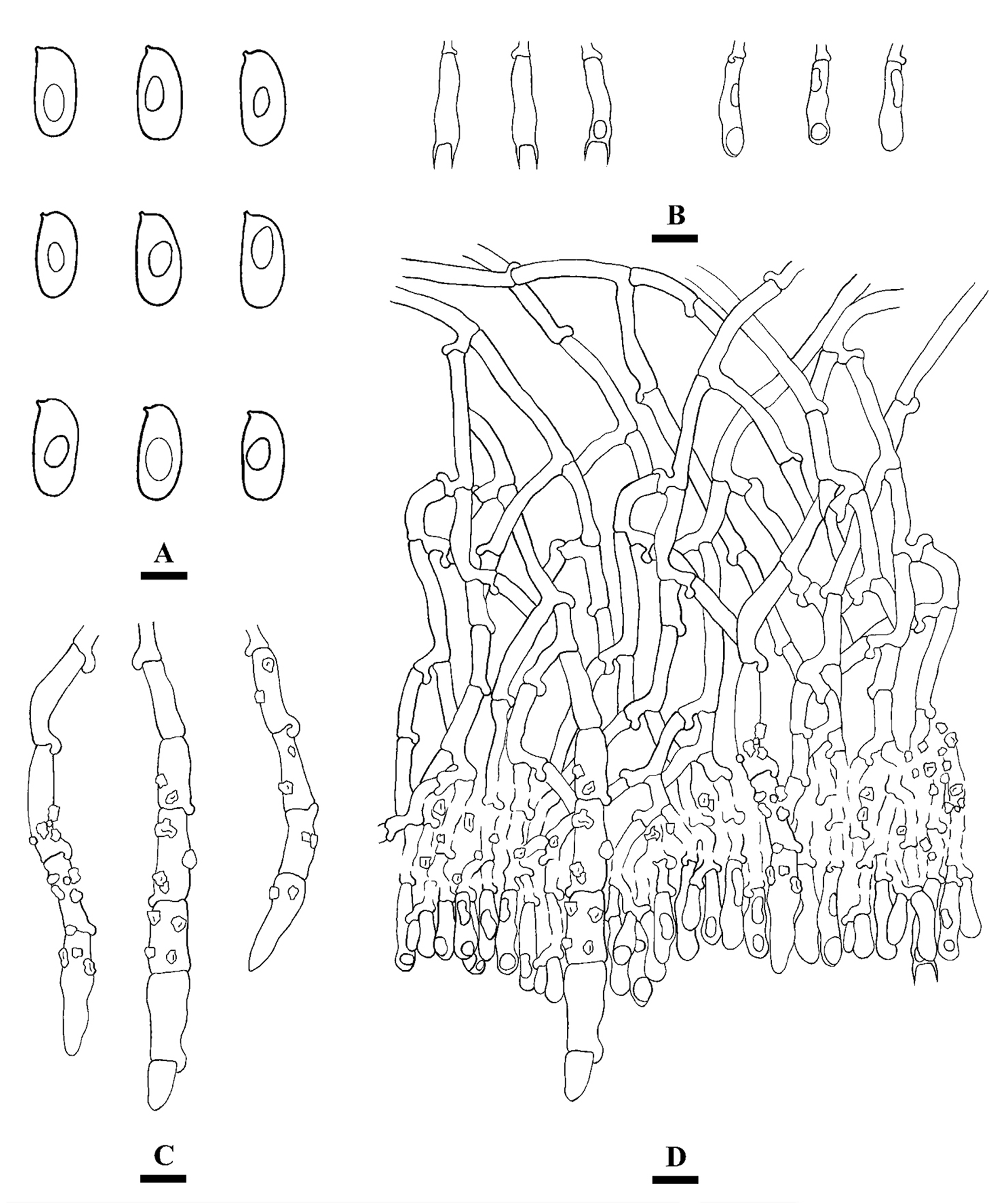
Hyphoderma yunnanense Z.Y. Duan & C.L. Zhao , sp. nov. Figs. 7 View FIGURE 7 , 8 View FIGURE 8
MycoBank no.: MB 847111
Etymology:— yunnanense (Lat.) refers to the province (Yunnan Province, China) of the specimen.
Holotype:— CHINA. Yunnan Province, Kunming, Southwest Forestry University , E 102°45′55″, N 25°04′01″, elev. 1939 m, on fallen angiosperm trunk, 31 June 2018, CLZhao 8845 ( SWFC). GoogleMaps
Fruiting body:— annual, resupinate, adnate, corneus when fresh, coriaceous upon drying, without odor and taste when fresh, and up to 8 cm long, 4 cm wide, and 50–150 µm thick. Hymenial surface tuberculate, slightly cream when fresh, cream to pale buff on drying. Margin sterile, narrow, white to pale cream, up to 1–2 mm wide.
Hyphal structure: — Monomitic; generative hyphae with clamp connections,colorless, thin-walled, frequently branched, interwoven, 2.5–4.5 µm in diam, IKI–, CB–, tissues unchanged in KOH.
Hymenium: — Cystidia cylindrical, with multiple clamped septa, encrusted with crystals, thin-walled, 63–124 × 7–10 µm; basidia clavate to subcylindrical, slightly sinuous, with 2 sterigmata and a basal clamp connection, 15.5–20 × 3.5–4.5 µm; basidioles in shape similar to basidia, but slightly smaller.
Basidiospores: — (9.5–)10–11.5(–12) × 4–5.5(–6) µm, L = 10.71 µm, W = 4.8 µm, Q = 2.23 (n = 30/1), ellipsoid to cylindrical, colorless, thin-walled, smooth, with oil drops inside, IKI–, CB–.
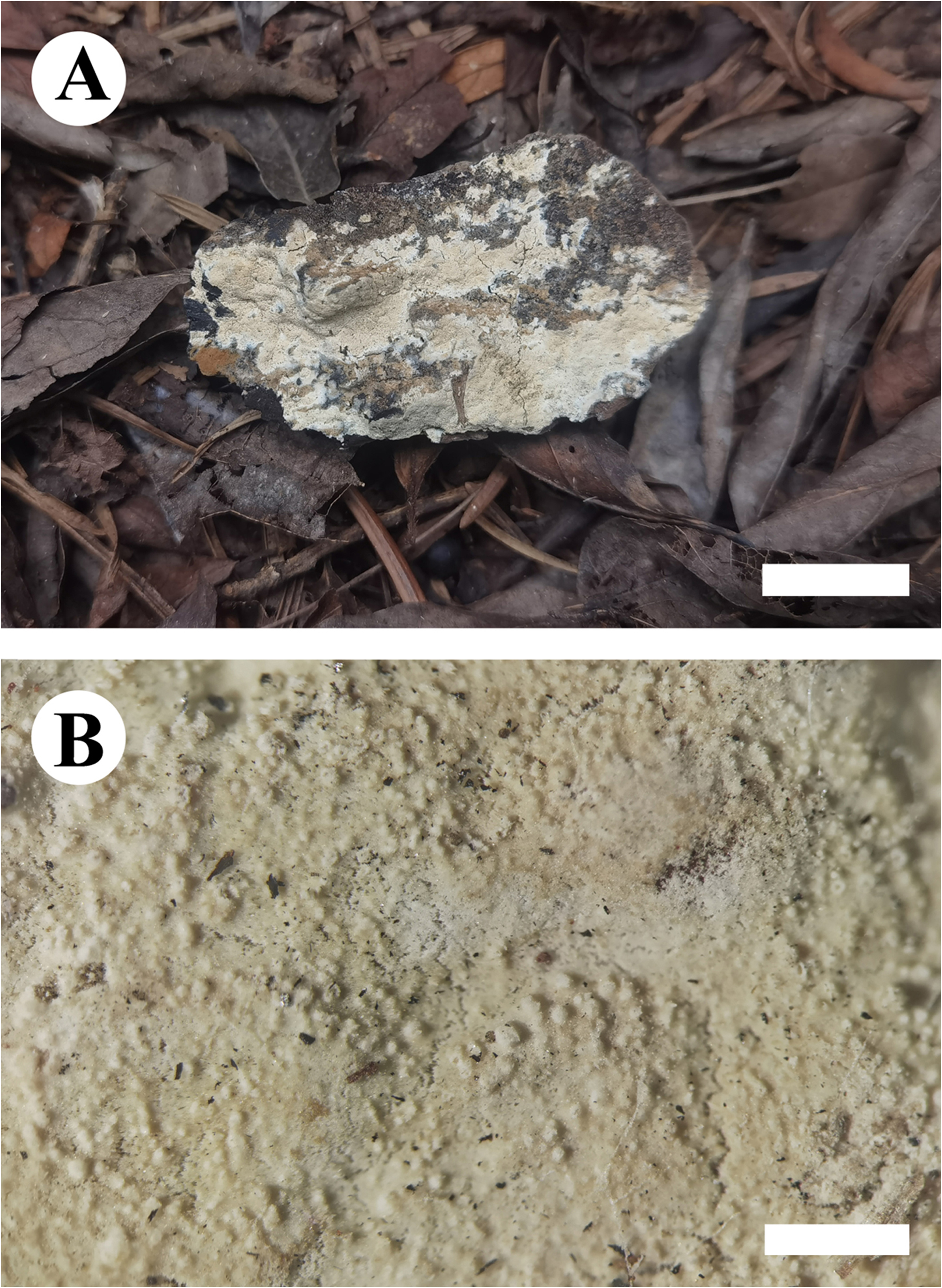
FIGURE 7. Basidiomata of Hyphoderma yunnanense. Bars: A = 1 cm, B = 1 mm (Holotype: CLZhao 8845). Photo plate by: Zi-Yan Duan.
No known copyright restrictions apply. See Agosti, D., Egloff, W., 2009. Taxonomic information exchange and copyright: the Plazi approach. BMC Research Notes 2009, 2:53 for further explanation.
|
Kingdom |
|
|
Phylum |
|
|
Class |
|
|
Order |
|
|
Family |
|
|
Genus |