
Calcigorgia japonica Dautova, 2007
|
publication ID |
https://doi.org/ 10.5852/ejt.2018.408 |
|
publication LSID |
lsid:zoobank.org:pub:D57D13DB-0AF8-4C2A-AD29-0805B324D727 |
|
DOI |
https://doi.org/10.5281/zenodo.5672421 |
|
persistent identifier |
https://treatment.plazi.org/id/321387DE-8D5B-0866-FDE3-850AA8340597 |
|
treatment provided by |
Plazi |
|
scientific name |
Calcigorgia japonica Dautova, 2007 |
| status |
|
Calcigorgia japonica Dautova, 2007 View in CoL
SEA OF JAPAN: 1 specimen, 39°35′ N, 135°01′ E, 832–736 m, 8 Aug. 1933, K.M. Deryugin leg. ( ZIN RAS 1/10706 ). GoogleMaps
Description
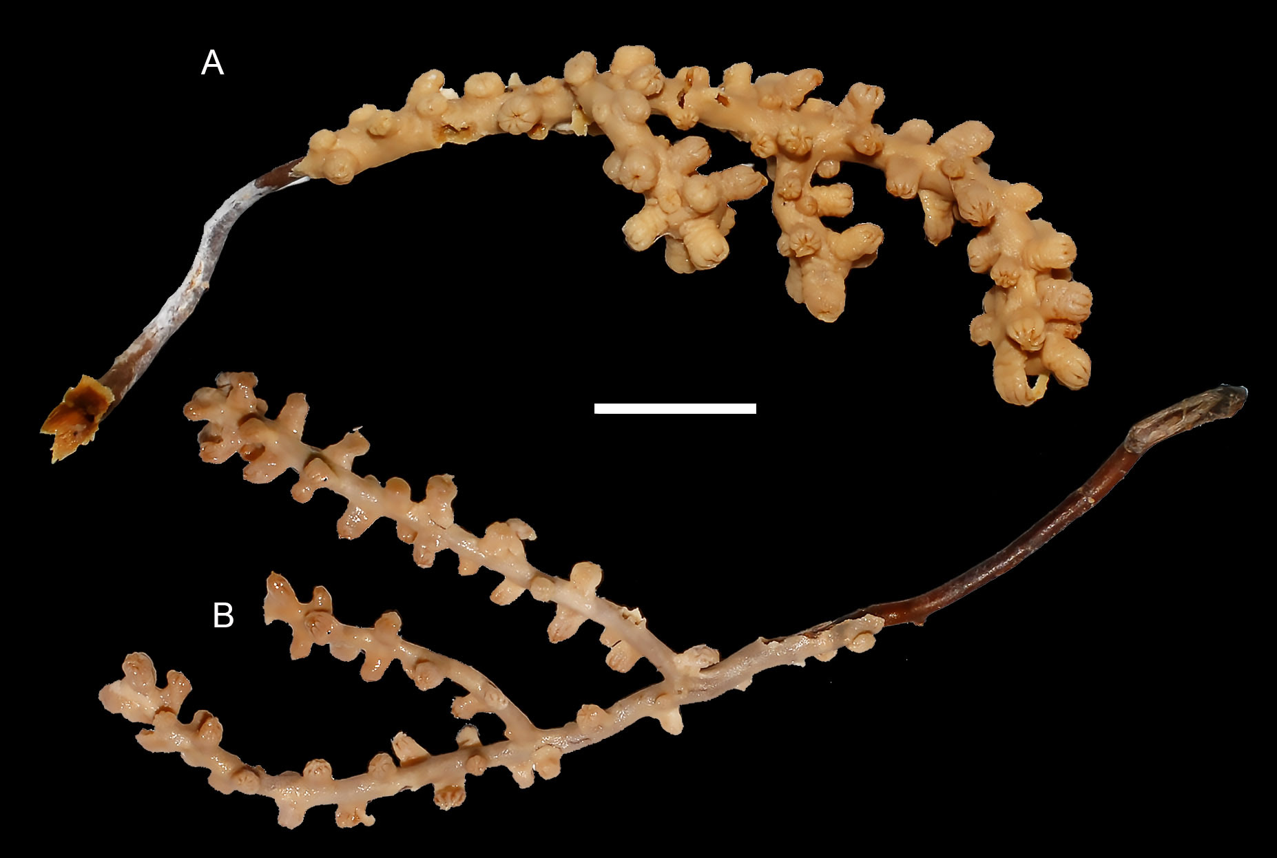
Scantily branched delicate colonies. Side branches placed irregularly. Holotype alcohol-preserved colony of 65 mm high and 25 mm wide ( Fig. 1A View Fig. 1 ). Paratype 75 mm high and 27 mm wide ( Fig. 1B View Fig. 1 ). Polyps up to 5 mm high and 2.2 mm wide, disposed irregularly at distances of 2–10 mm from each other. Tentacles folded over polyp and partly retracted to the inside. Polyp body is smooth and slightly widening downward. No polyps entirely retracted into coenenchyme.
Coenenchyme is a 0.3 mm thick and consists of two layers, a 0.15 mm thick outer layer, with smooth surface and a very thin, semi-transparent inner layer. Axis made of concentric layers. Axial canal in this specimen very narrow and, therefore, hardly visible. Sclerites unordered in tentacles, polyp body wall and coenenchyme.
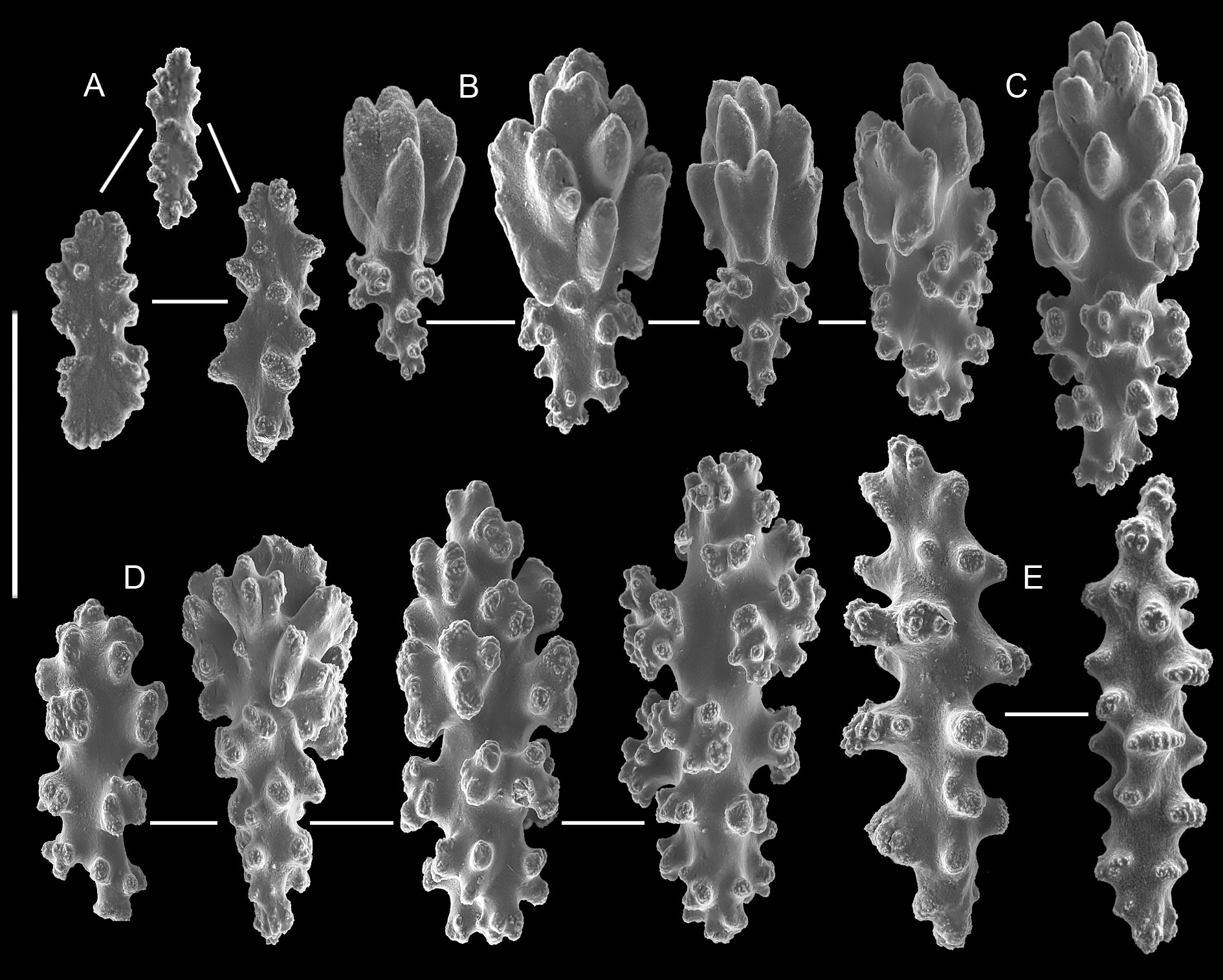
Tentacles contain flattened elongated bodies, clubs and spindles. Flattened bodies of irregular shape, mainly 0.7–0.9 mm long, covered by sparse tubercles (Fig. 2A). Clubs mainly 0.09–0.12 mm long, sometimes up to 0.14 mm, with a plump or elongated head, consisting of several leafy processes ( Fig. 2B View Fig. 2 ). Short handle straight or slightly curved, pointed or blunt, and ornamented with small warts. Some longer clubs, up to 0.18 mm, with staggered leafy processes on head (Fig. 2C) transitional to warty. Warty clubs mainly up to 0.12–0.15 mm long; their handles with well-developed warts tending to be girdled (Fig. 2D). Small spindles straight, usually up to 0.1–0.12 mm long. Some of these spindles up to 0.18 mm long, ornamented with tall hillocks tending to be girdled (Fig. 2E).
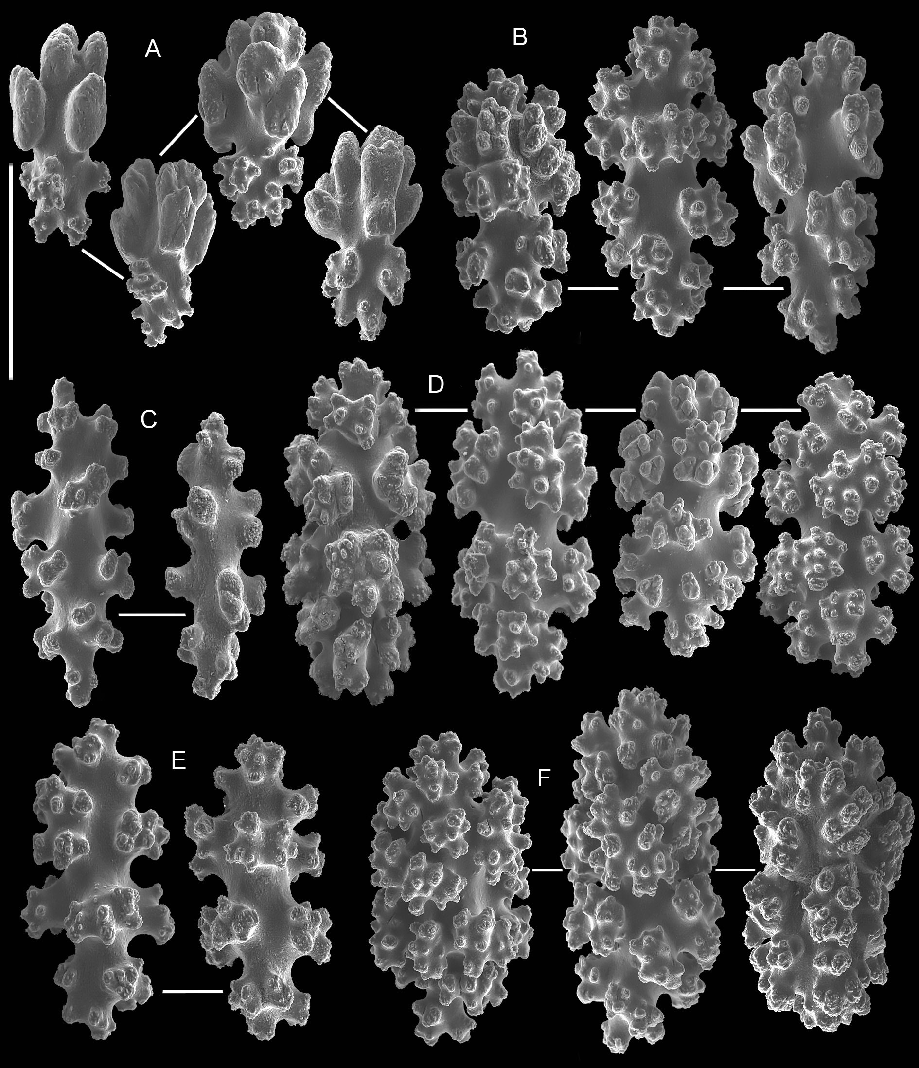
Numerous clubs in body wall of polyps, mainly 0.11–0.14 mm long, with a plump head consisting of leafy processes ( Fig. 3A View Fig.3 ). Handles of clubs mostly thick, with a girdle of well-developed warts. Longer clubs, up to 0.15 mm, with staggered leafy processes on head (Fig. 3B) are transitional to warty, mostly 0.12–0.15 mm long (Fig. 3C). Warty club-like spindles, up to 0.15 mm long, with well-developed warts arranged into 4–5 girdles (Fig. 3D). Warty clubs also coincide with capstans, mostly 0.15–0.17 mm long, with girdled warts and plump terminal tufts ( Fig. 3E View Fig.3 ), and ovals bearing some unordered processes (Fig. 3F). Spindles, mostly 0.15–0.17 mm long, also occur. These spindles have 4 girdles of more or less developed warts and plump terminal warts (Fig. 3G). Spindles and ovals common here.
External layer of coenenchyme comprises leafy clubs, mostly 0.08–0.09 mm long, with short blunt handle (Fig. 4A) and sparse warty clubs, mostly 0.13–0.15 mm. These clubs with well developed heads and blunt warty handles (Fig. 4B). Warty spindles, mostly 0.13 mm long, also occur here (Fig. 4C). Capstans, up to 0.13–0.15 mm, numerous; these are well-calcified 8-radiate sclerites with two girdles of warts and plump terminal processes (Fig. 4D). Some capstans, up to 0.10–0.13 mm long, less calcified, but with well-formed and girdled warts (Fig. 4E). Well-calcified capstans with very developed warts are transitional to the ovals, up to 0.15 mm long (Fig. 4F).
Internal layer of coenenchyme contains weakly calcified capstans, up to 0.13 mm long, of same shape as those in the outer layer ( Fig. 4E View Fig. 4 ).
Paratype and variations
Paratype MIMB 20724 colony shape and size similar to holotype (Fig. 1B). Sclerites composition (Fig. 5) coincides with that in holotype (Figs 2–4). The only difference is less calcification of sclerites in coenenchyme of paratype – it contains no ovals, but capstans and 8-radiate sclerites with two girdles of warts and plump terminal processes are present here (Fig. 5H). Paratype’s sclerites slightly smaller – leafy clubs in polyp and coenenchyme up to 0.1 mm long (Fig. 5A, D, G) vs up to 0.14 mm in holotype (Figs 2B–C, 3A–B). Capstans in polyp of paratype up to 0.15 mm long (Fig. 5F) vs up to 0.18 mm in holotype ( Fig. 3E View Fig.3 ).
Colour
In alcohol preserved material: polyps and coenenchyme creamy; colony axis black or deeply brown, sclerites colourless.
Distribution
This species is known for certain from the Kurile Islands, Sea of Okhotsk, Northwestern Pacific. Depth range is from 300 m to 900 m.
Remarks
This species should be identified as a member of the family Acanthogorgiidae , as it agrees well enough with the distinctive characters of the family: polyps are not divided into an anthocodia and anthostele; tentacles, when retracted, are folded above oral disk ( Bayer 1981; Fabricius & Alderslade 2001). Within the family Acanthogorgiidae , the examined specimen should be referred to as belonging to the genus Calcigorgia Broch, 1935 , as its distinctive characters agree with those of the latter: sclerites are small spindles, capstans and clubs that are irregularly distributed in the polyp ( Broch 1935; Bayer 1981).
The most obvious distinctive characters of C. matua sp. nov., differing from characters of other known members of the genus, are the shape of polyps (with a slender wider part, no folded peduncle in fixed specimens) and the presence of two different types of clubs among the sclerites. One of these types, the clubs with leaf-like processes on their heads and warty handle, has been described in neither C. japonica nor C. spiculifera .
All three species of Calcigorgia differ from each other in the composition of polyp sclerites (see Table 1 View Table 1 ). While the shape of warty clubs of the polyp and coenenchyme is the same in C. matua sp. nov., C. japonica and C. spiculifera , the size of these sclerites differs significantly (see Table 1). Thus, the morphology of polyps and composition and size of sclerites in polyp and coenenchyme are rather clearly different in the three species. This did not allow us to refer the examined specimen to one of the already known species of the genus Calcigorgia Broch, 1935 .

No known copyright restrictions apply. See Agosti, D., Egloff, W., 2009. Taxonomic information exchange and copyright: the Plazi approach. BMC Research Notes 2009, 2:53 for further explanation.
|
Kingdom |
|
|
Phylum |
|
|
Class |
|
|
Order |
|
|
Family |
|
|
Genus |