
Venonia spirocysta Cai, 1991
|
publication ID |
https://doi.org/10.5281/zenodo.4618989 |
|
persistent identifier |
https://treatment.plazi.org/id/2D258789-FF94-FFAF-C90B-FCF682C9FC83 |
|
treatment provided by |
Carolina (2021-03-18 18:43:19, last updated by Plazi 2023-11-02 02:04:38) |
|
scientific name |
Venonia spirocysta Cai, 1991 |
| status |
|
Venonia spirocysta Cai, 1991 View in CoL
( Figs. 41-44 View Figs )
Venonia spirocysta Cai View in CoL , in Chen & Zhang, 1991: 208, Figs. 211.1- 4; Cai, 1993: 60, Figs. 1-16 View Figs View Figs View Figs ; Yin et al., 1997: 50, Fig. 21 View Figs ; Song et al., 1999: 346, Figs. 202A, E.
Material examined. - 1 female, Kenting National Park , Pingtong County, Taiwan, Apr.2000, coll. Yu-Lung Hsieh (NMNS-THU-Ar- 02-0168) ; 1 male, Kenting National Park , Pingtong County, Taiwan, Apr.2000, coll. Yu-Lung Hsieh (NMNS-THU-Ar-01-0106) ; 2 males, Lugu Country , Nantou County, Taiwan, 25 Aug.1995, coll. Wen- Hao Chou (NMNS-THU-Ar-02-0157) .
Description. – Female: Total length 4.05: carapace length 1.90, width 1.35; abdomen length 2.27, width 1.53. Body with brown pubescence. Median band of carapace yellowishbrown, distinct, narrowing backward. Anterior eye row procurved, AME larger than ALE, AME-AME slight shorter than AME-ALE. Chelicerae with 3 teeth on both promargin and retromargin. Legs yellowish-brown, with brown annulations. Posterior margin of epigynal plate concave ( Figs. 41-42 View Figs ).
Male: Total length 2.57-3.37. One specimen: total length 2.70, carapace length 1.53, width 1.16; abdomen length 1.29, width 0.92. Similar to female in general shape and colour. Median band of carapace narrow, indistinct. Annulations of legs almost indistinguishable. Femur of leg I and pedipalp dark brown in colour. Tarsus of pedipalp without thick terminal setae, cymbium relatively large, genital bulb almost occupying the whole tarsus. Median apophysis pointing anteriorly ( Figs. 43-44 View Figs ).
Distribution. – Taiwan, Guangxi, Fujian, Jiangxi, Zhejiang, Hunan, Guizhou.
Cai, B. Q., 1993. A new species of the genus Venonia from China (Araneae: Lycosidae). Journal of Hunan Normal University (Natural Science), 21: 60 - 63.
Song, D. X., M. S. Zhu & J. Chen, 1999. The Spiders of China. Shijiazhuang: Hebei Science and Technology Publishing House. 640 pp.
Yin, C. M., X. J. Peng, L. P. Xie, Y. H. Bao & J. F. Wang, 1997. Lycosids in China. Changsha: Hunan Normal University Press. 317 pp.

Figs. 41-44. Venonia spirocysta Cai, 1991. 41-42. epigynum (41. ventral view; 42. dorsal view); 43-44. left pedipalp of male (43. ventral view; 44. retrolateral view). Scale bars: Figs. 41-44 = 0.10mm.

Figs. 1-5. Pardosa sp. 1-2. epigynum (1. ventral view; 2. dorsal view); 3-4. left pedipalp of male (3. ventral view; 4. retrolateral view); 5. terminal part of right pedipalp (ventral view). Abbreviations:ct: copulatory tube; cy: cymbium; em: embolus; h: hood; ma: median apophysis; ms: median septum; sp: spermatheca; st: subtegulum; t: tegulum. Scale bars: Figs. 1-2 = 0.15 mm; Figs. 3-5 = 0.20 mm.
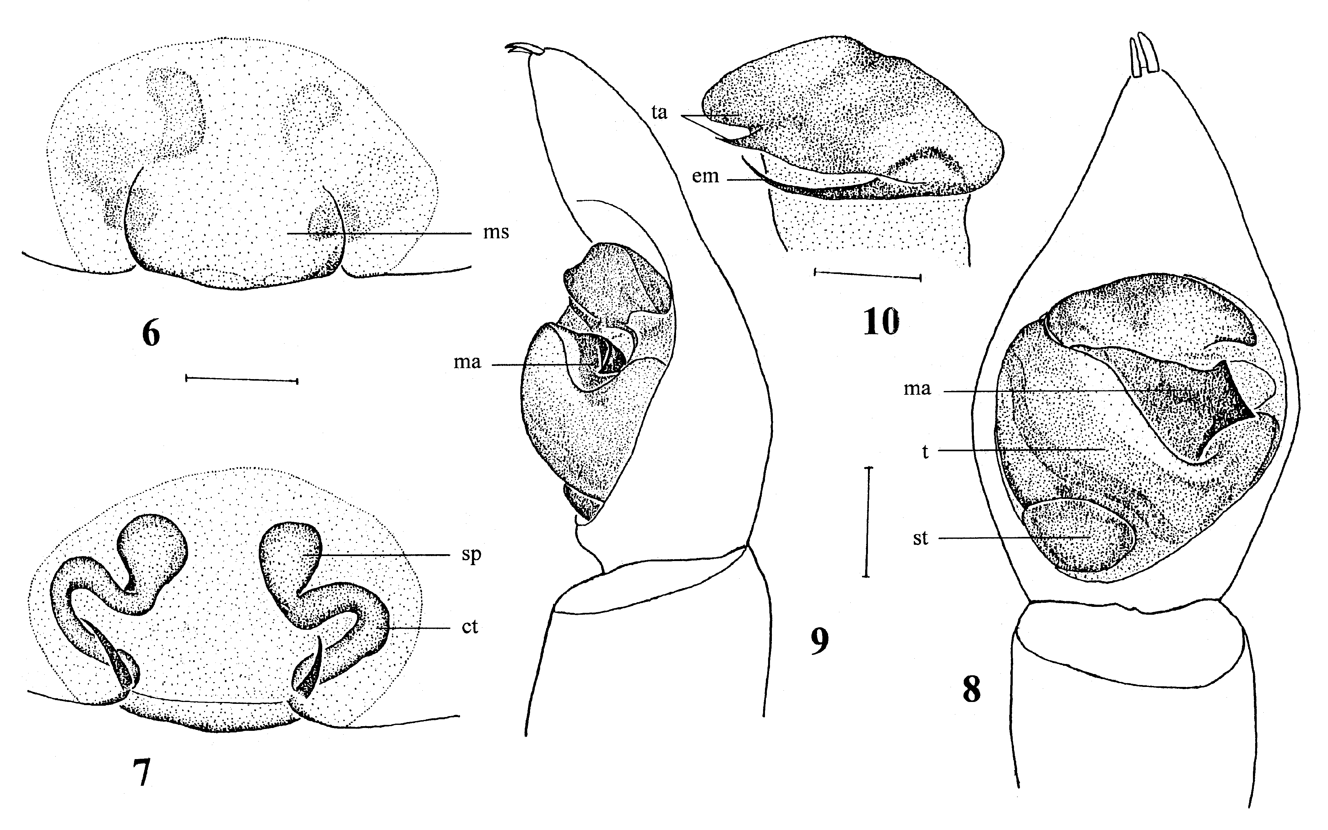
Figs. 6-10. Arcotosa labiata, new species. 6-7. epigynum (6. ventral view; 7. dorsal view); 8-9. left pedipalp of male (8. ventral view; 9. retrolateral view); 10. terminal part of right pedipalp (ventral view). Abbreviations:ct: copulatory tube; em: embolus; ma: median apophysis; ms: median septum; sp: spermatheca; st: subtegulum; t: tegulum; ta: terminal apophysis. Scale bars: Figs. 6-9 = 0.15mm; Fig. 10 = 0.10mm.
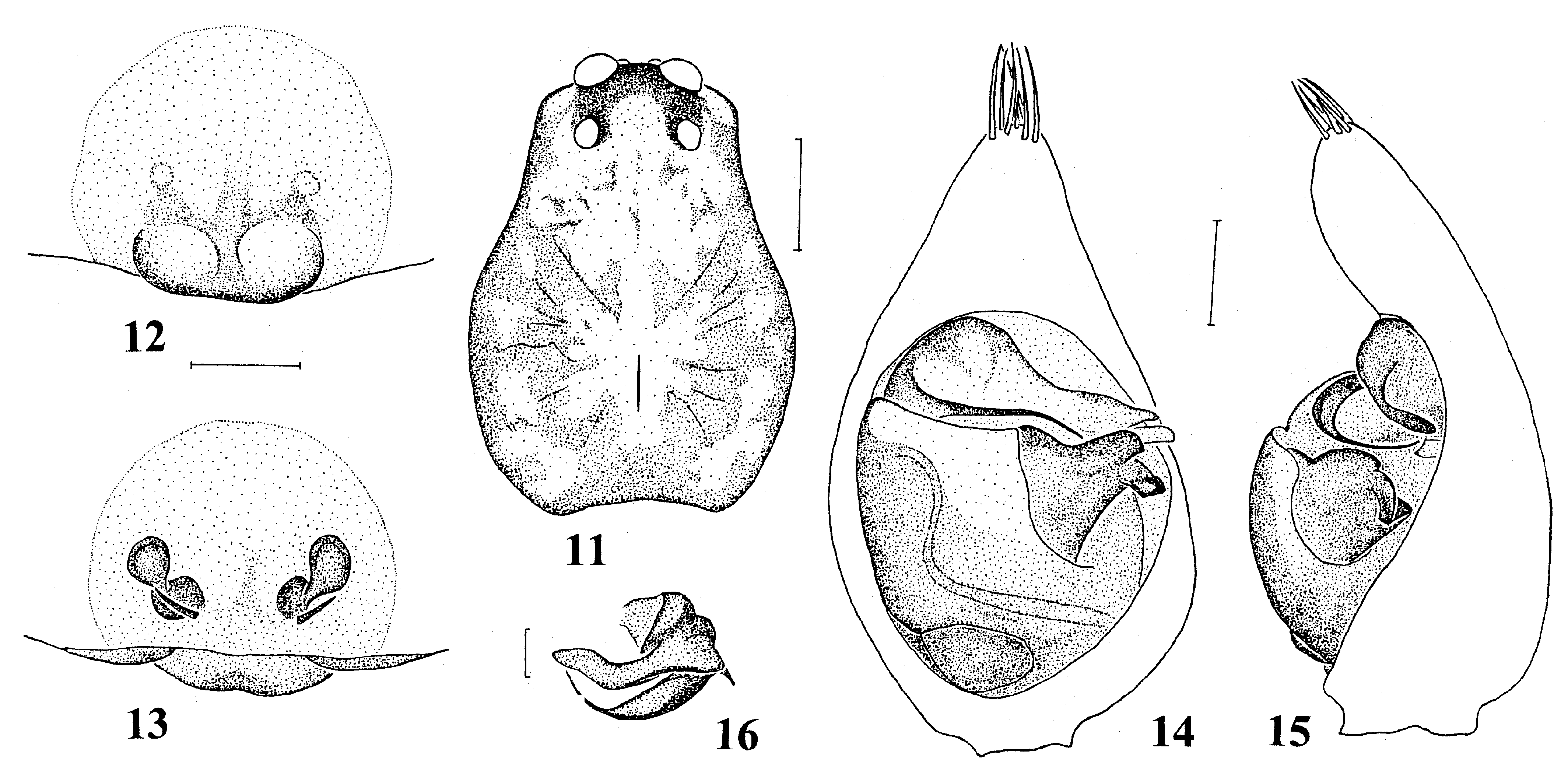
Figs. 11-16. Arcotosa truncata, new species. 11. carapace (female, dorsal view); 12-13. epigynum (12. ventral view; 13. dorsal view); 14- 15. left pedipalp of male (14. ventral view; 15. retrolateral view); 16. terminal part of right pedipalp (ventral view). Scale bars: Fig. 11 = 0.80mm; Figs. 12-15 = 0.15mm; Fig. 16 = 0.10mm.
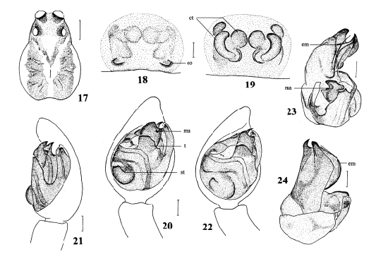
Figs. 17-24. Pirata digitatus, new species. 17. carapace (female, dorsal view); 18-19. epigynum (18. ventral view; 19. dorsal view); 20-22. left pedipalp of male (20. ventral view; 21. retrolateral view; 22. prolaterla view); 23-24. right pedipalp of male (expanded, 23. ventral view; 24. dorsal view). Abbreviations:co: copulatory opening; ct: copulatory tube; em: embolus; ma:median apophysis; st: subtegulum; t: tegulum. Scale bars: Fig. 17 = 0.40mm; Figs. 18-19 = 0.05mm; Figs. 20-24 = 0.10mm.
No known copyright restrictions apply. See Agosti, D., Egloff, W., 2009. Taxonomic information exchange and copyright: the Plazi approach. BMC Research Notes 2009, 2:53 for further explanation.