
Myzophyllobothrium narinari ( Shipley and Hornell, 1906 ) Jensen & Pen & Caira, 2021
|
publication ID |
https://doi.org/ 10.11646/zootaxa.4999.3.1 |
|
publication LSID |
lsid:zoobank.org:pub:051E68AE-6A5B-44AC-8F90-AEECB8997883 |
|
DOI |
https://doi.org/10.5281/zenodo.5118886 |
|
persistent identifier |
https://treatment.plazi.org/id/2A0987A5-FFCF-FFFD-FF7A-3F9A26F6FF3B |
|
treatment provided by |
Plazi |
|
scientific name |
Myzophyllobothrium narinari ( Shipley and Hornell, 1906 ) |
| status |
comb. nov. |
Myzophyllobothrium narinari ( Shipley and Hornell, 1906) n. comb.
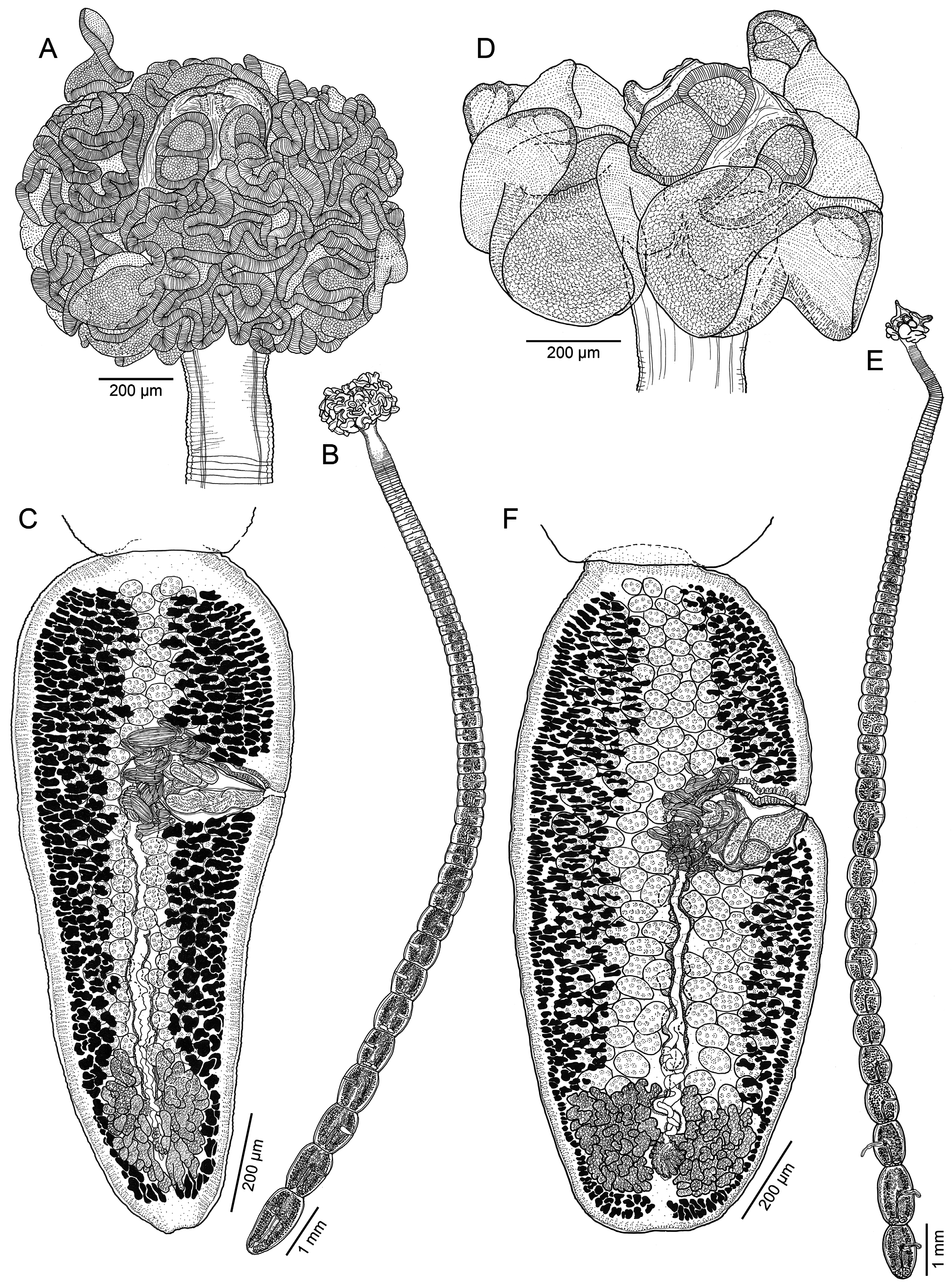
( Figs. 2A–C View FIGURE 2 , 3 View FIGURE 3 , 4B–E View FIGURE 4 )
(Syn. Myzocephalus narinari Shipley and Hornell, 1906 ; Myzocephalus sp. 1 of Caira et al. 2014).
Type and only known host: Whitespotted eagle ray, Aetobatus ocellatus (as Aetobatis [sic] narinari ) ( Myliobatiformes : Aetobatidae ).
Type locality: Dutch Bay , Sri Lanka (as Ceylon) .
Additional localities: Darwin (12°20’11”S, 130°54’39”E), Northern Territory, Australia (AU-41; see Caira et al. 2001, 2014); Sepuk Laut (00°12’51.60”S, 109°05’00.30”E), West Kalimantan, South China Sea, Indonesian Borneo (KA-304).
Site of infection: Spiral intestine.
Specimens deposited: Two mature worms ( BRT-P nos. 004 and 005); five mature worms ( USNM nos. 1655902– 1655906), scolex longitudinal ( USNM no. 1655908) and cross-section series ( USNM no. 1655907), and proglottid cross-section series ( USNM no. 1655909); five mature worms ( LRP nos. 10336–10340), scolex longitudinal section series ( LRP nos. 10341–10346), and two SEM strobilar vouchers ( LRP nos. 10347 and 10348); scolices examined with SEM retained with KJ at the University of Kansas.
Sequence data: KF685887 View Materials (hologenophore, LRP no. 8280) ex Aetobatus ocellatus (AU-41) of Caira et al. (2014) (as Myzocephalus sp. 1 ); MZ189003 View Materials and MZ189004 View Materials (hologenophores, LRP nos. 10349 and 10350, respectively) ex Aetobatus ocellatus (SL-2); MZ189002 View Materials (hologenophore, LRP no. 10361) ex Aetobatus ocellatus (KA-304).
Re-description. Based on 12 whole mature worms, cross-section series of one scolex, longitudinal-section series of two scolices, one cross-section series of mature proglottids, and two scolices examined with SEM).
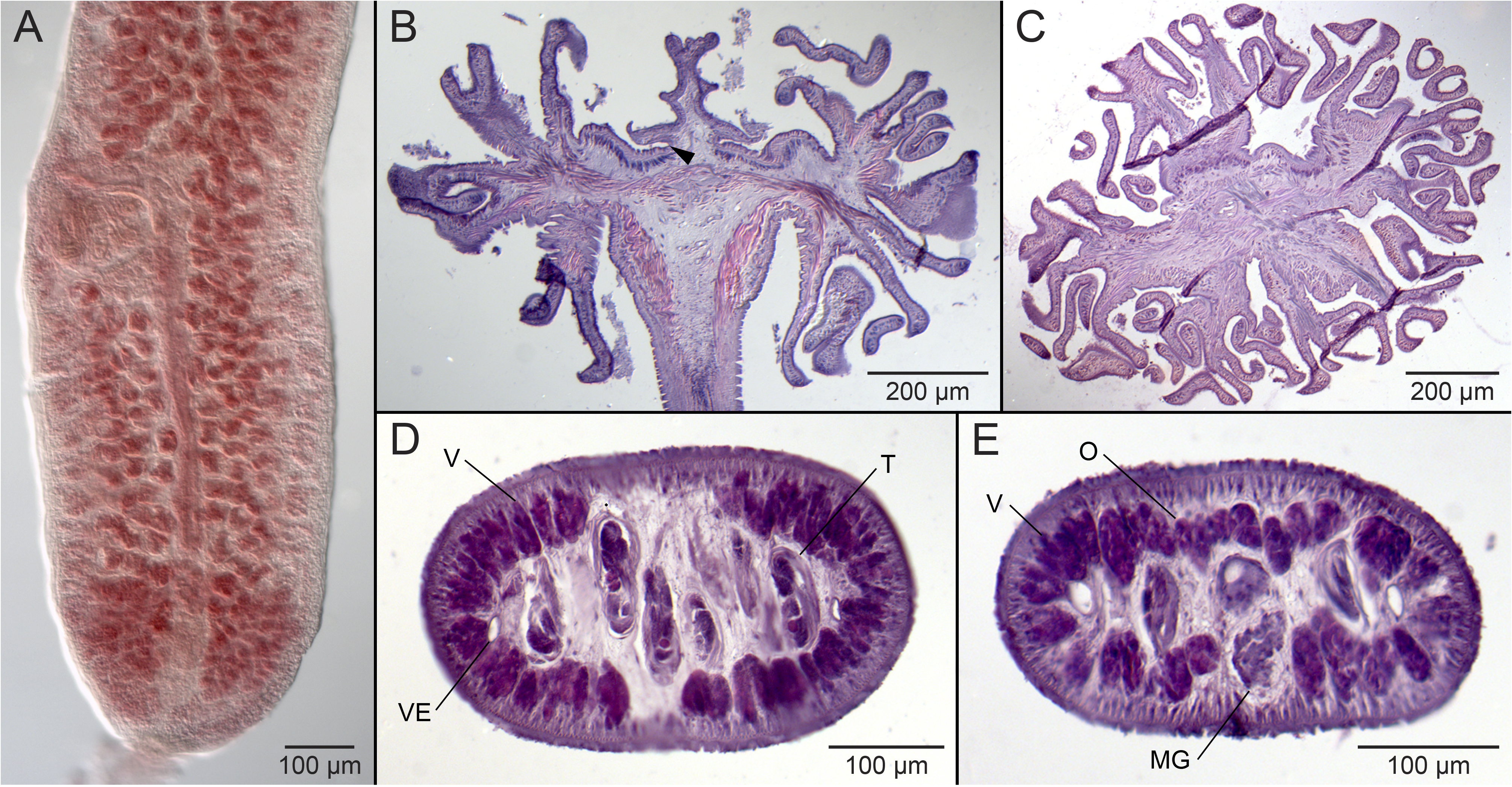
Worms euapolytic, slightly craspedote, 10.1–19.7 (14.9 ± 3; 12) mm long; proglottids 72–113 (93 ± 13; 12) in total number; maximum width at level of scolex ( Fig. 2B View FIGURE 2 ). Scolex globular ( Figs. 2A View FIGURE 2 , 3A View FIGURE 3 ), 748–1,103 (926 ± 113.1; 9) long, 961–1,532 (1,247 ± 179.3; 12) wide, consisting of four acetabula and cephalic peduncle. Scolex proper 350–502 (426 ± 49.7; 12) wide. Acetabulum bothridiate in form, 200–294 (247 ± 30.6; 6; 10) long, biloculate, sessile anteriorly, free posteriorly; anterior loculus undivided, 81–120 (100 ± 11.5; 10; 20) long, 100–160 (130 ± 13.0; 10; 24) wide, without postero-lateral extensions; posterior loculus undivided, 100–197 (149 ± 25.8; 7; 11) long, 88–156 (122 ± 20.1; 9; 13) wide. Cephalic peduncle bearing four stalked remi ( Figs. 3B View FIGURE 3 , 4C View FIGURE 4 ); region between bothridia and remi, and region posterior to remi short; stalk short; remus voluminous ( Fig. 3A, B View FIGURE 3 ), highly folded, wider than long, tapering terminally, with terminal primary areola ( Fig. 2A View FIGURE 2 , 3A, B View FIGURE 3 ); primary areola 56–99 (78 ± 13.6; 8; 11) long, 84–124 (104 ± 13.0; 6; 8); subterminal secondary areola not observed. Neck absent.
Apex of scolex proper ( Fig. 3C View FIGURE 3 ), distal surface of anterior loculus ( Fig. 3D View FIGURE 3 ) and posterior loculus ( Fig. 3E View FIGURE 3 ), proximal surface of anterior loculus and posterior loculus ( Fig. 3F View FIGURE 3 ), distal ( Fig. 3G View FIGURE 3 ) and proximal ( Fig. 3H View FIGURE 3 ) surfaces of remus, and distal surface of primary areola covered with slender gladiate spinitriches and capilliform filitriches of varying densities; proximal surface of primary areola ( Fig. 3I View FIGURE 3 ) covered with slender gladiate spinitriches and shorter capilliform filitriches; capilliform filitriches on rims of bothridia and remi conspicuous ( Fig. 3J View FIGURE 3 ). Strobila covered with capilliform filitriches ( Fig. 3K View FIGURE 3 ).
Immature proglottids wider than long, becoming slightly longer than wide with maturity ( Fig. 2B View FIGURE 2 ), 66–107 (87 ± 12; 12) in number; last immature proglottid 368–732 (550 ± 109.1; 12) long, 390–648 (519 ± 74.6; 12) wide. Mature proglottids becoming longer than wide posteriorly ( Fig. 2C View FIGURE 2 ), 2–6 (4 ± 1; 12) in number; terminal mature proglottid 1,010 –1,600 (1,222 ± 180.1; 10) long, 360–649 (486 ± 103.2; 10) wide, length to width ratio 1.7–3.5 (2.6 ± 0.5; 10):1. Gravid proglottids not observed. Testes in field extending from anterior margin of proglottid to ovarian bridge, arranged in multiple columns, one irregular layer deep in cross section ( Fig. 4D View FIGURE 4 ), 98–167 (133 ± 18; 12; 32) in total number, 21–38 (30 ± 4; 12; 32) in number in post-poral field, 19–41 (30 ± 5.4; 12; 36) long, 32–68 (50 ± 8.2; 12; 36) wide. Vas deferens coiled, essentially medial to cirrus sac. Cirrus sac pyriform, 155–267 (211 ± 31.4; 12) long, 108–160 (134 ± 18.9; 12) wide, thin-walled, containing coiled cirrus; cirrus armed with spinitriches. Genital pores irregularly alternating, 58–66% (62 ± 2.6; 10) of proglottid length from posterior end; genital atrium shallow. Vagina sinuous, extending from ootype along midline of proglottid, opening into common genital atrium anterior to cirrus sac. Ovary at posterior margin of proglottid, H-shaped in frontal view, 177–302 (232 ± 45.7; 10) long, 165–315 (229 ± 49.9; 10) wide, tetralobed in cross section ( Fig. 4E View FIGURE 4 ); ovarian margins lobulated. Vitellarium follicular; follicles 12–43 (27 ± 6.9; 12; 36) long, 28–65 (46 ± 8.5; 12; 36) wide, arranged in two lateral bands; each band consisting of multiple irregular columns of follicles, encroaching in midline of proglottid, extending throughout length of proglottid, interrupted by terminal genitalia, uninterrupted by ovary. Uterus ventral, medial, extending from ootype region to level of cirrus sac; uterine duct sinuous, entering uterus at midpoint. Excretory vessels four, arranged in one dorsal and one ventral pair on each lateral margin of proglottid.
Remarks. We have been unable to locate the type specimens of this species. Although this species is clearly recognizable based on the original description of the scolex and accompanying illustrations by Shipley and Hornell (1906), these authors were unable to provide a detailed description of the proglottid anatomy due to limitations of the material at hand, and thus presented only measurements of the main features of the species (total length, scolex width, and mature proglottid width). The specimens on which this re-description is based are generally consistent with the original description, however, they are smaller in several respects than those described by Shipley and Hornell (1906) (10.1–19.7 vs. 25 mm in total length; 748–1,103 vs. 2 mm in scolex width; 360–649 vs. 1mm in terminal proglottid width). Nonetheless, given this material came from the type host near the type locality, we have little doubt about its conspecificity with M. narinari and attribute the differences observed to intraspecific variation, or differences in fixation and/or variation in measurements taken from live versus preserved material. This re-description expands the concept of this species of Shipley and Hornell (1906) to include new information on scolex morphology. These authors (pg. 47) described this species to bear “four slipper shaped bothridia each divided by a horizontal ridge into two areolas” and a “ruff formed of four immensely crumpled lateral extensions”. After viewing these highly mobile portions of the scolex in live worms, they contemplated whether this worm exhibited “a double set of bothridia”. The terminology of their otherwise highly accurate account of the scolex of this species is revised here such that the lateral extensions are referred to as remi as defined by Jensen and Caira (2006). Histological sections ( Fig. 4B View FIGURE 4 ) confirm that these represent extensions of the cephalic peduncle separate from the bothridia. In addition, as seen in other members of the genus, each of the bothridia of this species was found to bear a terminal primary areola. The re-description also provides the first detailed information on the proglottid anatomy and the microthrix pattern of this species.
Myzophyllobothrium narinari differs from M. rubrum , M. chongi , M. gambangi , M. limae , and M. myliobatidis in possessing remi that are highly folded, rather than moderately or weakly folded. In addition, it is a smaller worm than M. rubrum (10.1–19.7 vs. 80 mm in total length [TL]). It is a larger worm than M. chongi , M. gambangi , M. limae , and M. myliobatidis (10.1–19.7 vs. 1.4–1.8, 1.1–2.1, 2.4–5, 1.4–3.4 mm in TL, respectively), with a greater number of testes (98–167 vs. 22–37, 22–36, 28–43, and 27–38, respectively). This is the species referred to by Caira et al. (2014) in their molecular phylogenetic analysis and in GenBank ( KF685887 View Materials ) as Myzocephalus sp. 1 .
Although not included in the re-description, specimens of this species collected from the type host off West Kalimantan on the island of Borneo (GenBank no. MZ189002 View Materials ) and off the Northern Territory in Australia ( KF685887 View Materials ) were included in our phylogenetic analysis. Despite slight differences in morphology (e.g., total length and scolex size) these specimens were found to be identical in sequence for 28S and are considered herein to be conspecific with the specimens of M. narinari from the type locality of Sri Lanka .

| USNM |
Smithsonian Institution, National Museum of Natural History |
No known copyright restrictions apply. See Agosti, D., Egloff, W., 2009. Taxonomic information exchange and copyright: the Plazi approach. BMC Research Notes 2009, 2:53 for further explanation.
|
Kingdom |
|
|
Phylum |
|
|
Class |
|
|
Order |
|
|
Family |
|
|
Genus |