
Myzophyllobothrium nagasawai, Jensen & Pen & Caira, 2021
|
publication ID |
https://doi.org/ 10.11646/zootaxa.4999.3.1 |
|
publication LSID |
lsid:zoobank.org:pub:051E68AE-6A5B-44AC-8F90-AEECB8997883 |
|
DOI |
https://doi.org/10.5281/zenodo.5118890 |
|
persistent identifier |
https://treatment.plazi.org/id/930A6E47-571D-411F-9377-2DD6E8351AE1 |
|
taxon LSID |
lsid:zoobank.org:act:930A6E47-571D-411F-9377-2DD6E8351AE1 |
|
treatment provided by |
Plazi |
|
scientific name |
Myzophyllobothrium nagasawai |
| status |
sp. nov. |
Myzophyllobothrium nagasawai n. sp.
( Figs. 2D–F View FIGURE 2 , 5A–M View FIGURE 5 , 6 View FIGURE 6 )
urn:lsid:zoobank.org:act:930A6E47-571D-411F-9377-2DD6E8351AE1
Type and only known host: Naru eagle ray, Aetobatus narutobiei ( Myliobatiformes : Aetobatidae ).
Type locality: Seto Inland Sea , Japan .
Site of infection: Spiral intestine.
Type specimens: Holotype (complete mature worm; MPM no. 21757) and four paratypes (one incomplete mature worm, one detached pre-gravid proglottid, and one detached gravid proglottid [ MPM no. 21758], and one egg preparation [ MPM no. 21759]); six paratypes (three complete mature worms [ USNM nos. 1655910–1655912], two detached gravid proglottids [ USNM nos. 1655913 and 1655914], and one proglottid cross-section series [ USNM no. 1655915]); five paratypes (two complete mature worms [ LRP nos. 10351 and 10352], two detached gravid proglottids [ LRP nos. 10353 and 10354], and one proglottid cross-section series [ LRP nos. 10355–10358]) and two SEM strobilar vouchers ( LRP nos. 10359 and 10360); two scolices and one gravid proglottid examined with SEM retained with KJ at the University of Kansas.
Sequence data: None.
Etymology: This species is named in honor of Kazuya Nagasawa of Hiroshima University, Japan, for his contributions to marine parasitology and for collecting the specimens on which this description was based.
Description. Based on six complete mature worms, one incomplete mature worm, one detached pre-gravid proglottid, five detached gravid proglottids, two cross-section series of mature proglottids, two scolices and one detached gravid proglottid examined with SEM, and one egg preparation.
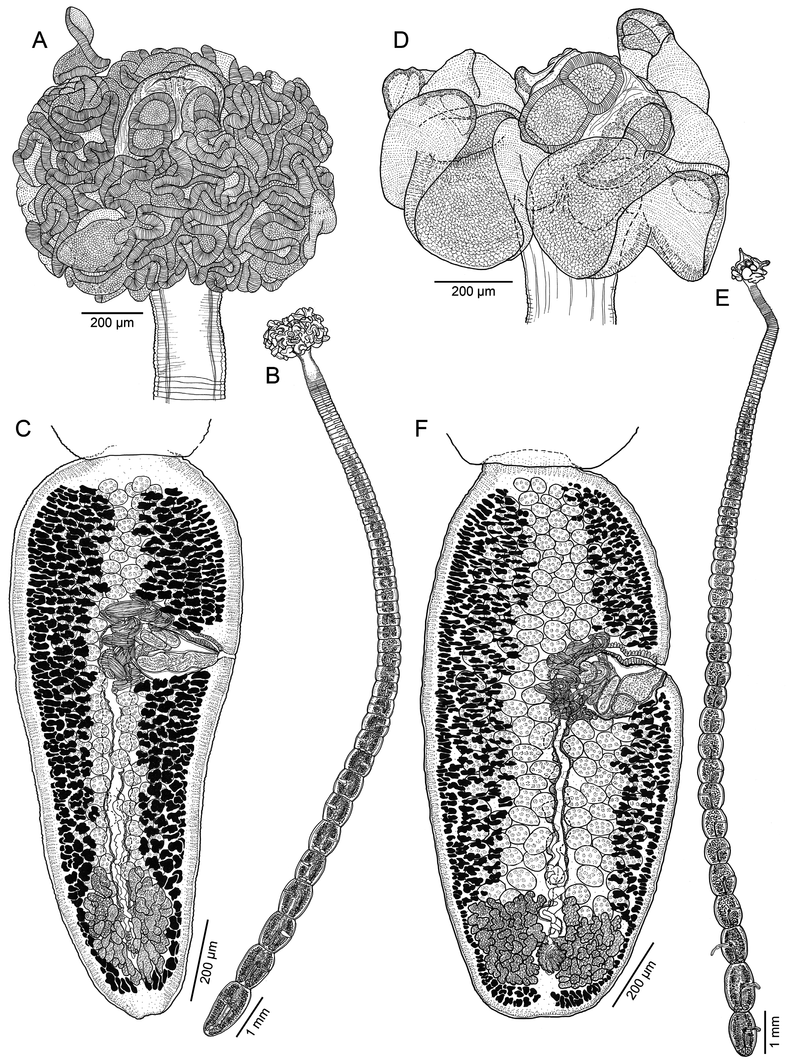
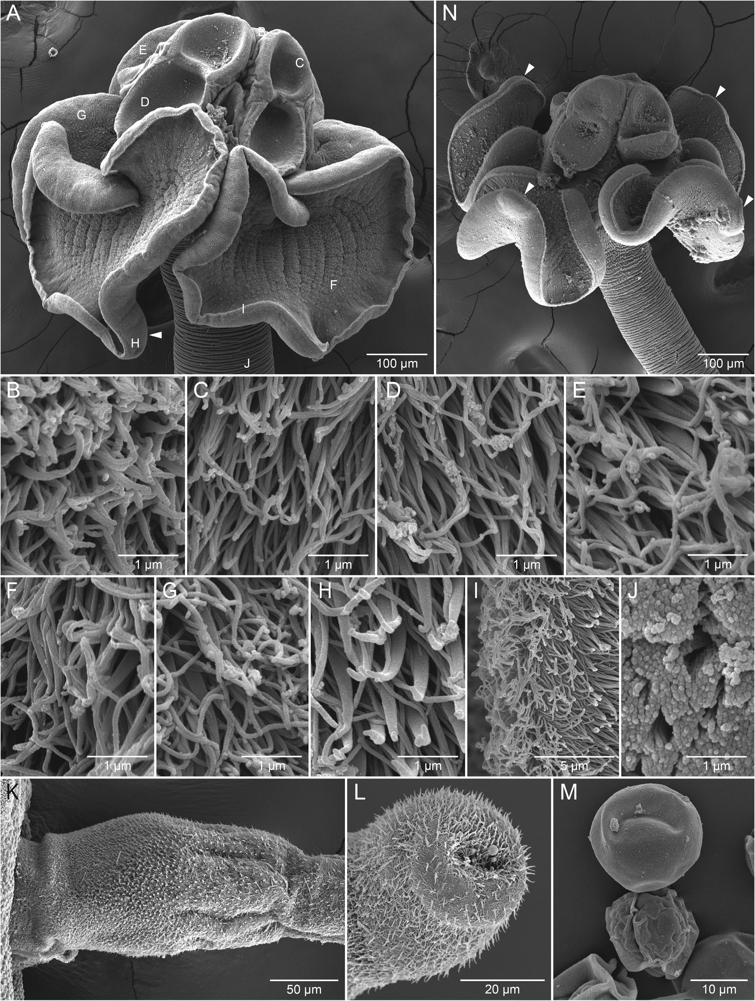
Worms euapolytic, slightly craspedote, 16.6–24.3 (20.0 ± 2.8; 6) mm long; proglottids 90–118 (99 ± 11; 6) in total number; maximum width at level of scolex ( Fig. 2E View FIGURE 2 ). Scolex 601–1,019 (762 ± 171.9; 7) long, 915–1,299 (1,092 ± 123.7; 7) wide, consisting of four acetabula and cephalic peduncle. Scolex proper 367–405 (390 ± 15.5; 5) wide. Acetabulum bothridiate in form, 255–374 (324 ± 41.7; 6; 12) long, biloculate, sessile anteriorly, free posteriorly; anterior loculus undivided, 98–163 (134 ± 18.3; 7; 14) long, 130–216 (164 ± 31.1; 7; 14) wide, without posterolateral extensions; posterior loculus undivided, 156–226 (196 ± 24.9; 6; 12) long, 135–201 (164 ± 19.6; 6; 11) wide. Cephalic peduncle bearing four stalked remi ( Figs. 2D View FIGURE 2 , 5A View FIGURE 5 ); region between bothridia and remi, and region posterior or remi short; stalk 62–179 (116 ± 30.7; 6; 12) long, 144–234 (183 ± 28.5; 6; 11) wide; remus without stalk, weakly folded, slightly longer than wide, 455–880 (597 ± 185.6; 4; 6) long, 386–610 (494 ± 71.1; 6; 11) wide, tapering terminally, with terminal primary areola and inconspicuous subterminal secondary areola; primary areola 48–65 (56 ± 5.5; 6; 11) long, 84–125 (105 ± 14.3; 6; 11) wide. Neck absent.
Apex of scolex proper ( Fig. 5B View FIGURE 5 ), distal surface of anterior loculus ( Fig. 5C View FIGURE 5 ) and posterior loculus ( Fig. 5D View FIGURE 5 ), proximal surface of anterior loculus and posterior loculus ( Fig. 5E View FIGURE 5 ), distal ( Fig. 5F View FIGURE 5 ) and proximal ( Fig. 5G View FIGURE 5 ) surfaces of remus, and distal surface of primary areola covered with slender gladiate spinitriches and capilliform filitriches of varying densities; proximal surface of primary areola ( Fig. 5H View FIGURE 5 ) covered with slender gladiate spinitriches and shorter capilliform filitriches; filitriches on rims of bothridia and remi conspicuous ( Fig. 5I View FIGURE 5 ). Strobila covered with capilliform filitriches ( Fig. 5J View FIGURE 5 ).
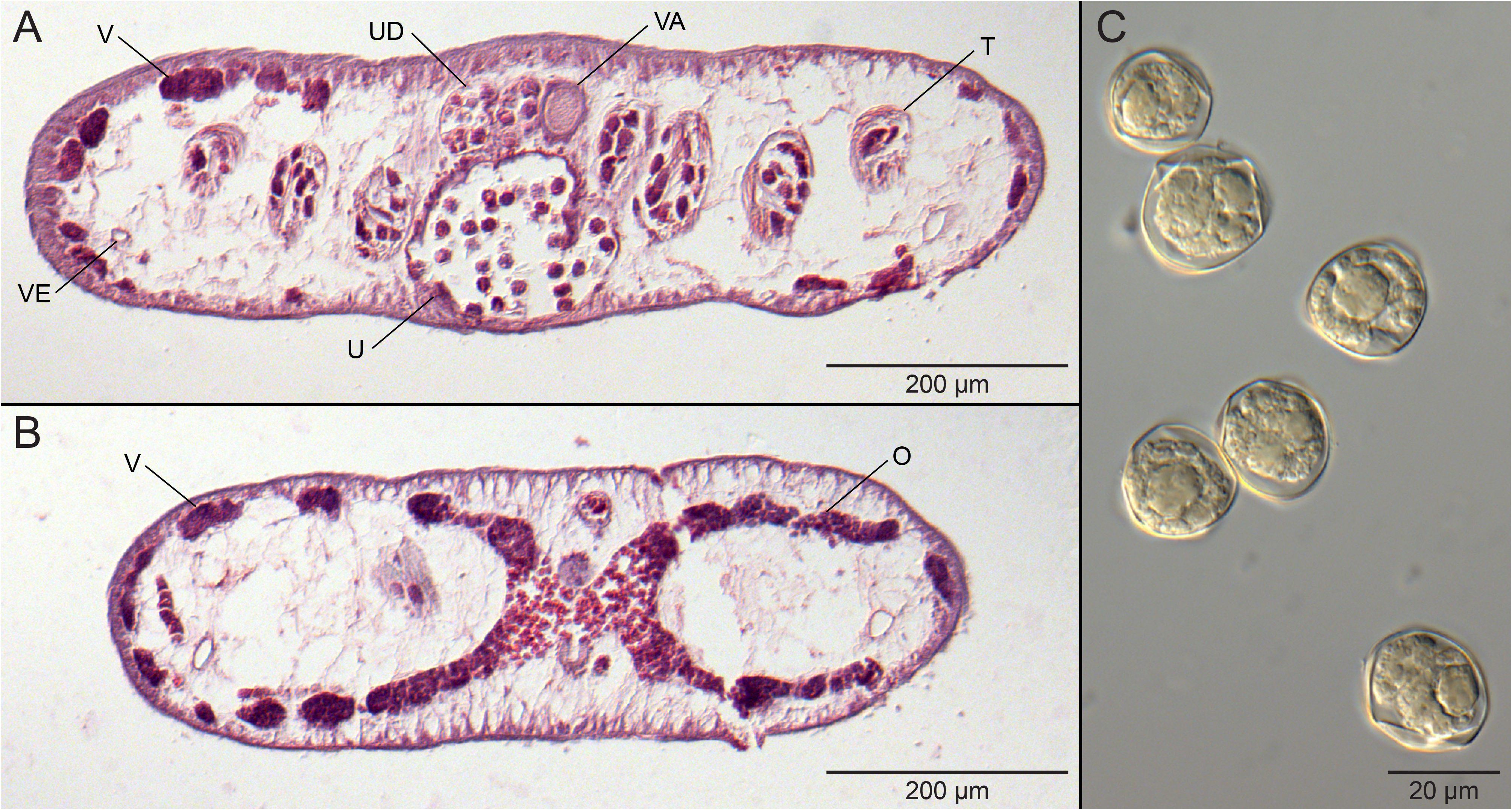
Immature proglottids wider than long, becoming approx. as long as wide with maturity ( Fig. 2E View FIGURE 2 ), 76–111 (89 ± 14; 6) in number; terminal immature proglottid 552–628 (601 ± 27.2; 6) long, 482–670 (567 ± 74.6; 6) wide. Mature proglottids becoming longer than wide posteriorly ( Fig. 2F View FIGURE 2 ), 5–12 (8 ± 2; 6) in number; terminal proglottid 966–1,707 (1,257 ± 279.3; 6) long, 477–682 (586 ± 85.5; 6) wide, length to width ratio 1.6–3.6 (2.2 ± 0.7; 6):1. Detached gravid proglottids 2,406 –3,629 (2,805 ± 483.6; 5) long, 1,000 –1,242 (1,110 ± 117.0; 5) wide. Testes in field extending from anterior margin of proglottid to ovarian bridge, arranged in multiple columns, one irregular layer deep in cross section ( Fig. 6A View FIGURE 6 ), 131–224 (177 ± 28; 6; 18) in total number, 28–50 (38 ± 7; 6; 18) in number in post-poral field, 21–49 (34 ± 7.9; 6; 18) long, 38–90 (57 ± 15.1; 6; 8) wide. Vas deferens coiled, essentially medial to cirrus sac. Cirrus sac pyriform, 204–260 (228 ± 20.5; 6) long, 102–155 (117 ± 19.3; 6) wide, thin-walled, containing coiled cirrus; cirrus armed with coniform spinitriches and capilliform filitriches ( Fig. 5L View FIGURE 5 ), spinitriches denser at base ( Fig. 5K View FIGURE 5 ), 77–88 (n=2) wide at base, at least 158 long (n=1). Genital pores irregularly alternating, 57–62% (59 ± 2.1; 6) of proglottid length from posterior end; genital atrium shallow. Vagina weakly sinuous, extending from ootype along midline of proglottid, opening into common genital atrium anterior to cirrus sac. Ovary at posterior margin of proglottid, H-shaped in frontal view, 173–337 (221 ± 65.8; 6) long, 186–405 (324 ± 73.5; 6) wide, tetralobed in cross section ( Fig. 6B View FIGURE 6 ); ovarian margins lobulated. Vitellarium follicular; follicles 7–20 (12 ± 3.7; 6; 18) long, 16–53 (31 ± 10.1; 6; 18) wide, arranged in two lateral bands; each band consisting of multiple irregular columns of follicles, encroaching in midline of proglottid, extending throughout length of proglottid, interrupted by terminal genitalia, uninterrupted by ovary. Uterus ventral ( Fig. 6A View FIGURE 6 ), medial, extending from ootype region to level of cirrus sac; uterine duct sinuous, entering uterus approximately at midpoint. Excretory vessels four, arranged in one dorsal and one ventral pair on each lateral margin of proglottid. Eggs spherical ( Figs. 5M View FIGURE 5 , 6C View FIGURE 6 ), single, 18–23 (21 ± 1.4; 18) in diameter.
Remarks. This new species differs from M. chongi , M. gambangi , M. limae , and M. myliobatidis in that its remi are wider than long, rather than longer than wide. It is also a much larger worm than all four of these species (16.6–24.3 vs. 1.4–1.8, 1.1–2.1, 2.4–5, 1.4–3.4 mm in TL, respectively). The remi of M. nagasawai n. sp. are weakly folded, rather than highly folded and voluminous, as seen in M. narinari and, whereas the remi of M. narinari taper conspicuously posteriorly, those of M. nagasawai n. sp. are wider and more bluntly rounded at their posterior-most point. This new species most closely resembles M. rubrum in that its remi are both wider than long and weakly folded. It conspicuously differs from the latter species in that it is substantially shorter in total length (4.6–24.3 vs. 80 mm).
Specimens of M. nagasawai n. sp. from its type host, Aetob. narutobiei , off Japan, fixed in ethanol for molecular work were not available for study. Two specimens taken from the type host in the Gulf of Tonkin off Viet Nam were included in our phylogenetic analysis. Although the scolex morphology of the more than 25 specimens from two host specimens from Viet Nam was highly consistent with that of specimens from Japan ( Fig. 5N View FIGURE 5 vs. 5A), the strobila of specimens from Viet Nam was much more delicate and smaller than that of the specimens from Japan. However, the specimens from Viet Nam were also all immature. We have referred to these specimens as M. cf. nagasawai until mature worms from Viet Nam can be examined and their identity confirmed. The strobila voucher of the scolex of the specimens from Viet Nam examined with SEM was deposited in the LRP (LRP no. 10367).
No known copyright restrictions apply. See Agosti, D., Egloff, W., 2009. Taxonomic information exchange and copyright: the Plazi approach. BMC Research Notes 2009, 2:53 for further explanation.
|
Kingdom |
|
|
Phylum |
|
|
Class |
|
|
Order |
|
|
Family |
|
|
Genus |