
Protoblepharon mccoskeri, Ho, Hsuan-Ching & Johnson, David, 2012
|
publication ID |
https://doi.org/ 10.5281/zenodo.212880 |
|
publication LSID |
lsid:zoobank.org:pub:2501EB81-95BC-4D9A-AA45-0615D63197E3 |
|
DOI |
https://doi.org/10.5281/zenodo.5622712 |
|
persistent identifier |
https://treatment.plazi.org/id/DEF0F7A3-0EB5-48C7-BC0D-3F010A31996F |
|
taxon LSID |
lsid:zoobank.org:act:DEF0F7A3-0EB5-48C7-BC0D-3F010A31996F |
|
treatment provided by |
Plazi |
|
scientific name |
Protoblepharon mccoskeri |
| status |
sp. nov. |
Protoblepharon mccoskeri View in CoL sp. nov.
New English name: Taiwanese flashlight fish Figures 1 View FIGURE 1 A–C, 2, 3, 4, 5A, 6A–B, 7, 8, Table 1–3
Holotype. NMMB-P 10807, 305 mm SL, Fugang Fishing port, Taitung, E. Taiwan, ca. 22°47.5’N, 121°11.6’E, ca. 300 m depth, 25 Dec. 2010, hook and line, purchased by H.-C. Ho.
Diagnosis and comparison. A species of Protoblepharon differing from its only congener, P. rosenblatti , in having the following: body deeper (2.3 in SL vs. 2.8 in SL in P. rosenblatti ); dorsal profile more strongly convex; pelvic fin origin anterior to (vs. posterior to) vertical through origin of dorsal fin, prepelvic length and pectoral fin much shorter, the appressed fin falling short of (vs. reaching) lateral line; pelvic fin shorter, the appressed fin falling short of (vs. reaching well beyond) midpoint between pelvic and anal fin origins; eye and light organ smaller; longest dorsal-fin spine the second (vs. the seventh); first, second and sixth dorsal-fin spines longer; seventh dorsal-fin spine much shorter; pseudobranch filaments 38 (vs. ca. 27); scale rows along vertical between origin of dorsal fin and lateral line ca. 30 (vs. ca. 18); scale rows along body axis over 260 (vs. ca. 145); enlarged scales surrounding anal fin base 37 (vs. 21 at anal fin base); single row of enlarged cycloid, plate-like scales along the margin of gill cavity (vs. 2–3 irregular rows of small scales).
Description. Counts and measurements of the holotype compared to those of P. rosenblatti are provided in Tables 1–3.
Head length 2.7 in SL, 1.1 in body depth; body depth 2.3 in SL; body width 4.8 in SL, 1.8 in HL; snout length 4.0 in HL; eye diameter 5.9 in HL; orbital diameter 4.4 in HL, 1.2 in interorbital width; interorbital width 3.7 in HL; light organ length 7.5 in HL; pre-dorsal length 2.3 in SL; pre-pelvic length 2.3 in SL; pre-anal length 1.3 in SL; pectoral fin length 2.0 in HL; dorsal fin height 2.4 in HL; 1st dorsal-fin spine length 5.1 in HL; 2nd dorsal-fin spine length 4.5 in HL; 3rd dorsal-fin spine length 5.4 in HL; 4th dorsal-fin spine length 5.4 in HL; 5th dorsal-fin spine length 7.5 in HL; 6th dorsal-fin spine length 8.2 in HL; 7th dorsal-fin spine 6.7 in HL; pelvic fin length 2.4 in HL; pelvic-fin spine length 3.9 in HL; anal fin height 3.4 in HL; 1st anal-fin spine length 11.8 in HL; 2nd anal-fin spine 6.5 in HL; caudal peduncle depth 3.8 in HL; caudal peduncle length 2.1 in HL; caudal fin length 1.8 in HL.
Body compressed but relatively thick (width 2.0 in depth) and deep (depth at origin of dorsal fin 1.1 times head length, 1.5 in length without head). With mouth open, profile sloping strongly from occiput to snout and convex in region of mesethmoid, then dropping slightly to symphysis of upper jaw ( Fig. 1 View FIGURE 1 A); upper-jaw symphysis below level of horizontal through middle of eye ( Fig. 1 View FIGURE 1 B). Nostrils anterior, completely dorsal to eye with mouth open, the anterior with thickend posterior rim. Jaws originating anteriorly below horizontal through middle of eye, maxilla extending posteriorly to vertical through anterior of eye. Posterior supramaxilla ovoid, anterodorsal surface with small pointed process extending anteriorly along posterodorsal egde of small anterior supramaxilla. Posterior supramaxilla covering most of posterior portion of maxilla, posteroventral corner of maxilla exposed and covered with tiny black papillae. Distinct notch at symphysis of premaxillae, presumably accommodating small dentigerous knob at symphysis of dentaries when mouth closed.
P. mccoskeri sp. nov. P. rosenblatti NMMB-P10807 AMS I.24275-001
Dorsal fin spines and rays VI–I, 14 VI –I,14 Anal fin spine and rays II, 11 II, 11
Pectoral fin rays ii+15+i ii+15+i Pelvic fin rays I, 5 I, 5
Caudal fin rays 11, 10+9, 9 10, 10 +9, 9 Gill rakers on 1st gill arch 2+4+13+2=21 2+5+12+2=21 Vertebrae 14+16=30 14+16=30 Scale rows between dorsal-fin base and Lateral line scales ca. 30 ca. 18 Lateral scale rows 59 & 61 59 & 60 Body scale rows>260 ca. 145 Abdominal scutes 13 13
Enlarged scales on dorsal-fin base 100 97
Enlarged scales on anal-fin base 37 22
Premaxillae, including most of lateral and medial surface, covered with bands of villiform teeth; no teeth at symphyseal notch ( Fig. 1 View FIGURE 1 C). Each dentary with narrow band of villiform teeth posteriorly, patch of slightly larger teeth covering entire symphysis, except for small median notch ( Fig. 1 View FIGURE 1 C). Vomer edentulous, palatines with welldeveloped, J-shaped patch of villiform teeth (anterior end of patch “hooked.”) Bones of head and pectoral girdle covered with numerous, rugose to minutely serrate ridges. Cleithrum with large exposed surface posteriorly, margin smooth. Supracleithrum almost covered by opercle ( Fig. 1 View FIGURE 1 B, 2). Anterior infraorbitals enlarged, covering anteroventral corner of orbit, and slightly flared laterally, forming medially sloping plate. Laterosensory canals of head appearing as channels of dark skin surrounded by bone, skin covered with small black papillae and perforated frequently by pores.
Eye relatively small, diameter 5.9 in HL. Notch at posterodorsal corner of orbit ( Fig. 1 View FIGURE 1 B). No fleshy papillae on posterior rim of orbit. Light organ ( Fig. 3 View FIGURE 3 ) relatively narrow and small, length 7.5 in HL. Light organ free posteriorly, with a relatively wide cup supporting its anterior end and extending posteriorly along entire ventral surface of light organ as a thin ovoid plate. Strongly elevated ridge on anterior portion of cup creating deep groove between itself and anterior portion of light organ. Anterior portion of fibrocartilaginous stalk continuous with its contralateral member across snout, with no attenuation at commissure. Posterior portion of stalk twisted, passing through small gap between lacrimal and nasal. Many tiny black papillae on outer surface of stalk followed by some large black column-like papillae at anterodorsal corner of light organ complex. Organ capable of being rotated downward into pocket below eye and medial to infraorbitals. When occluded, dorsal margin of light organ well below infraorbital rim. Black elastic shutter membrane attached along outer margin of suborbital pocket, erectible with rotation of light organ ( Figs. 4 View FIGURE 4 A–C).
Scales mostly spinoid type of Roberts (1993), except for series of enlarged smooth scales on gill chamber near isthmus; enlarged sculptured scales along lateral line ( Fig. 2 View FIGURE 2 ); midventral area posterior to pelvic-fin base with series of 13 enlarged, keeled scutes followed by 9 enlarged scales surrounding anus ( Fig. 5 View FIGURE 5 ), and many enlarged scales along bases of dorsal and anal fins ( Fig. 6A View FIGURE 6. A – C ). Lateral line covered by 59 (right side) and 61 (left side) enlarged spinoid scales; 100 (48 right, 52 left) enlarged scales along dorsal-fin base and 37 (18 right, 19 left) enlarged scales along anal-fin base, all variable in size. Row of enlarged irregular shaped cycloid scales along outer margin of gill chamber ( Fig. 6B View FIGURE 6. A – C ). Large area of slightly enlarged spinoid scales posterior to gill chamber between lateral line and pectoral fin base, followed by numerous small spinoid scales ( Fig. 2 View FIGURE 2 ). About 30 irregular rows of spinoid scales between dorsal-fin base and lateral line; about 40 irregular rows of spinoid scales between lateral line and origin of anal fin; more than 260 rows of scales along body axis (all difficult to count because of irregular distribution of scales). Head mostly scaleless, a few thick, heavily sculptured scales at anterodorsal corner of opercle, scales coalescing on cheek to form strong bony covering ( Figs. 1 View FIGURE 1 B–C). Gular region naked, with low, pigmented, transverse ridges.
P. mccoskeri sp. nov. P. rosenblatti
NMMB-P10807 AMS I.24275-001
Standard length 305 mm 220 mm
Spinous and soft dorsal fins continuous, deeply notched. First dorsal fin with six spines, second spine longest, gradually shorter to sixth. Length of first spine 1.1 in that of second, third 1.2 (measured with a broken piece connected by membrane), fourth 1.2, fifth 1.7, sixth 1.8; length of second spine 1.9 in that of longest third dorsalfin soft ray. Second dorsal fin with one spine (i.e., seventh dorsal spine) and 15 soft rays, length of spine 1.5 in second spine, third soft ray longest, gradually shorter to last ray. First dorsal-fin soft ray unbranched, its length about two-thirds of longest ray; remaining soft rays all branched, the last split to base. Anal fin shallower than dorsal fin, with 2 spines and 11 branched soft rays, length of second (longest) soft ray about 1.4 in that of dorsal fin, last ray spilt to base. Caudal fin deeply forked, with 11 procurrent and 10 principal rays (uppermost unbranched) in upper lobe and 9 procurrent and 9 principal rays (lowermost one unbranched) in lower lobe. Pectoral-fin base nearly horizontal, with 18 rays (2 simple rays +15 branched rays + 1 simple ray), sixth ray longest, appressed fin not reaching lateral line. Pelvic fin inserting anterior to vertical from dorsal-fin origin, relatively small, with one short spine and 5 branched soft rays, first soft ray longest, appressed fin not reaching mid-point between pelvic and anal fin origins.
Gill rakers rod-shaped with plate-like base or flat tooth plates, all bearing numerous tiny teeth; three rows on first to third arches and two rows on fourth: counts in Table 3 View TABLE 3 . A very large tooth patch on each side of third pharyngobranchial, absent from the rest pharyngobranchials. A very large tooth patch on each side of fifth ceratobranchial. Basibranchials edentulous.
Axial and caudal skeleton: Fourteen abdominal vertebrae and 16 caudal vertebrae, including second ural centrum. Dorsal fin supported by 21 anteroposteriorly expanded pterygiophores (seven spinous, 14 soft ray); one supraneural anterior to the first and second neural spines. Supraneural and pterygiophore insertion formula 0/0/1+1/1/1/1/1/1/1/1/1/2/ 1/2/3/2. Anal fin supported by 12 anteroposteriorly expanded pterygiophores, first three inserting anterior to haemal spine of first caudal (15th) vertebra.
Neural spine of fourth preural vertebra extends dorsally to base of first procurrent ray. Neural spine of third preural vertebra extends dorsally to support fifth procurrent ray and haemal spine extends posteriorly to support third to ninth procurrent rays. Neural spine of second preural vertebra low, haemal spine of normal length. Three epurals; one parhypural. six hypurals. ( Fig. 7 View FIGURE 7 ).
Otolith morphology: Sagitta otolith fusiform, large in size, 14.9 and 15.4 mm in width respectively ( Fig. 8 View FIGURE 8 A– B). Distal surface concave, with three ridges on dorsal portion and one ridge on rostrum. Proximal surface slightly convex; dorsal area of narrow, about same width with ventral area; dorsal depression and ventral depression absent; crista superior not well-developed; dorsal margin lobed, with three lobes, the posterior lobe a short protraction; ventral rounded in outline, with a crenate margin; anterior and posterior surfaces irregular. Excrisura a nearly right angle gape; antirostrum less developed, with a flat or slightly convex margin; rostrum a large, pointed protraction extending well beyond the antirostrum. Pseudo-excrisura a deep concave; pseudo-rostrum a large triangular protraction. Sulcus groove very deep and wide, heteromorph; collum absent; a narrow and elongated ostium (anterior sulcus) connected to the very wide and oval cauda (posterior sulcus) directly. Colliculum heteromorphy, well-developed in cauda and less developed in ostium.
Coloration. Uniformly reddish black when fresh and dark black when preserved. Gular region and outer margin of gill chamber pale with pigmented ridges; lateral-line scales grayish.
Etymology. Named after Dr. John E. McCosker, senior scientist of the California Academy of Sciences, in recognition of his interest in and contribution to our knowledge of flashlight fishes.
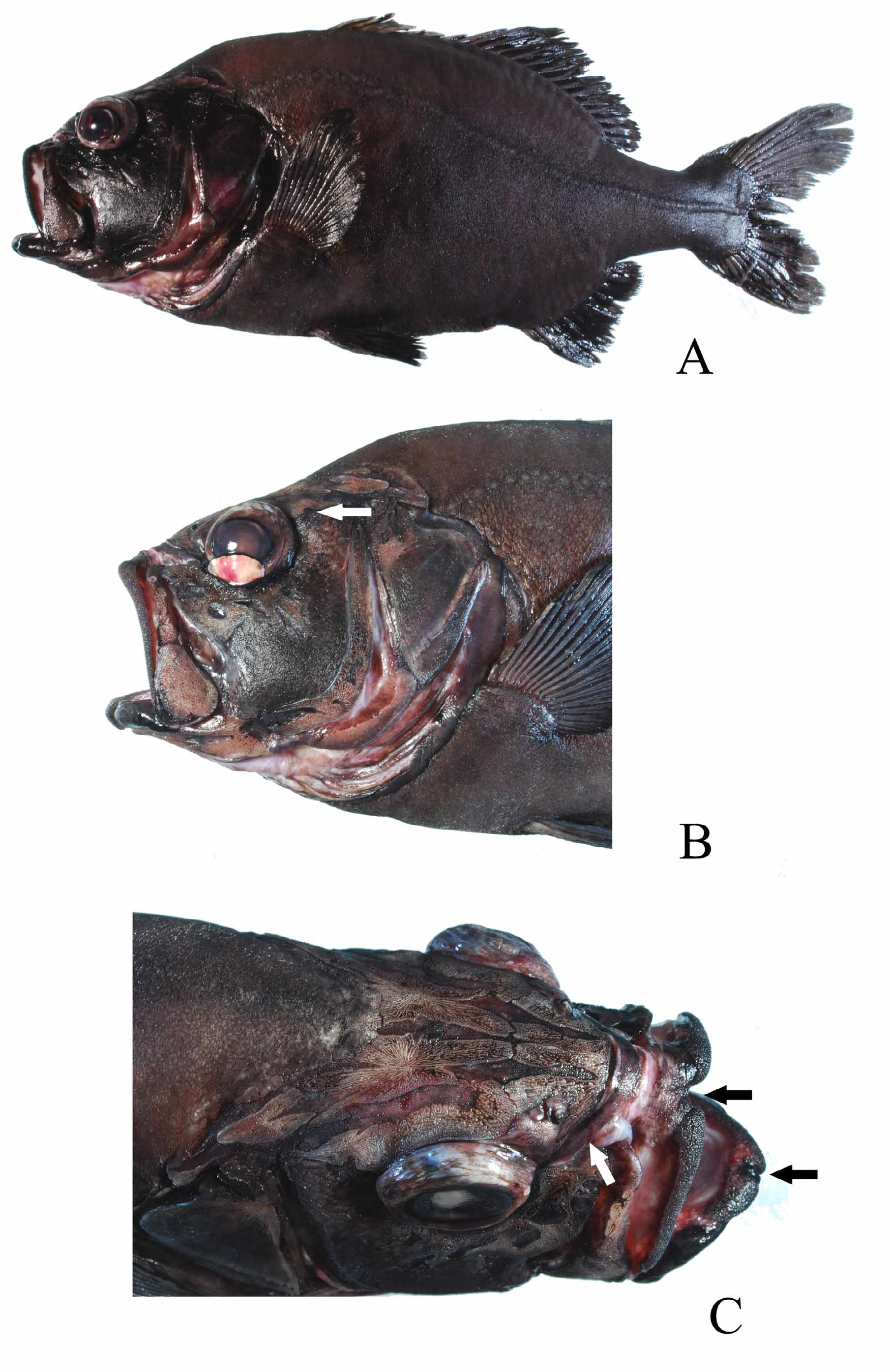
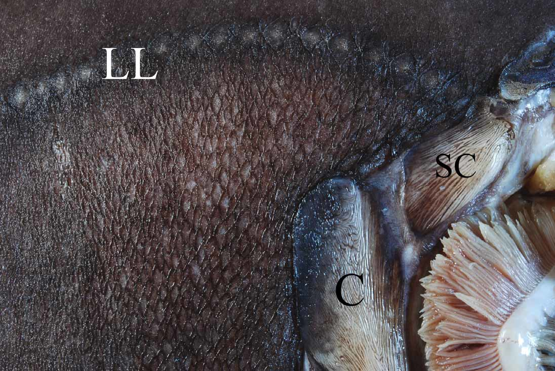
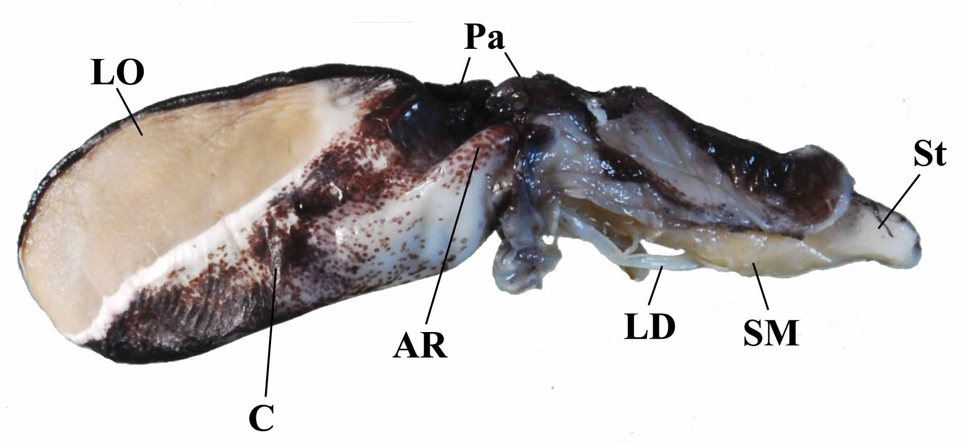
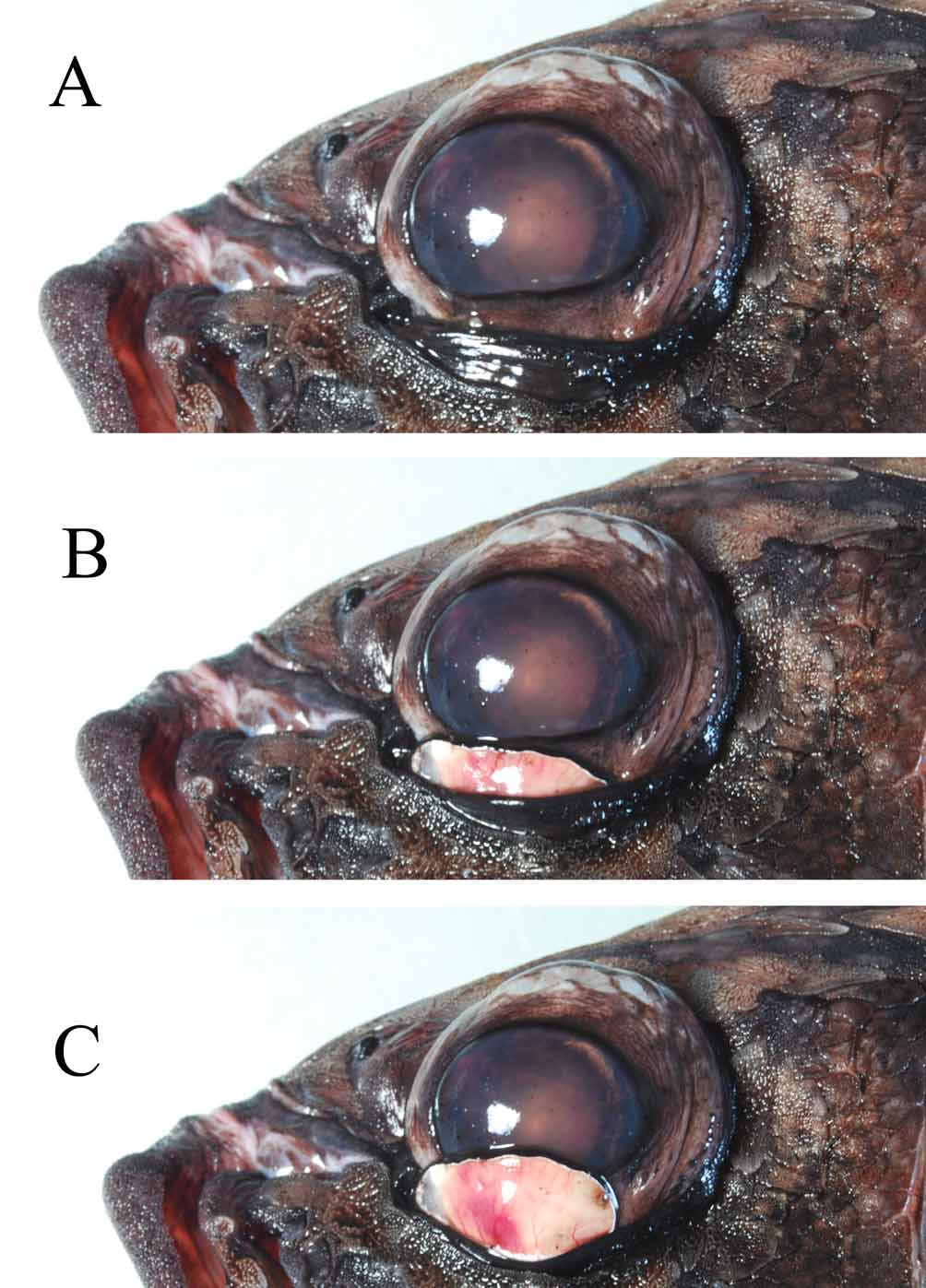
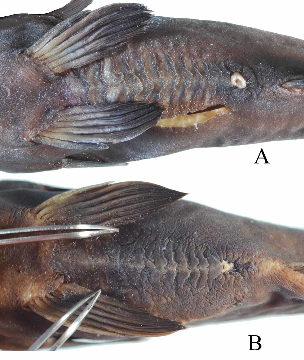
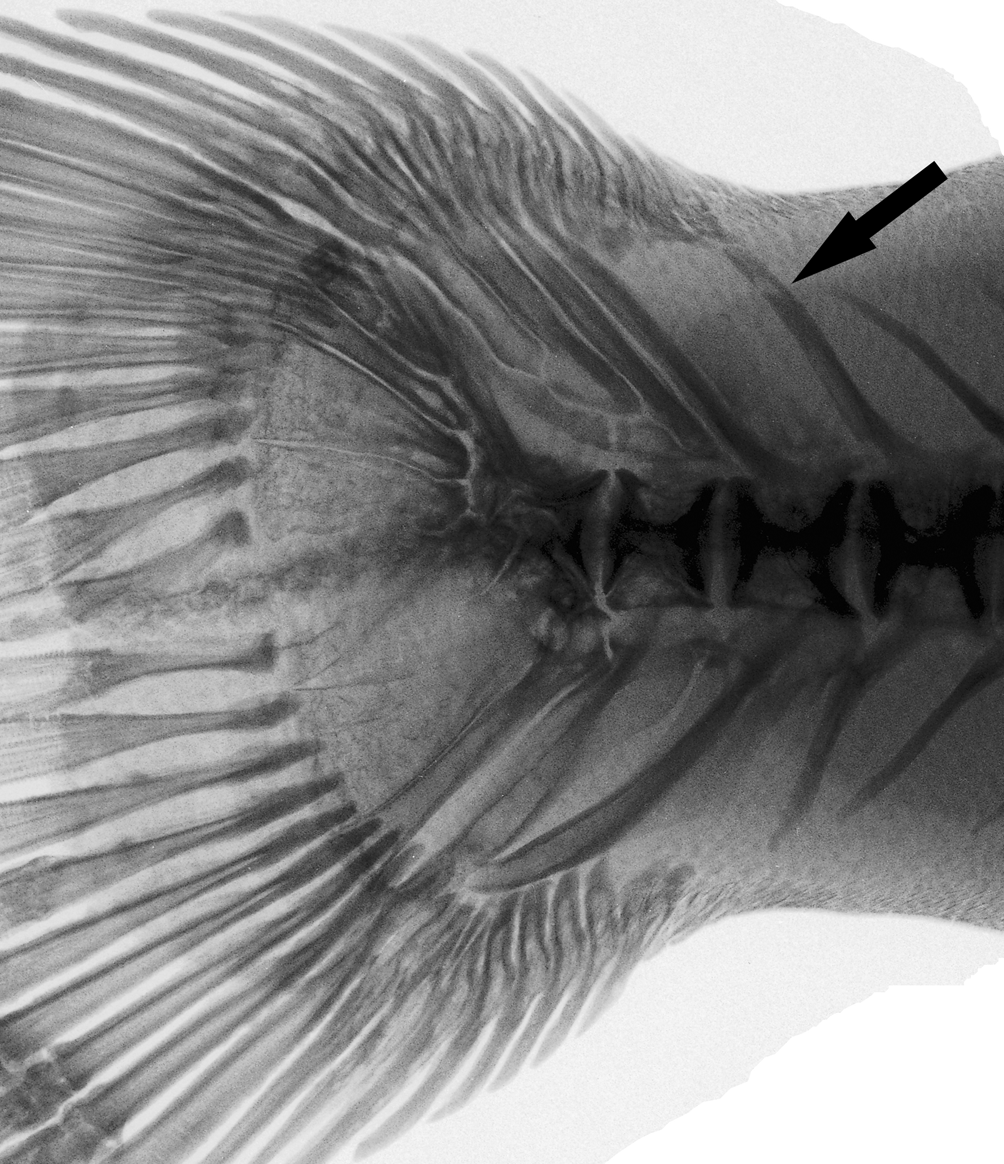
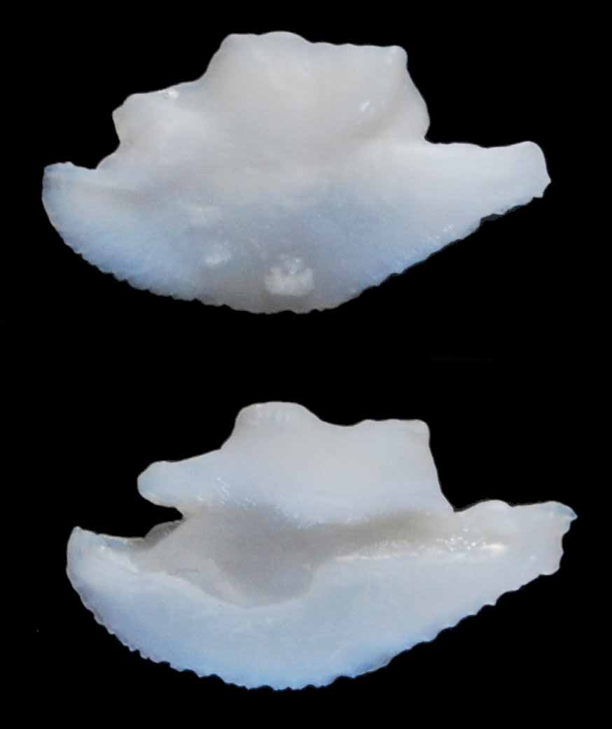
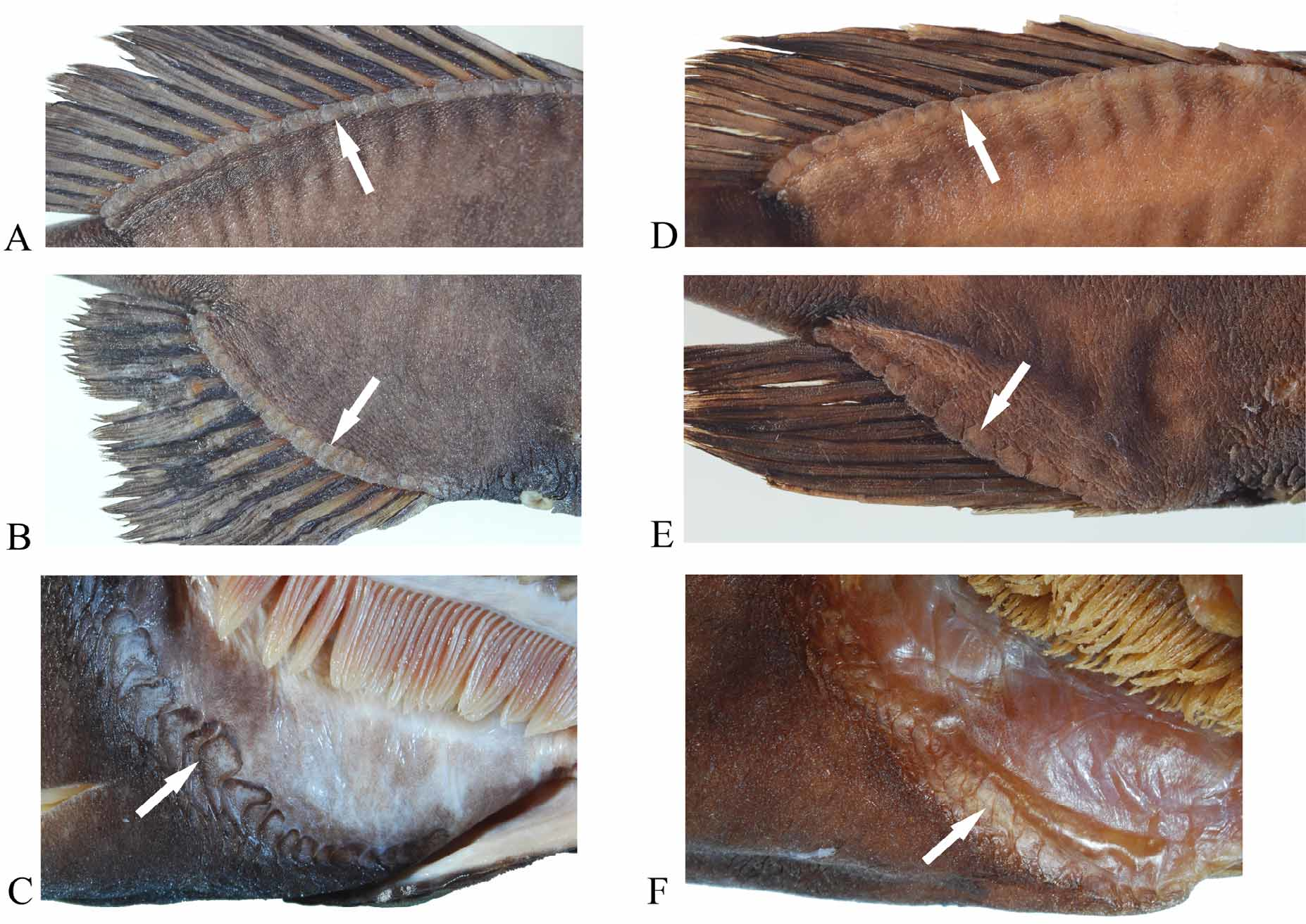
TABLE 3. Counts of the gill rakers of two species of Protoblepharon. The numbers are toothed plates + rakers. Question marks for P. rosenblatti are rakers obscured by reverted stomach.
| Upper limb Outer row | Median row | Inner row | Lower limb Outer row | Median row | Inner row | |
|---|---|---|---|---|---|---|
| P. mccoskeri sp. nov. 1st Gill arch 2nd Gill arch 3rd Gill arch | 2+4 5+2 4+0 | 4+0 3+0 absent | 1+1 3+1 2+0 | 2+13 5+11 3+10 | 10+0 13+0 11+0 | 2+12 0+11 3+7 |
| 4th Gill arch | absent | absent | absent | 5+0 | absent | 2+0 |
| P. rosenblatti 1st Gill arch | 2+5 | absent | 2+1 | 2+12 | absent | 0+14 |
| 2nd Gill arch 3rd Gill arch | ?+3 5+1 | absent absent | 0+2 2+1 | 3+12 3+11 | 11+0?+0 | 1+11 3+9 |
| 4th Gill arch | ? | ? | ? | ? | ? | ? |
No known copyright restrictions apply. See Agosti, D., Egloff, W., 2009. Taxonomic information exchange and copyright: the Plazi approach. BMC Research Notes 2009, 2:53 for further explanation.
|
Kingdom |
|
|
Phylum |
|
|
Class |
|
|
Order |
|
|
Family |
|
|
Genus |