
Odontanthias xanthomaculatus (Fourmanoir & Rivaton)
|
publication ID |
https://doi.org/ 10.11646/zootaxa.5222.2.3 |
|
publication LSID |
lsid:zoobank.org:pub:39825DC3-F184-44E7-9ED3-5E947E93BADA |
|
DOI |
https://doi.org/10.5281/zenodo.7456541 |
|
persistent identifier |
https://treatment.plazi.org/id/261187E8-A22B-E66B-FF5F-FF672216DDF1 |
|
treatment provided by |
Plazi |
|
scientific name |
Odontanthias xanthomaculatus (Fourmanoir & Rivaton) |
| status |
|
Odontanthias xanthomaculatus (Fourmanoir & Rivaton)
Figures 1–3 View FIGURE 1 View FIGURE 2 View FIGURE 3 ; Tables 1–2 View TABLE 1 View TABLE 2
Anthias xanthomaculatus Fourmanoir & Rivaton 1979: 414 View in CoL , fig. 8 (type locality, passe Bulari, New Caledonia); Bauchot et al. 1984: 10 (list).
Pseudanthias xanthomaculatus View in CoL ; Heemstra & Randall 1999: 2466, 2471 (key, list); Randall & Pyle 2001: 34 (list); Fricke et al. 2011: 387 (list); Anderson 2018: 49 View Cited Treatment (list).
Odontanthias grahami Randall & Heemstra 2006: 17 View in CoL , figs. 1H, 6, 7, pl. 4B (type locality, off northern New South Wales, Australia); White 2011: 27 (key); Anderson 2018: 23 View Cited Treatment (list); Gill & Russell 2019: 182, figs. 1, 2 (probable synonymy with Anthias xanthomaculatus Fourmanoir & Rivaton View in CoL ); Parenti & Randall 2020: 21 (list).
Odontanthias xanthomaculatus ; Gill & Russell 2019: 176, figs. 1, 2 (redescription of holotype; assignment to Odontanthias View in CoL and probable synonymy with O. grahami View in CoL ); Parenti & Randall 2020: 22 (list); Anderson 2022: 569 View Cited Treatment (list).
Diagnosis. A species of Odontanthias with the following combination of characters: dorsal-fin rays X,14; pectoralfin rays 15–17, usually 16; lateral-line scales 36–41; gill rakers 12–13 + 26–30 = 38–43.
Description. Dorsal-fin rays X,14, all segmented rays branched; anal-fin rays III,7, all segmented rays branched; pectoral-fin rays 15–17, all rays branched except upper 2; pelvic-fin rays 1,5, all segmented rays branched; principal caudal-fin rays 9 + 8; branched caudal-fin rays 6 + 7; upper procurrent caudal-fin rays 4–7 (6 or 7 in all but two smallest specimens); lower procurrent caudal-fin rays 4–7 (6 or 7 in all but two smallest specimens); total caudal-fin rays 25–31 (29–31 in all but two smallest specimens); lateral-line scales 36–41; scales above lateral-line to origin of dorsal fin 6–7; scales above lateral-line to base of fifth dorsal-fin spine 2–4; scales below lateral line to origin of anal fin 15–18; circumpeduncular scales 18–24; gill rakers 12–13 + 26–30 = 38–43; branchiostegal rays 7; pseudobranch filaments 16–26 (increasing in number with size).
Vertebrae 10 + 16; supraneurals 2; anterior dorsal pterygiophore formula S+S//3/1+1/1/1/1/1/1, S/ S/3/1+1/1/1/1/1/1 or /S+S/3/1+1/1/1/1/1/1; main shaft (proximal component) of first dorsal pterygiophore angled slightly anterordorsally; dorsal-fin pterygiophores in interneural spaces 9–13 1/1/1/1/1+1 or 1/1/1/1+1/1; no trisegmental pterygiophores associated with dorsal and anal fins; ribs present on vertebrae 3 through 10; epineurals present on vertebrae 1 through 10–12; no parapophyses on first caudal vertebra; parhypural and hypurals autogenous; well-developed hypurapophysis on parhypural; epurals 3; single uroneural (posterior uroneural absent); ventral tip of cleithrum with well-developed posteroventral process ( Gill & Russell, 2019: figs. 2–3).
Morphometric values are summarised in Table 2 View TABLE 2 .
Mouth large, oblique, posterior margin of maxilla reaching to point ranging from vertical through posterior edge of pupil to vertical through posterior edge of eye; mouth terminal, lower jaw projecting slightly; no supramaxilla; premaxilla with 1 or 2 enlarged recurved canine anterolaterally, a band of small conical teeth about 6 or 7 rows wide at symphysis reducing to 1–4 rows on sides of jaw, with outer row teeth larger and curved inwards (anteriorly) or forwards (posteriorly); a few teeth in band nearest symphysis enlarged and caniniform; dentary with 1–3 enlarged recurved canines anterolaterally, followed by a band of small conical teeth about 4–12 rows wide, reducing to about 2 or 3 rows on sides of jaw, then a single row posteriorly, last-mentioned larger and curved; a few teeth in band nearest symphysis enlarged and caniniform; sides of lower jaw with 1–3 enlarged, caniniform teeth, these sometimes preceded by narrow edentate area; vomer with an inverted, chevron to roughly heart-shaped patch of small conical teeth (see Randall & Heemstra 2006: figs. 1H and 1O), sometimes pointed anteriorly; palatine with curved band of small conical teeth, band broadest anteriorly, narrowing to 1 or 2 rows of teeth posteriorly; endopterygoid (mesopterygoid) and ectopterygoid edentate; tongue pointed with large oval patch of small conical or granular teeth, sometimes imbedded and difficult to see.
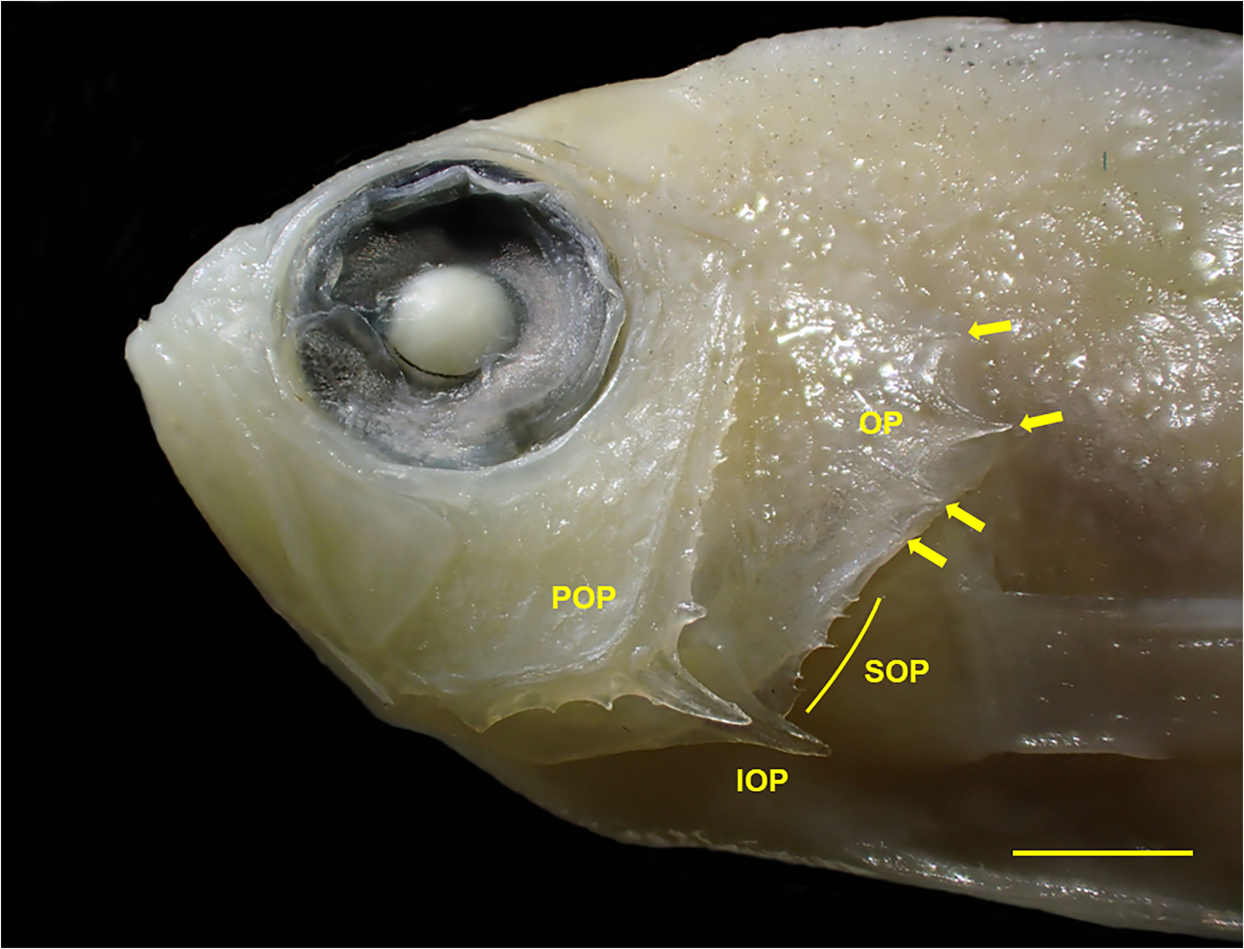
Opercle with 3–5 spines; when 3 opercle spines present, upper spine smallest, well separated from other spines and partially concealed by scales, middle spine largest, positioned immediately above dorsal edge of subopercle (portion concealed by opercle), lower spine below internal junction with subopercle; increase in number of opercle spines results from doubling of tips on middle or lower spine, and in juveniles from presence of additional small spine below lower spine ( Fig. 1 View FIGURE 1 ); posterior edge of preopercle with 8–26 fine serrations, increasing in number with size of specimen; serrations increasing in size ventrally, with prominent spine at angle, which is enlarged in juveniles and mirrored by similar spine on interopercle ( Fig. 1 View FIGURE 1 ); ventral edge of preopercle with 1–5 serrations; posterior edge of interopercle of juveniles with enlarged spine, which becomes smaller or disappears in larger specimens, and usually preceded by 1–5 weak to strong serrations; subopercle with 1–7 weak to strong serrations; posttemporal with 1–6 weak to strong serrations; supracleithrum of juveniles with 1–3 serrations, serrations absent in larger specimens. Anterior nostril positioned at middle of snout, tubular with small flap on posterior rim, flap reaching or almost reaching posterior nostril when adpressed; posterior nostril at mid-upper, anterior border of orbit, with slightly raised anterior rim. No papillae on posterior rim of orbit.
Scales ctenoid with peripheral cteni ( Roberts 1993); lateral line broadly arched over pectoral fin following body contour to end of dorsal fin, then sharply horizontal on caudal peduncle, extending to caudal-fin base; head fully scaled except lips, narrow area behind upper lip, area in front of nostrils, most or all of first infraorbital, chin and branchiostegal membranes; no auxiliary scales, except sometimes a few on head and nape; posterior portion of spinous dorsal fin with low sheath of small scales; soft portion of dorsal fin with low scaly sheath, and wedge of small scales between rays, extending up to basal half of rays; anal fin with low scaly sheath and wedge of small scales between segmented rays; caudal fin with scaly basal sheath, with small scales extending over almost all of fin, except for fin tips and posterior part of membranes of middle rays; pectoral fins with basal, wedge-shaped sheath of small scales.
Origin of dorsal fin above or slightly in front of vertical through first lateral-line scale; first dorsal-fin spine 1.5–1.9 in second spine; fourth or fifth dorsal-fin spine longest in small specimens, third spine longest in specimens larger than about 90 mm SL; tips of first 7–12 segmented dorsal-fin rays filamentous, third or fourth ray longest; anal-fin origin beneath vertical through base of second to fourth segmented dorsal-fin ray; first anal-fin spine 1.7–2.1 in second spine; second or third anal-fin spine longest; second segmented anal-fin ray longest, with filamentous tip; caudal fin deeply emarginate, lobes moderately broad and not pointed, most branched rays with short filamentous tips; pectoral fin pointed, nineth ray longest, reaching to point ranging from above anus to above anterior third of anal-fin base; second segmented pelvic-fin ray longest and filamentous.
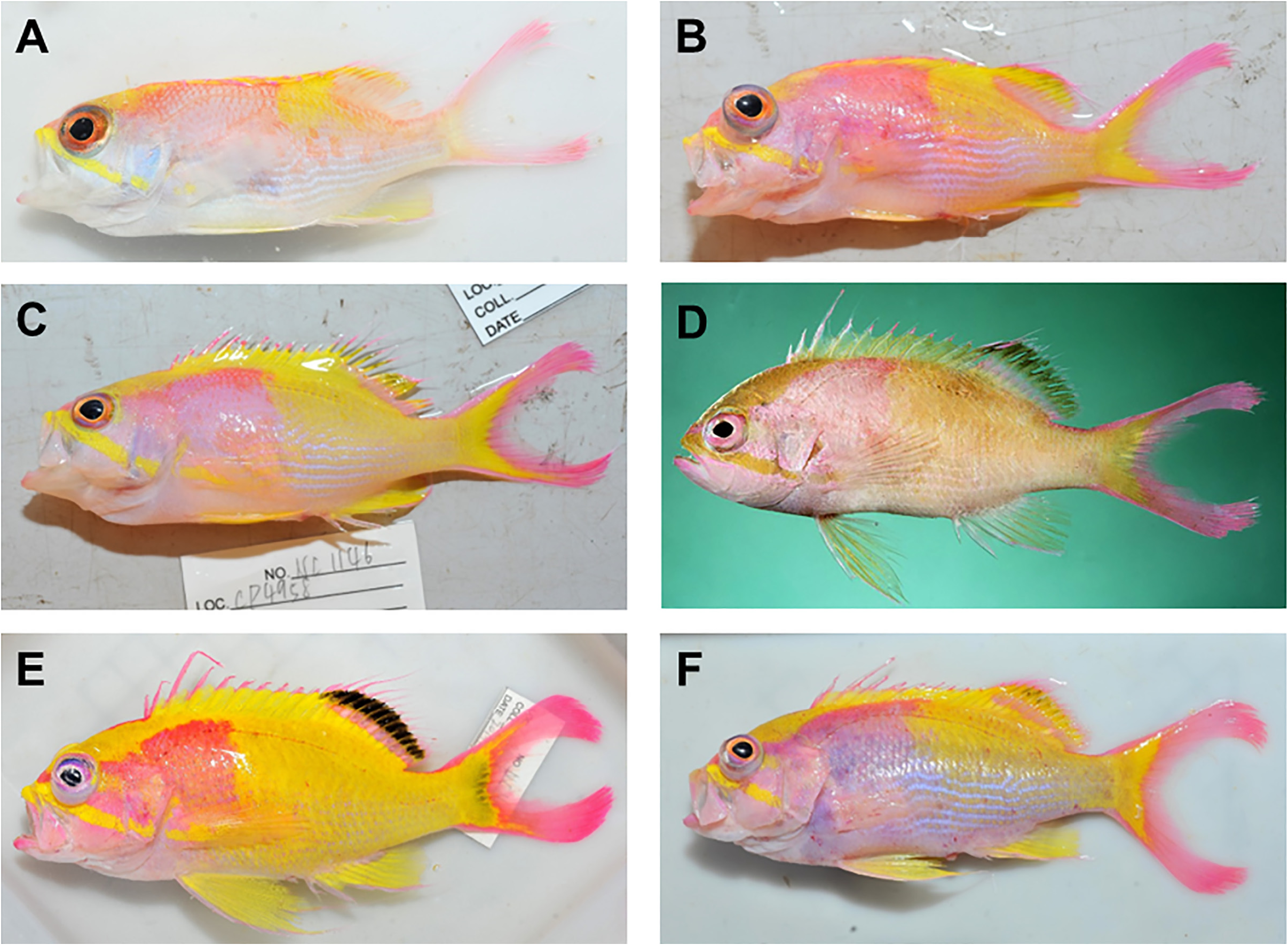
Colour in life (based on colour notes provided in Fourmanoir & Rivaton 1979, and colour photos of specimens from northern New South Wales, southern Queensland and the Lord Howe Rise; Fig. 2 View FIGURE 2 ): head pale pink to bright pink, becoming paler to white ventrally; dorsal part of head and nape bright yellow; bright yellow oblique stripe extending from tip of upper lip beneath eye to lower part of subopercle; larger specimens with diffuse yellow stripe extending form mid-posterior edge of orbit to upper part of preopercle, sometime extending farther on to opercle; iris pink to orange, sometimes bright blue on periphery; body pink, paler pink to white ventrally; juveniles with large yellow spot dorsally beneath posterior few dorsal-fin spines and anterior few segmented dorsal-fin rays, yellow area becoming more extensive in larger specimens, sometimes including entire body except breast and area above basal half of pectoral fin; area between horizontal scale rows on lower part of posterior body and caudal peduncle sometimes white to bright blue, forming narrow irregular stripes; pectoral-fin base pink, sometimes with bright yellow stripe from beneath eye extending on to base; spinous dorsal fin greenish yellow to bright yellow, anterior few spines yellow to bright yellow, tips of remaining spines pale to bright pink; juveniles with dorsal fin yellow where adjacent to large yellow spot on upper body, remainder of fin pinkish hyaline with red to orange rays; larger specimens with soft portion of dorsal fin pinkish to bright pink, basal part of fin narrowly to broadly bright yellow, and distally with submarginal bright yellow to yellowish grey or black stripe, and distal tips of fin rays bright pink; caudal fin bright pink, base of fin pale yellow to white in juveniles, becoming bright yellow in adults, sometimes overlain with dusky grey; upper and lower margins of caudal fin white in juveniles, becoming bright pink in adults; spine and leading edge of pelvic fins white to pale pink, remainder of fin pale yellow to bright yellow; pectoral fins hyaline to pinkish or yellowish hyaline, sometimes with bright yellow stripe from pectoral-fin base extending well on to fin.
TABLE 2. (Continued)
Colour in preservative: head and body pale tan; lower yellow stripe on head sometimes persists, becoming pale brown; indistinct greyish brown blotch sometimes present on upper body beneath middle of dorsal fin; dusky grey to black areas on dorsal and caudal fins of large specimens remain, becoming dusky brown to black; other fins tan, without markings.
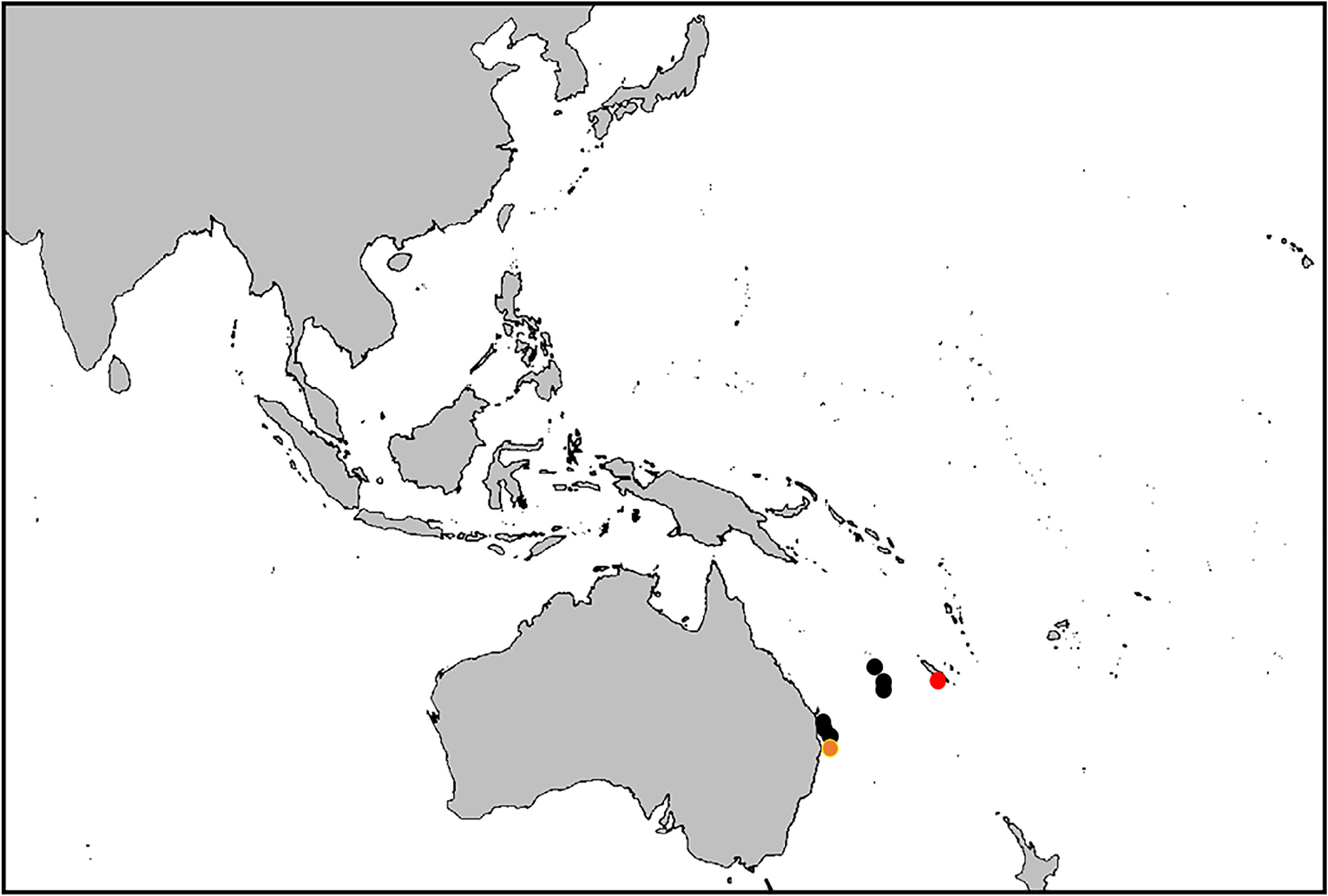
Habitat and distribution. The holotype of Odontanthias xanthomaculatus was collected at a depth of 200 m, from Passes de Bulari, which lies south of Nouméa, New Caledonia. The Australian specimens, including the holotype of O. grahami , were collected in 110–210 m off southern Queensland and northern New South Wales. The new specimens were collected from an intermediate area in New Caledonia in the vicinity of the Chesterfield Islands and Nova and Argo Banks on the Lord Howe Rise ( Fig. 3 View FIGURE 3 ), and extend the depth range to 330 m. It is likely the species is more widely distributed in the southern Coral Sea and northern Tasman Sea.
Remarks. Gill & Russell (2019) noted several differences between O. grahami (i.e., Australian specimens of O. xanthomaculatus ) and the holotype of O. xanthomaculatus : head spination (development of large spines on the preopercle angle and interopercle; number of preopercular serrations; number of spines on the opercle; presence or absence of serrations on the supracleithrum), relative length of the third dorsal-fin spine (elongate versus not elongate), and certain live coloration details. All these differences can be attributed to ontogenetic variation, as detailed above and in Table 2 View TABLE 2 and Figs 1–2 View FIGURE 1 View FIGURE 2 , and we therefore consider Odontanthias grahami Randall & Heemstra a junior synonym of Anthias xanthomaculatus Fourmanoir & Rivaton. Ontogenetic variation in these features renders some characters problematic in keys to Odontanthias species published by Randall & Heemstra (2006) and White (2011), such as the length of the third dorsal-fin spine relative to other dorsal-fin spines, the size of the preopercular spine, and the coloration in life of the dorsal fin. However, we do not here offer a revised key, pending the completion of a more extensive morphological study of species in the genus, which will also address the possible inclusion of species currently assigned to Sacura Jordan & Richardson, 1910 and Meganthias Randall & Heemstra, 2006 ( Gill & Russell 2019; Zajonz et al. 2020). The combination of X,14 dorsal-fin rays, 15–17 (usually 16) pectoral-fin rays and 36–41 tubed lateral line scales distinguishes O. xanthomaculatus from all species in the three genera. Rare specimens of O. elizabethae Fowler, 1923 , may have this combination of characters (though usually with 18 pectoral-fin rays), but that species is readily distinguished in having more numerous gill rakers (13–15 + 30–32 = 44–47 versus 12–13 + 26–30 = 38–43), a differently shaped vomerine tooth patch (arrow shaped, with tip pointing posteriorly versus chevron to inverted heart-shaped, sometimes pointed anteriorly), and a markedly different live coloration ( Randall & Heemstra 2006: pl. 3a versus Fig. 2 View FIGURE 2 ).
Material Examined. Australia. AMS I.32142-001, 93.0 mm SL (holotype of Odontanthias grahami Randall & Heemstra ), New South Wales, off Brunswick Heads (28°27′S, 153°50′E to 28°24′S, 153°49′E), 126–130 m, trawl, K.J. Graham, FRV Kapala Cruise 1, Station 32, 16 Feb 1991; QM I.21166, 91.7 mm SL, Queensland, east of Stradbroke Island (27°35′S, 153°50′E), 210 m, trawl, Queensland Fisheries Service, 5 Dec 1982; QM I.38666, 83.3 mm SL, Queensland, off Cape Moreton (27°03′S, 153°33′E), 110–114 m, trawl, S. McCulloch, 10 Feb 2010; QM I.38967, 71.8 mm SL, Queensland, east of Coolum (26°32′S, 153°36′E), 115 m, trawl, Queensland Fisheries Service, 19 Oct 2010. New Caledonia. MNHN IC- 1978-0477, 42.0 mm SL (holotype of Anthias xanthomaculatus Fourmanoir & Rivaton ), Passes de Bulari, 200 m, trap, P. Fourmanoir; NTUM 13743, 3: 55.7–125.0 mm SL, Station CP4958, 23°08′S, 159°31′E, 280 m, Argo Bank, R / V Alis, French beam trawl, Kanadeep expedition, 6 Sep 2017; NTUM 13744, 127.0 mm SL, Station CP5002, 22°30′S, 159°25′E, Nova Bank, 320–330 m, R / V Alis, French beam trawl, Kanadeep expedition, 18 Sep 2017; NTUM 13745, 2: 25.7–48.5 mm SL, station CP5034, 19°48′S, 158°28′E, 240–260 m, Chesterfield Plateau, Coral Sea, R / V Alis, French beam trawl, Kanadeep expedition, 22 Sep 2017.
| QM |
Queensland Museum |
| MNHN |
Museum National d'Histoire Naturelle |
| R |
Departamento de Geologia, Universidad de Chile |
| V |
Royal British Columbia Museum - Herbarium |
No known copyright restrictions apply. See Agosti, D., Egloff, W., 2009. Taxonomic information exchange and copyright: the Plazi approach. BMC Research Notes 2009, 2:53 for further explanation.
|
Kingdom |
|
|
Phylum |
|
|
Class |
|
|
Order |
|
|
Family |
|
|
Genus |
Odontanthias xanthomaculatus (Fourmanoir & Rivaton)
| Gill, Anthony C. & Tang, Chi-Ngai 2022 |
Odontanthias xanthomaculatus
| Anderson, W. D. Jr 2022: 569 |
| Parenti, P. & Randall, J. E. 2020: 22 |
| Gill, A. C. & Russell, B. C. 2019: 176 |
Odontanthias grahami
| Parenti, P. & Randall, J. E. 2020: 21 |
| Gill, A. C. & Russell, B. C. 2019: 182 |
| Anderson, W. D. Jr 2018: 23 |
| White, W. T. 2011: 27 |
| Randall, J. E. & Heemstra, P. C. 2006: 17 |
Pseudanthias xanthomaculatus
| Anderson, W. D. Jr 2018: 49 |
| Fricke, R. & Kulbicki, M. & Wantiez, L. 2011: 387 |
| Randall, J. E. & Pyle, R. L. 2001: 34 |
| Heemstra, P. C. & Randall, J. E. 1999: 2466 |
Anthias xanthomaculatus
| Bauchot, M. - L. & Desoutter, M. & Randall, J. E. 1984: 10 |
| Fourmanoir, P. & Rivaton, J. 1979: 414 |