
Megalomma sp. 1
|
publication ID |
https://doi.org/ 10.3853/j.0067-1975.61.2009.1529 |
|
persistent identifier |
https://treatment.plazi.org/id/2202BF4F-FFDD-5B6C-FED8-5647FB03FCAB |
|
treatment provided by |
Felipe |
|
scientific name |
Megalomma sp. 1 |
| status |
|
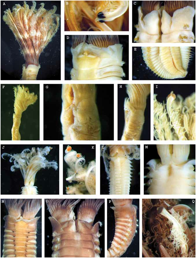
Figs 2N–Q View Figure 2 , 4I,J View Figure 4 , 5E View Figure 5
Material examined. Queensland. AM W30022 (1 spec.), Abbott Point, near Bowen, 19°53'S 148°05'E, coll. by CRC Reef Research GoogleMaps Centre Ltd for Queensland Ports Survey , from pylon scraping, 8 Jul. 1998, 7 m.
Description. Single specimen complete, measures 74 mm long (18 mm crown) and 5 mm wide. Crown longer than thorax, with 22 pairs of radioles in each lobe, arranged in a semicircle. External margin of radioles quadrangular at the base and rounded towards the tips ( Fig. 2N,O,Q View Figure 2 ), without lateral flanges. Tips of radioles shorter than pinnules ( Fig. 2Q View Figure 2 ). Radiolar skeleton with 15–20 cells in transverse section at the base of radioles ( Fig. 5E View Figure 5 ). More than half (18–19) of the radioles with single subdistal compound eyes. Dorsalmost pair of radioles with large eye almost surrounding the tip of radiole, not spiral ( Fig. 2Q View Figure 2 ); on adjacent radioles bearing subdistal eyes, eyes small and spherical, similar in size. Dorsal lips with radiolar appendages as long as two thoracic chaetigers, lateral lamellae not reaching the tip of appendage, three dorsal pinnular appendages on each lip. Ventral lips rounded and well developed; parallel ventral lamellae and ventral sacs present. Caruncle absent. Smooth keel arising dorsally and directed ventrally between dorsal lips ( Fig. 2 O View Figure 2 ). Dorsal margins of collar fused to the faecal groove, with dorsolateral V-shaped notches on both sides and shallow dorsolateral pockets becoming a shallow groove more dorsally ( Fig. 2 O View Figure 2 ); lateral margins smooth ( Fig. 2P View Figure 2 ), triangular non-overlapping ventral lappets, with a complete midventral incision ( Fig. 2N View Figure 2 ). Ventral shields quadrangular with indented margins laterally, slightly diminishing in width posteriorly, in contact with the neuropodial tori, except in anterior chaetigers ( Fig. 2N View Figure 2 ). First ventral shield 1.5 times longer than the rest, with an m-shaped anterior margin ( Fig. 2N View Figure 2 ). First chaetiger with superior and inferior elongate narrowly hooded notochaetae, superior row longer than inferior. Rest of thoracic chaetigers with superior elongate narrowly hooded notochaetae arranged in irregular rows and inferior broadly hooded notochaetae (type B) in single row. Notopodia of thoracic chaetigers with a large lamellate lobe separating inferior and superior fascicles of notochaetae. Neuropodial tori becoming slightly shorter posteriorly. Thoracic uncini with several rows of small teeth, all similar in size above main fang; uncinus with well developed breast and handle about twice the length of the distance from breast to main fang ( Fig. 4I View Figure 4 ). Companion chaetae with asymmetrical membrane. Abdominal neuropodia with broadly hooded chaetae. Notopodial uncini similar to thoracic uncini but with handles half their length ( Fig. 4J View Figure 4 ). Posterior chaetigers regenerating, pygidium papilla-like, with scattered eyespots present.
Reproductive features. Eggs present in anterior abdominal segments.
Colour pattern. Crown and thorax with purple pigmentation after fixation. There are some conspicuous dark spots located on the ventral margin of the thoracic tori which we do not consider to be eyespots, as the patches are not discrete (pigment dilutes dorsally along tori edges) and they are also not interramal as in other sabellids that display segmental eyespots.
Remarks. This specimen does not fit the description of any previously described species but it is premature to describe it as new until more specimens are found and intraspecific variability can be assessed. The combination of such characters as margins of collar not fused to the faecal groove (although there is a ridge that continues from the end of the dorsal margins to where the middorsal faecal groove ends, which could be interpreted as fusion with the faecal groove), U-shaped dorsolateral incisions of the collar, the presence of low collar pockets, and subdistal eyes on most radioles makes the specimen similar to several other species previously described from the Indo-Pacific: M. multioculatum , from Thailand and M. pacificum from Gilbert Islands. However, M. multioculatum is described as having collar margins fused to the faecal groove and pockets present, and the latter species has been possibly referred to Demonax by Fitzhugh (2002). The specimen described above differs from M. multioculatum also in the development of the collar, as the ventral lappets are shorter, not visibly longer than lateral margins, but are elongate in M. multioculatum , in which the dorsal margins of collar are clearly fused to the faecal groove and there are shallow pockets present on both sides. Also, the specimen from Queensland has inferior thoracic chaetae arising in a single row and with progressively tapering tips (type B) while M. multioculatum has chaetae with broader and more slender tips (type A). The handles of thoracic uncini are also shorter in the Queensland specimen.
According to Tovar-Hernández and Salazar-Vallejo (2008), there are three species described as having the dorsal margins of collar not fused to the faecal groove, pockets present and radiolar eyes in most radioles (which have been previously placed in Group 2B). One of these, M. neapolitanum Claparède, 1868 , described from Italy, has been recently synonymized with M. lanigera (Grube, 1846) (Giangrande & Licciano, 2008) . The other two, M. heterops and M. perkinsi Tovar-Hernández & Salazar- Vallejo, 2006, were described from Florida. The specimen described above differs from M. heterops in the shape of the inferior thoracic chaetae which are broader in the base and thinner in the tip, bottle-shaped, and the thoracic uncini handles which are significantly shorter. According to the drawings and original description, M. perkinsi has lateral and dorsal anterior margins of the collar similar in length, and the collar fused to the faecal groove, features not shared by the specimen described above.


| AM |
Australian Museum |
No known copyright restrictions apply. See Agosti, D., Egloff, W., 2009. Taxonomic information exchange and copyright: the Plazi approach. BMC Research Notes 2009, 2:53 for further explanation.
|
Kingdom |
|
|
Phylum |
|
|
Class |
|
|
Order |
|
|
Family |
|
|
Genus |