
Arcella guadarramensis, 2022
|
publication ID |
https://doi.org/10.1093/zoolinnean/zlab074 |
|
publication LSID |
lsid:zoobank.org:pub:53637D76-285D-4AB8-9E52-6CDB6F6738D3 |
|
DOI |
https://doi.org/10.5281/zenodo.6461317 |
|
persistent identifier |
https://treatment.plazi.org/id/1C22923F-294E-0972-FC6D-4E0AFD48FC49 |
|
treatment provided by |
Plazi (2022-04-13 07:07:28, last updated 2024-11-25 17:34:24) |
|
scientific name |
Arcella guadarramensis |
| status |
sp. nov. |
ARCELLA GUADARRAMENSIS GONZÁLEZ- MIGUÉNS & LARA , SP. NOV.
( FIG. 8 View Figure 8 )
Z o o b a n k r e g i s t r a t i o n: u r n: l s i d: z o o b a n k. org:act: 57A3452C-3C46-44F2-A5C9-48496E34BB57.
Holotype: MA-Algae11251 .
Specific diagnosis: Test diameter: clade L: 114.60– 125.90 µm, average 120.30 µm (N = 6); aperture 19.60– 30.00 µm, average 23.63 µm. clade M 141.50–149.95 µm, average 146.06 µm (N = 4); aperture 31.25–34.50 µm, average 33.08 µm. Besides a difference in size, both clades have an identical morphology. Colour ranges from transparent to yellow-orange. Subhemispherical test shape, with flattened edges and dimples in the surface that gives the test a golf ball shape. No ribs or keels on the aboral side. Hexagonal building units are visible, which gives the test a rough appearance; little pores can be seen at the vertices. Building units can also be appreciated at the oral side of the test, with pores at the vertices and a central aperture. The aperture is invaginated outwards forming a short ring or lip.
Intraspecific variability: The building units may vary slightly in shape. Some building units may be collapsed, giving a rough surface. There may be certain deformations in the test that prevent it from having a perfectly circular morphology.
Diagnosis with closely related species: Arcella guadarramensis can be diagnosed by its specific sequences of the mtDNA markers and by its phylogenetic placement. Arcella guadarramensis differs morphologically from similar-looking G. succelli by (1) its morphometric differences with G. succelli (see Morphometrics and morphology; Fig. 2 View Figure 2 ), both, clade N and O, are notably smaller than G. succelli and (2) the rough outlook of the test.
Habitat: Wet Sphagnum moss, in a fen.
Type locality: Spain, Madrid, Puerto de Canencia (40°52’N 3°45’W).
Etymology: The name is derived from River Guadarrama, a river with a name of Arabic roots: wadi, river, and ar-rama, sandy. We propose this name as a reference to the type locality in ‘Sierra de Guadarrama’, a mountain range named after this river.
We provide a key (Supporting Information, Table S3) and a new figure (Supporting Information, Fig. S3 View Figure 3 ) to facilitate the identification of the new species.
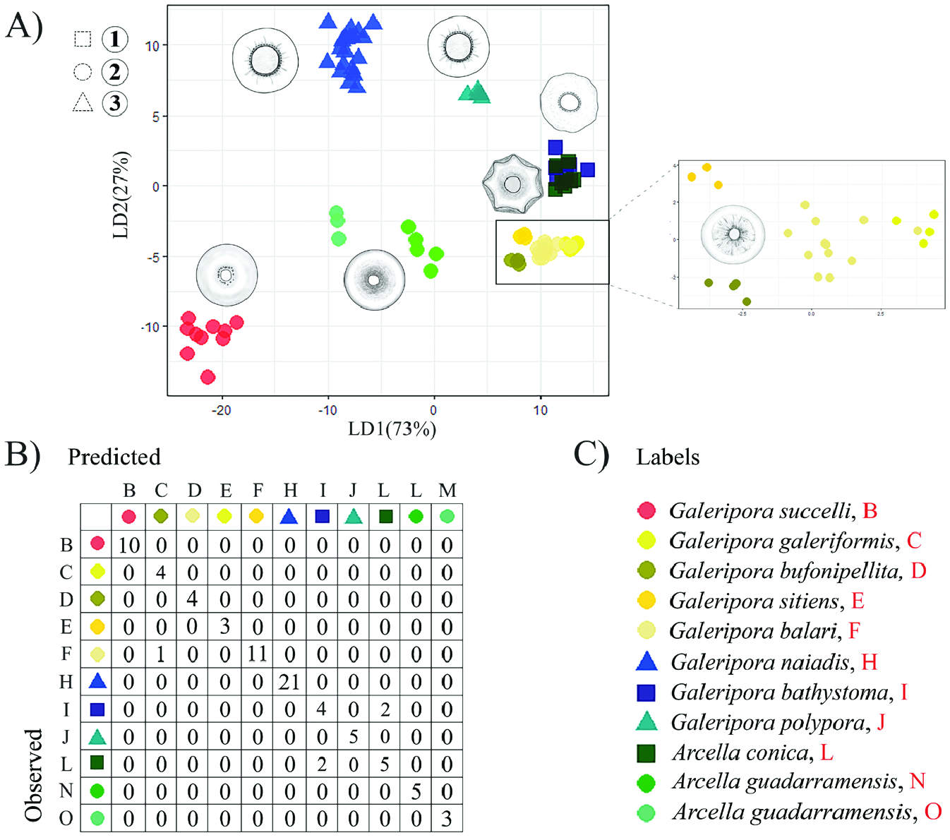
Figure 2. A, scatterplot of the scores of linear discriminants with x-axis representing discriminant function 1 (LD1) and y-axis representing discriminant function 2 (LD2). Colours represent the different mitochondrial clades and symbols refer to the different sections after Deflandre (1928): squares are for Section 1 ‘Vulgares’, circles for Section 2 ‘Carinatae’ and triangles for Section 3 ‘Aplanatae’. The drawings represent the different morphotypes. B, the table represents the results of a linear discriminant analysis which determines the relationship between predicted and observed specimens cells for each mitochondrial clade.
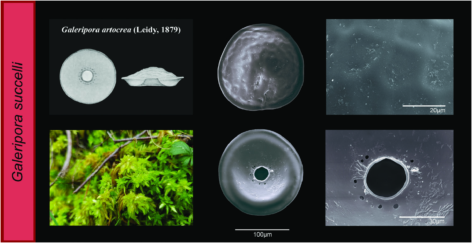
Figure 3. Galeripora succelli: scanning electron micrographs of the aboral and oral sides of the test. The images on the right represent a detail of the test and the structure of the aperture. Below left, a photograph of a typical habitat for the species, a peat bog. Above left, original drawing of the closest known resembling species, Galeripora artocrea (Leidy, 1879).
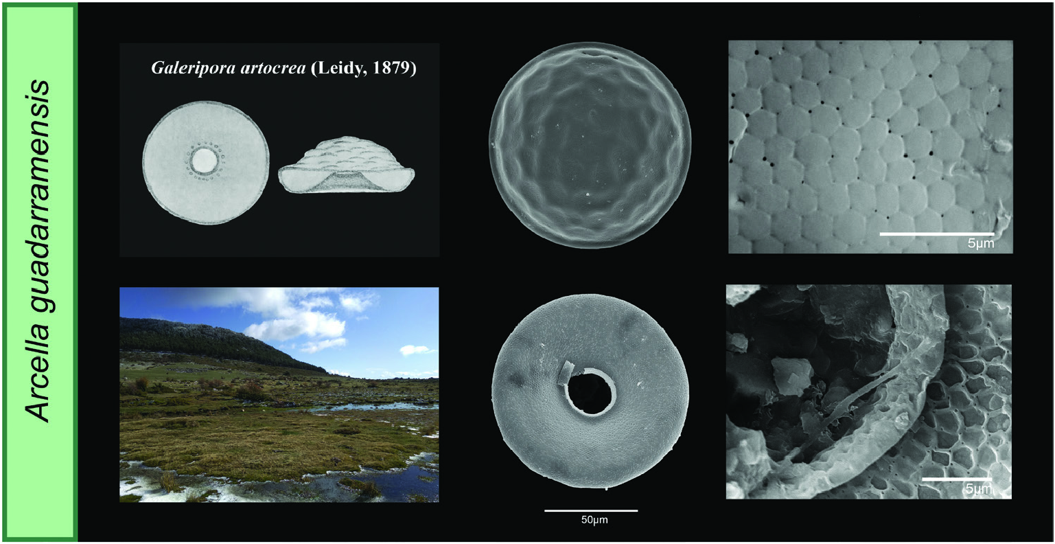
Figure 8. Arcella guadarramensis: scanning electron micrographs of the aboral and oral sides of the test. The images on the right represent a detail of the test and the structure of the aperture. On the left, a photograph of a typical habitat for this species, and original drawing of the closest resembling species, Galeripora artocrea (Leidy, 1879).
No known copyright restrictions apply. See Agosti, D., Egloff, W., 2009. Taxonomic information exchange and copyright: the Plazi approach. BMC Research Notes 2009, 2:53 for further explanation.
|
Kingdom |
|
|
Phylum |
|
|
Class |
|
|
Order |
|
|
SubOrder |
Glutinoconcha |
|
InfraOrder |
Sphaerothecina |
|
Family |
|
|
Genus |