
Milteliphaster woodmasoni Alcock 1893
|
publication ID |
https://doi.org/ 10.11646/zootaxa.4539.1.1 |
|
publication LSID |
lsid:zoobank.org:pub:2C72727B-79C5-407F-BD92-B12F98196800 |
|
DOI |
https://doi.org/10.5281/zenodo.5990799 |
|
persistent identifier |
https://treatment.plazi.org/id/193787A0-FFDD-FFDF-F4CB-FEE744BEC8BF |
|
treatment provided by |
Plazi |
|
scientific name |
Milteliphaster woodmasoni Alcock 1893 |
| status |
|
Milteliphaster woodmasoni Alcock 1893 View in CoL
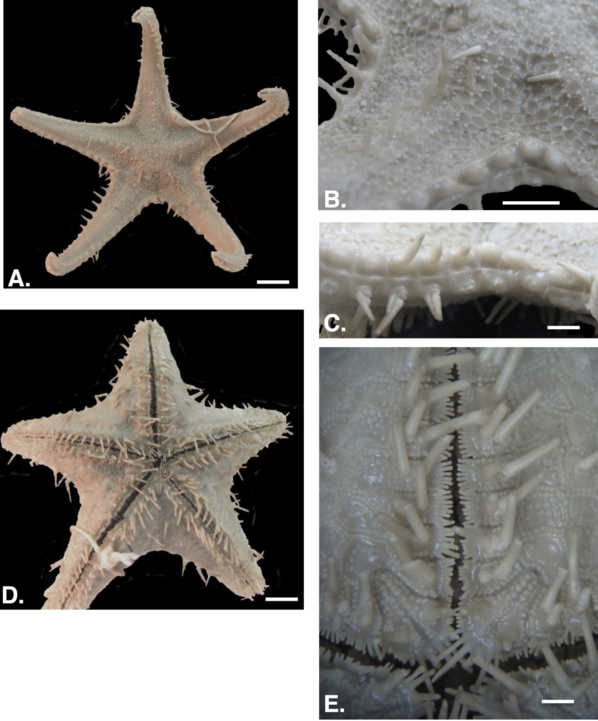
Figure 24 View FIGURE 24 A–E
Alcock 1893: 91; Macan 1938: 391; James 1983: 89 (checklist); Clark 1993: 264 (checklist); Sastry 2005: 29 (checklist) (as M. woodmasoni )
Diagnosis. Body strongly stellate (R/r=3.17), arms upturned, strap-like ( Fig. 24A View FIGURE 24 ). Abactinal plates abutted, homogeneous in shape, size with smooth surface extending to arm tip. Large, conical, sharp spines present along disk and carinal plate series ( Fig. 24B View FIGURE 24 ). Marginal plates forming distinct lateral border, occupying only ~13–17% of the total “r” distance on the disk ( Fig. 24C View FIGURE 24 ).
Comments. Milteliphaster woodmasoni displays marginal plate characters and body skeleton morphology that are substantially different from expressions in other known Calliaster species. Thus, pending further understanding of diversity within the group, Miltelphaster is retained as a separate genus. The lack of shared characters and overall dissimilarity between the species listed by Rowe and Gates (1995) do not support the addition of all post- Alcock (1893) species to Milteliphaster .
Thus, “ Milteliphaster ” wanganellensis should be assigned to Calliaster , and Calliaster spinosus and Calliaster regenerator are misplaced as Milteliphaster and should be retained in Calliaster . Calliaster represents a highly diverse genus and further revision is needed in order to determine the extent of further subdivisions. Given Alcock’s (1893) relatively short description, a re-description and figures of the species based on the Paris syntype is provided below.
Occurrence. Andaman Sea. 420– 530 m.
Description. Body stellate (R/r=3.17), arms elongate, tapering triangular in shape ( Fig. 24A View FIGURE 24 ). Interradial arcs acute. Arms strongly upturned.
Abactinal surface flattened. Abactinal plates round to polygonal, largest plates present proximally, smaller and more irregular ones occurring interradially and more distally adjacent to superomarginal contact ( Fig. 24B View FIGURE 24 ). Abactinal plates occur along arm to terminus with multiple plate series occurring along arm, narrowing to four or six distally. Plate surface smooth, with accessory structures absent from each plate surface. Distinct periphery of quadrate to polygonal granules, four to 15 mostly eight to 12, present around each plate. Each plate surface slightly sunken below the plane formed by the peripheral granules. A minority of proximal carinal plates on the arm base and near the primary circlet on the disk bearing sharp, very distinct spines, ( Fig. 24B View FIGURE 24 ) cylindrical in cross section with length of each approximately 7.0–9.0 mm above the abactinal surface. Spine bearing plates strongly convex with smooth surface around spine base. Spines in series occur variably with most in direct sequence at disk/arm region but others alternating every one to three plates. Spines disappear from carinal series on plates from approximately half way along arm to arm tip. Spines absent interradially and on adradial arm series. Papulae swollen, balloon-like in shape, abundantly present on abactinal surface, four to six present around each plate. Madreporite quadrate in shape, flanked by approximately six to seven abactinal plates.
No pedicellariae.
Superomarginals and inferomarginals, approximately 46–48 arm tip to arm tip with plate series primarily showing lateral facing ( Fig. 24B, C View FIGURE 24 ). Individual plates quadrate to elongate in outline with angular edges forming very narrow edge along abactinal periphery forming approximately (0.3–0.4 /2.3) ~13–17% of the total “r” distance on the disk. Superomarginal plates with mostly one or exceptionally two sharp, conical spines per plate. Each spine, approximately 0.3–0.4 mm tall, seated on a smooth, bare swollen base per plate ( Fig. 24C View FIGURE 24 ). Interradial plates, especially inferomarginals with greatest number of two-spined plates with single-spined plates present along most of disk and arm distance. Marginal plates each surrounded by quadrate to polygonal granules, approximately 20–60, mostly 40–50 with approximately 10x15 per side. Superomarginals much more strongly convex distally, especially adjacent to terminal plate. Terminal plate triangular, smooth with two broken bosses. Most marginal plates smooth, devoid of surficial accessories but one or two inferomarginals demonstrate 10–20 small round to polygonal granules on surface.
Actinal plates in two to three full series in chevron like formation ( Fig. 24D View FIGURE 24 ). Plate morphology varies from quadrate to polygonal to elongate trapezoidal in outline. Actinal plate surface mostly bare but with about 25% of surface covered by three to 15 round granules, identical to those forming granular periphery, present on distal corners of each plate. One to six actinal plates with large, pointed, conical spine similar to those on abactinal and marginal plates (about 0.3 to 0.4 mm tall) ( Fig. 24D View FIGURE 24 ). Actinal plates present only on disk, not extending far onto arm.
Furrow spines 10–11, blunt, pointed, closely arranged in straight to weakly curved series. Adambulacral plates with two, large blunt spines in transverse series ( Fig. 24E View FIGURE 24 ). These subambulacral spines set off from furrow spines by discrete space, at least 5x as thick and as along as furrow spines. Surface of adambulacral plate covered by close-set, polygonal granules 10–12. No pedicellariae observed. Oral plates with 9–10 furrow spines. Each plate bearing two to three elongate but blunt spines, 3x to 6x the length of the furrow spines projecting into the mouth. Oral plate surface with a single large, blunt conical spine sitting on each half of the oral plate. Each half of the plate with 10–12 angular granules present along edge of the oral plate sulcus sitting between plates. Oral plate covered by 12–18 angular granules present on plate surface ( Fig. 24E View FIGURE 24 ).
Material Examined. Syntype. IE-2014-164 , Andaman Sea. 420–530 m. Coll. Investigator 1 wet spec. R=7.3 r=2.3.
No known copyright restrictions apply. See Agosti, D., Egloff, W., 2009. Taxonomic information exchange and copyright: the Plazi approach. BMC Research Notes 2009, 2:53 for further explanation.
|
Kingdom |
|
|
Phylum |
|
|
Class |
|
|
Order |
|
|
Family |
|
|
Genus |