
Microporella protea Winston, 2005
Judith L Winston, 2016, Bryozoa of Floridan Oculina reefs, Zootaxa 4071 (1), pp. 1-81 : 56
|
publication ID |
https://doi.org/10.11646/zootaxa.4071.1.1 |
|
publication LSID |
lsid:zoobank.org:pub:D47C792F-E91D-40A6-ABB7-FA7810578562 |
|
DOI |
https://doi.org/10.5281/zenodo.6084823 |
|
persistent identifier |
https://treatment.plazi.org/id/19362D2E-2022-FF87-BBA5-FD1AFCB3F8EE |
|
treatment provided by |
Plazi |
|
scientific name |
Microporella protea Winston, 2005 |
| status |
|
Microporella protea Winston, 2005 View in CoL
( Fig. 31 View FIGURE 31 ; Table 30 View TABLE 30 )
Porellina ciliata : Smitt 1873: 26, pl. 6, figs 128–129.
? Microporella ciliata personata: Osburn 1947: 36 .
Microporella ciliata: Long & Rucker 1970: 20 , fig. 4:3.
Microporella protea Winston, 2005: 78 , figs 211–213, 215, 217–223.
Material examined. VMNH no. 70644, 70645; USNM no. 1283255.
Description. Colony unilaminar to multilaminar, encrusting ( Fig. 31 View FIGURE 31 A). Zooids irregularly rhomboidal ( Fig. 31 View FIGURE 31 B, D), with bumpy granular frontal shields penetrated by irregularly shaped pores of various sizes, which become increasingly obscured as secondary calcification increases. Zooidal boundaries delimited by sinuous or scalloped shallow grooves at junctions of intercalary cuticles of adjacent zooids. Primary orifice roundly semicircular, high-arched, with weakly concave proximal rim that may appear beaded as secondary calcification develops. Bases of 3–5 (usually 4) hollow spines on distal rim of orifice ( Fig. 31 View FIGURE 31 E, F). Ascopore crescentic, denticulate, located just proximal to orifice, surrounded by slightly raised collar. Avicularium single, large, broad distolaterally directed, with complete crossbar and triangular mandible, proximolateral to orifice on one side, subjacent to ascopore at about zooidal mid-length ( Fig. 31 View FIGURE 31 B). Ooecia ( Fig. 31 View FIGURE 31 D) globose, with same thick, granular to pustulose porous calcification as frontal shields, their rims slightly ribbed.
Remarks. Use of SEM has led to the discrimination of new species of Microporella based on ultrastructural differences in the number and nature of orificial spines, shape of orifice, presence of condyles, nature of proximal edge of orifice (smooth, serrated, beaded, etc.), morphology and position of ascopore, size, shape and position of avicularia, calcification of frontal shield, and form of ooecia. Microporella protea is only one of several species that occur along the Floridan coast.
Distribution. Cape Hatteras to Florida. Caribbean?
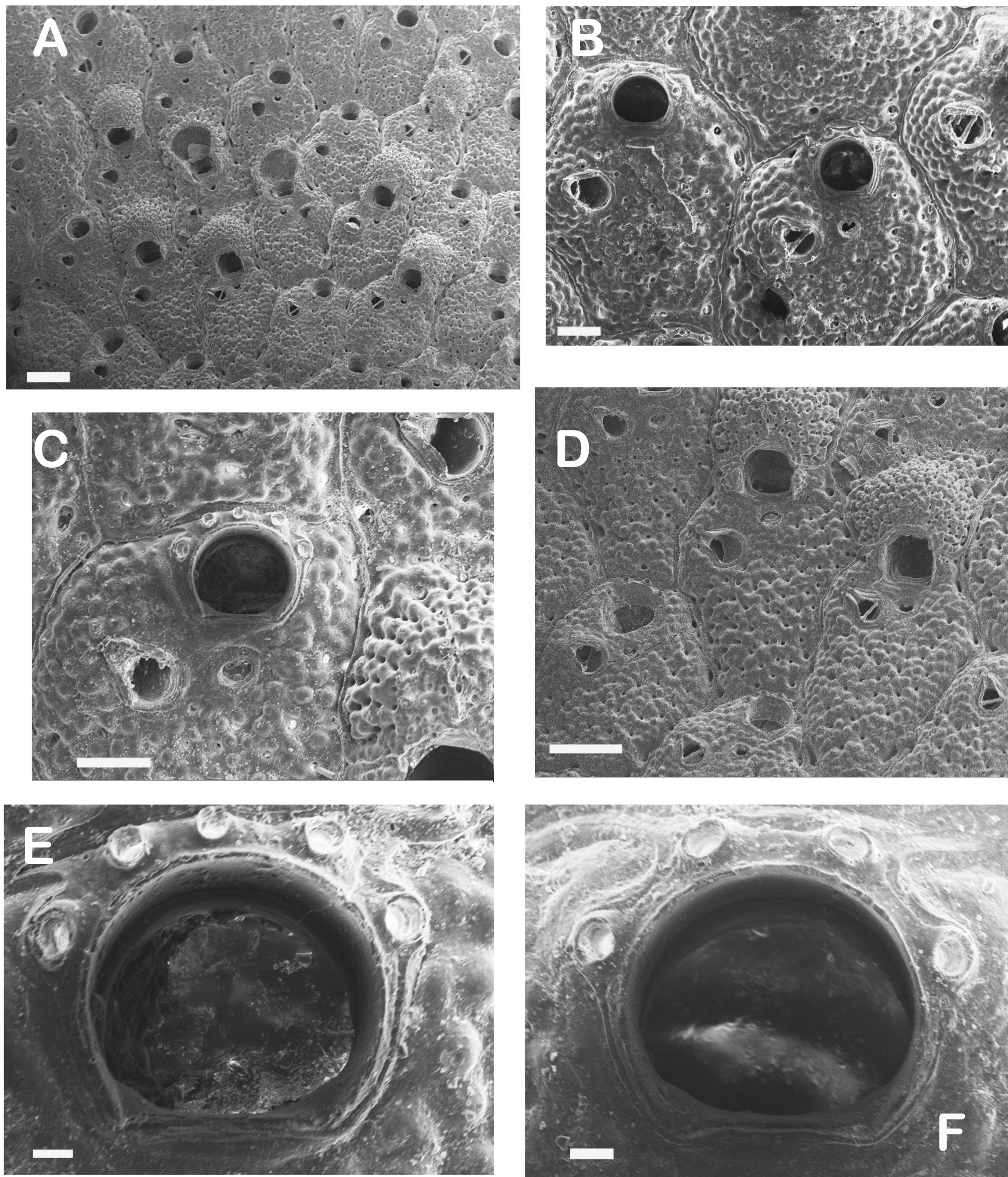
TABLE 30. Measurements in mm of Microporella protea Winston, 2005.
| Lz | Wz | Lo | Wo | Lov | Wov | Lav | Wav | |
|---|---|---|---|---|---|---|---|---|
| N | 18 | 18 | 18 | 18 | 18 | 18 | 18 | 18 |
| Mean | 0.743 | 0.572 | 0.107 | 0.128 | 0.282 | 0.337 | 0.141 | 0.088 |
| SD | 0.064 | 0.063 | 0.013 | 0.017 | 0.024 | 0.029 | 0.022 | 0.007 |
| Min | 0.630 | 0.486 | 0.090 | 0.090 | 0.252 | 0.270 | 0.108 | 0.072 |
| Max | 0.864 | 0.684 | 0.126 | 0.162 | 0.342 | 0.378 | 0.180 | 0.099 |
No known copyright restrictions apply. See Agosti, D., Egloff, W., 2009. Taxonomic information exchange and copyright: the Plazi approach. BMC Research Notes 2009, 2:53 for further explanation.
|
Kingdom |
|
|
Phylum |
|
|
Class |
|
|
Order |
|
|
SubOrder |
Flustrina |
|
Family |
|
|
Genus |