
Cypridopsis vidua (O.F. Müller, 1776)
|
publication ID |
https://doi.org/10.11646/zootaxa.5323.2.2 |
|
publication LSID |
lsid:zoobank.org:pub:998911D1-EF22-4100-8648-EC4B0AEF24C1 |
|
DOI |
https://doi.org/10.5281/zenodo.8221778 |
|
persistent identifier |
https://treatment.plazi.org/id/186FFE5E-FFE9-5C4E-0D9E-0D0DFD31F85E |
|
treatment provided by |
Plazi |
|
scientific name |
Cypridopsis vidua (O.F. Müller, 1776) |
| status |
|
Cypridopsis vidua (O.F. Müller, 1776) View in CoL View at ENA
( Figs 12‒15 View FIGURE 12 View FIGURE 13 View FIGURE 14 View FIGURE 15 )
Synonymies. Meisch et al. (2019) listed 19 synonymies of this species.
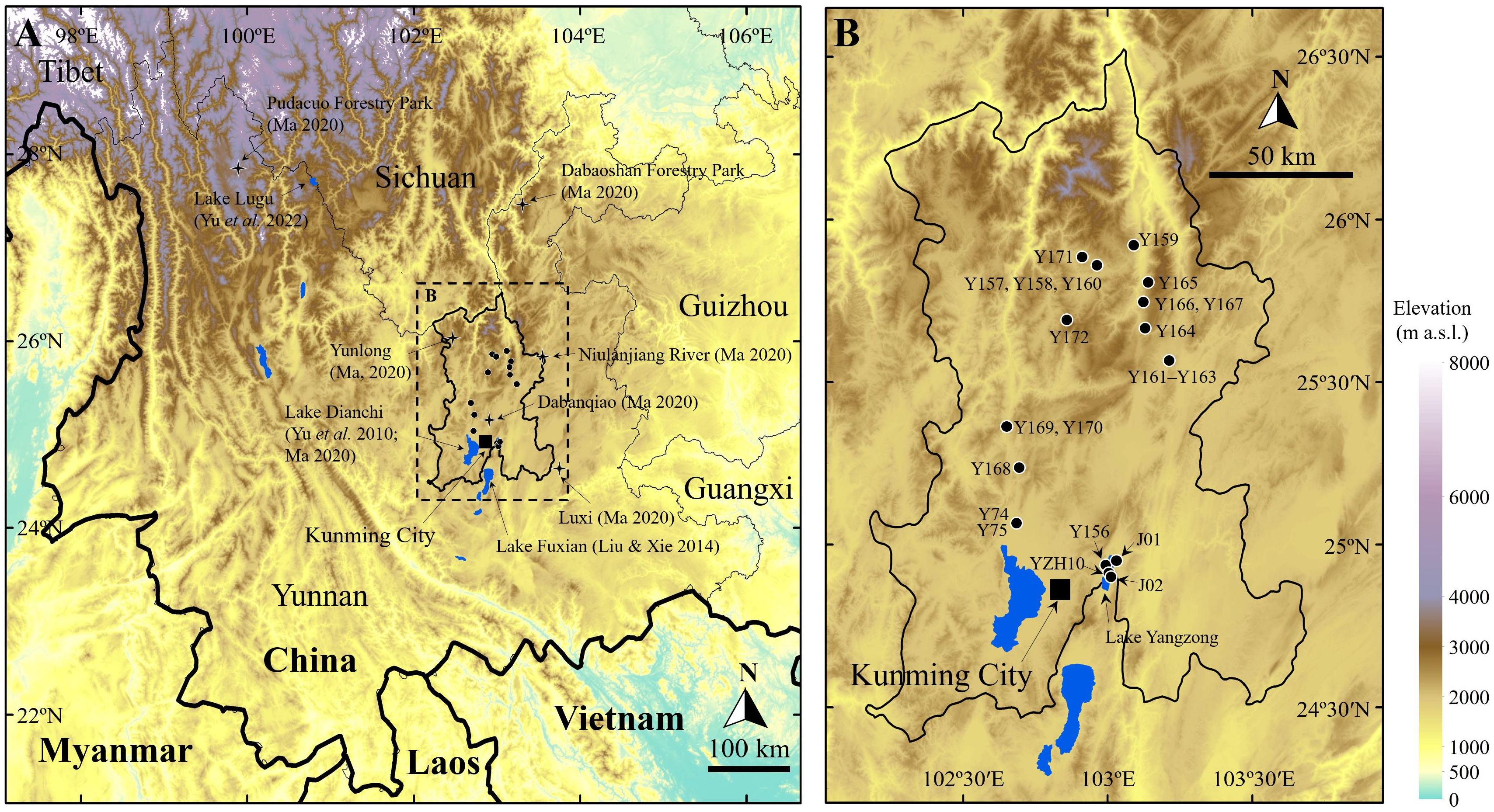
Material examined. One dissected male ( WOC 104) and two dissected females ( WOC 103, WOC105) from site YZH10. Two dissected males ( WOC 107, WOC108) and one dissected female ( WOC 106) from site Y156. One dissected male ( WOC 109) from site J01. Two dissected females ( WOC 128, WOC130) from site Y162. One dissected male ( WOC 139) and one dissected female ( WOC 138) from site Y169 ( Fig. 1B View FIGURE 1 ; Table 1 View TABLE 1 ).
Dimensions. Male LV length 0.54‒0.67 mm, averaging 0.59 mm, H/L ratio 0.63‒0.66. Female LV length 0.61‒0.64 mm, averaging 0.62 mm, H/L ratio 0.60‒0.66.
Diagnosis. See Martens et al. (2023).
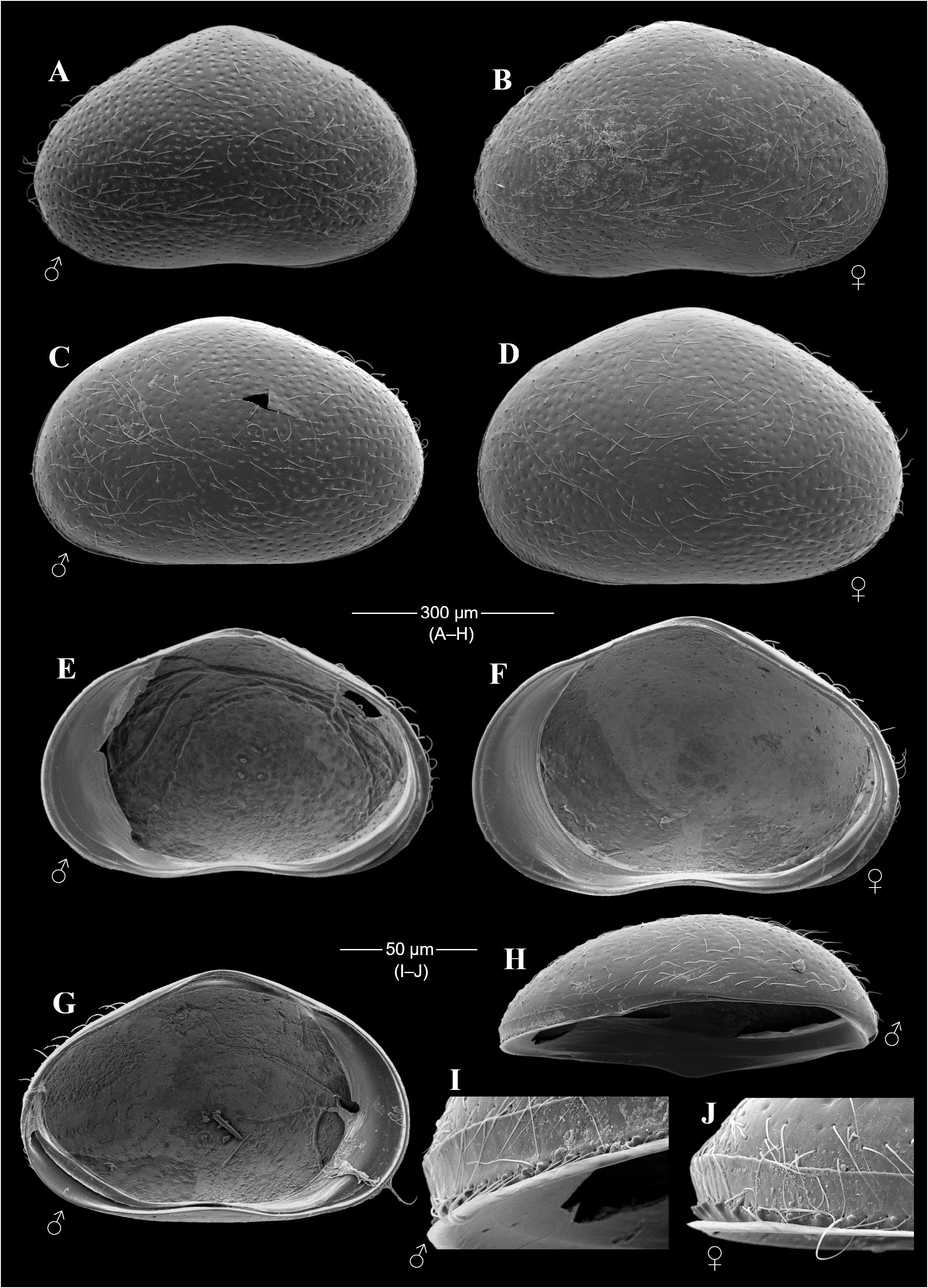
Brief description of C. vidua from the Kunming area. (Features for both sexes unless otherwise noted.) Carapace morphology similar to those described elsewhere (e.g. Meisch 2000; Zhai & Zhao 2014; Martens et al. 2023). Male ( Fig. 12A, C, E & G View FIGURE 12 ) average size somewhat smaller than female ( Fig. 12B, D, F & H View FIGURE 12 ). Both sexes with characteristic dark transverse bands on carapace, and with small denticles on anterior part of RV ( Fig. 12H‒J View FIGURE 12 ). Sex difficult to determine without dissection.
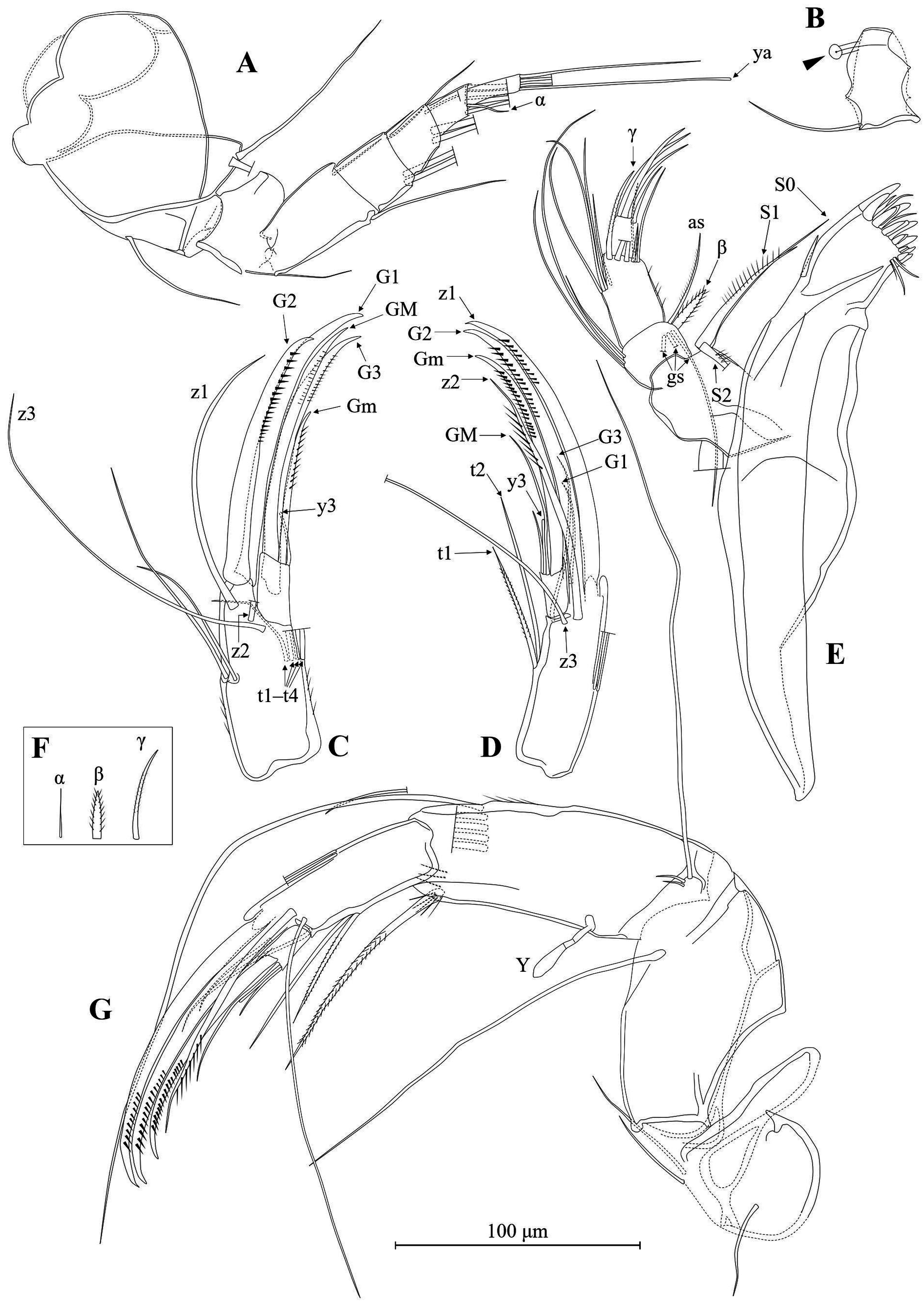
A1 ( Fig. 13A & B View FIGURE 13 ) with podomere lengths generally typical of Cyprididae and seta formula typical of Cypridoidea ( Candoninae excluded). RO very weakly sclerotized, with distal end spherically enlarged.
A2 ( Fig. 13C, D & G View FIGURE 13 ) with longest exopodal seta extending beyond terminal segment. Swimming setae extending beyond terminal claws. Penultimate segment undivided in both sexes. Chaetotaxy of last two segments with obvious sexual dimorphism. With four t-setae in female while two in male. Female, G 1, G2, and G3 claws thick, with G1 longest, smooth. G2 more strongly serrated. Male, both G1 and G3 reduced into seta forms, extending to mid-way of G2. Female, z-setae not transformed, although z1 thicker than z2 and z3. Male, z1 transformed into longest claw, z2 and z3 seta-form. Female, GM (situated to interior of Gm) longer and thicker but less conspicuously serrated than Gm. Male, GM shorter and slenderer than Gm, smooth.
Md ( Fig. 13E & F View FIGURE 13 ) with seta α very slim, smooth. Seta β thick, plumose. Number of grouped setae: 3. Seta γ plumose (small setules sometimes need to observe under 1000×).
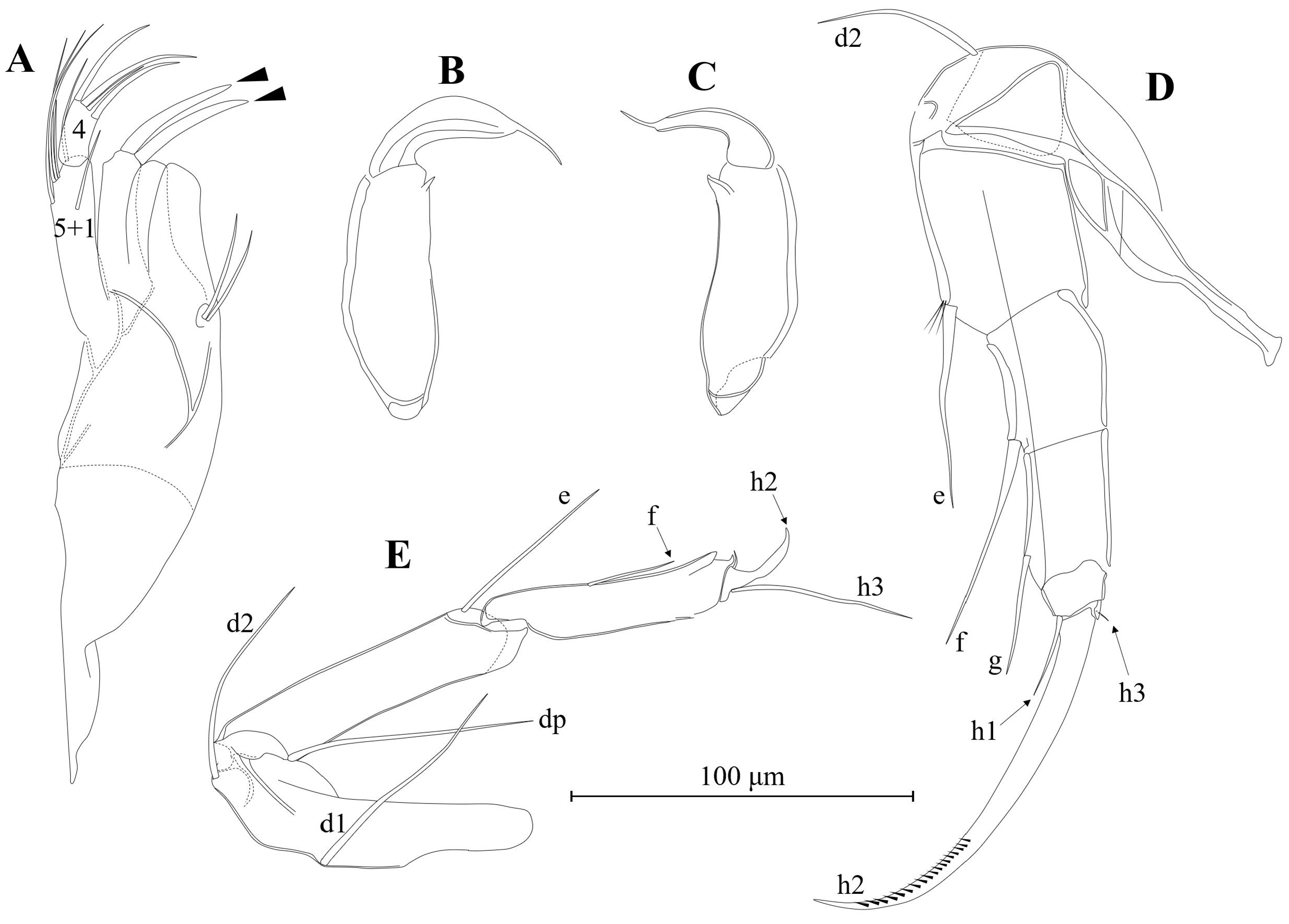
Mx ( Fig. 14A View FIGURE 14 ) palp 2-segmented. First segment with 5 + 1 setae. Second segment with four setae. Tooth bristles on distal gnathobasic endite smooth.
Male L5 palps ( Fig. 14B & C View FIGURE 14 ) transformed into claspers, slightly asymmetric, with left clasper ( Fig. 14B View FIGURE 14 ) wider than right ( Fig. 14C View FIGURE 14 ). Both claspers with soft, weakly sclerotized distal end.
L6 ( Fig. 14D View FIGURE 14 ) with setae d2, e, f, g, h1, and h3. Seta h3 tiny, difficult to see. Claw h2 thick, distally serrated.
L7 ( Fig. 14E View FIGURE 14 ) typical of Family Cyprididae .
Ur ( Fig. 15D View FIGURE 15 ) consisting of triangular base and distal filament. Right Ur slightly shorter than left. Short seta present on base of filament.
Ventral part of GL with pointed, sclerotized structure (arrowed in Fig. 15D View FIGURE 15 ).
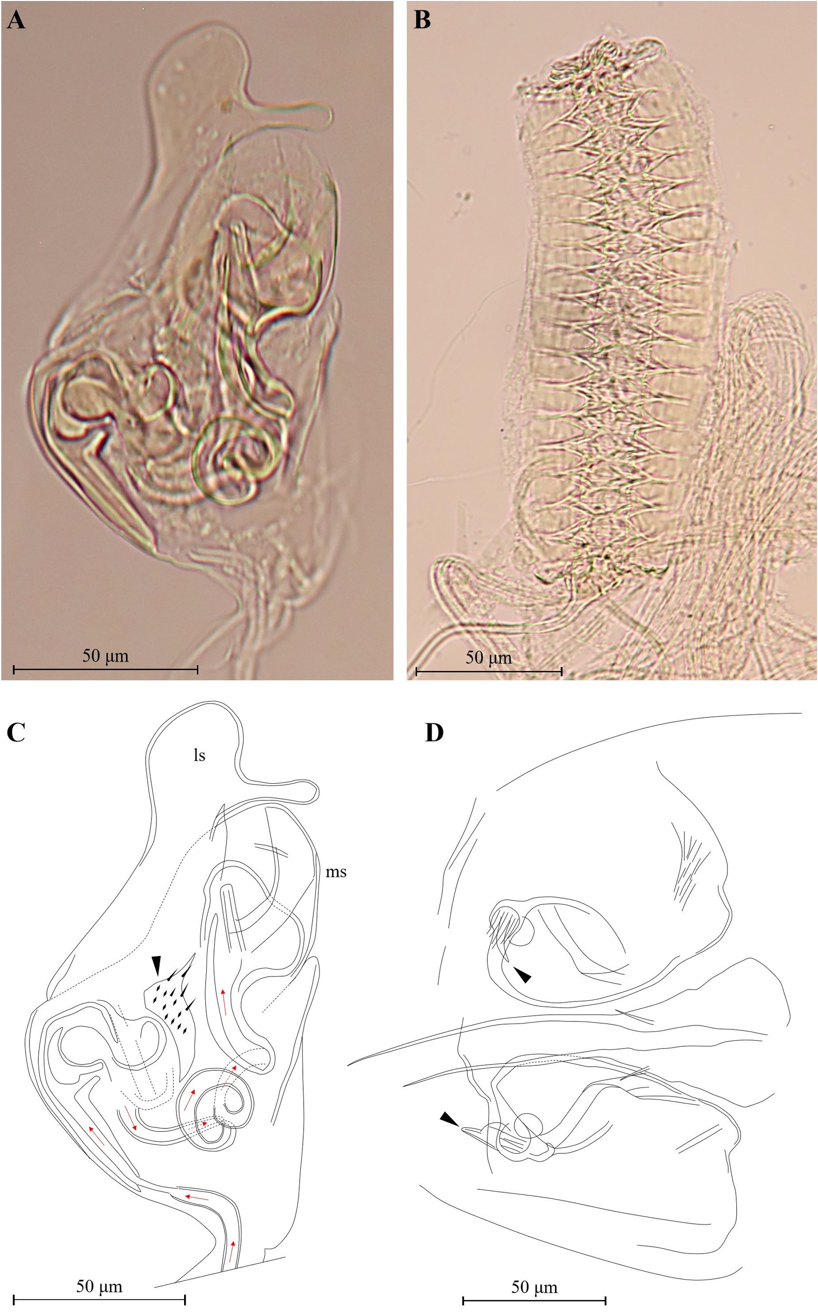
Hp ( Fig. 15A & C View FIGURE 15 ) with greatest width slightly proximal to mid-length. ms broadly rounded. ls longer than ms, distally rounded, intero-subapically with finger-like transverse process touching distal end of ms. Internal structures typical of Cypridopsinae . Labyrinth stout, accompanied by irregular-shaped, spinous, sclerotized structure (arrowed in Fig. 15C View FIGURE 15 ). Post-labyrinthal spermiduct with two full coils and thick-walled descending tube leading to bursa-copulatrix.
ZO ( Fig. 15B View FIGURE 15 ) with 15‒16 rosettes (can be unequal between right and left sides).
Comparison. The most distinct intra-species variation in the chaetotaxy structure of C. vidua may be the length of the longest exopodal seta of the A2. In both the present specimens and those described from Beijing in northern China (Zhai & Zhao 2014), this seta is rather long, extending beyond the terminal segment, sometimes to mid-way of the terminal claws. In the specimens from Europe and Massachusetts of the USA ( Meisch 2000; Martens et al. 2023), it is only somewhat longer than the first endopodal segment. The G2 claw of the female A2 has large serration teeth that are much more conspicuous than other claws in the European and Beijing specimens ( Meisch 2000; Zhai & Zhao 2014), but this feature is not pronounced in the females from Kunming ( Fig. 13C View FIGURE 13 ). Within the only two verified male records of C. vidua (the present specimens and those reported from Massachusetts of the USA by Martens et al. 2023), the soft distal sections of both L5 claspers are longer and finer in the present specimens than those of the USA specimens. The tiny seta h3 of the L6, which is present in the present specimens and that illustrated by Meisch (2000), is absent in Zhai & Zhao (2014) and Martens et al. (2023).
Külköylüoğlu et al. (2022) described Cypridopsis schwartzi from Texas of the USA, which include males. The morphologies of those specimens come close to C. vidua , and it is uncertain if C. schwartzi should be a synonym of C. vidua ( Martens et al. 2023) . If it is conspecific to C. vidua , then the lack of transverse bands on the carapace, the bulb-shaped RO of the A1, the length of the longest exopodal seta of the A2 (extending to about the end of the first endopodal segment), the smooth seta γ of the Md palp, and the spherically enlarged proximal end of the ZO, which Külköylüoğlu et al. (2022) observed in C. schwartzi , would become part of the intra-species morphological variability of C. vidua . By contrast, if C. schwartzi can be confirmed as a separate species, then the above features could be helpful for distinguishing it from C. vidua .
No known copyright restrictions apply. See Agosti, D., Egloff, W., 2009. Taxonomic information exchange and copyright: the Plazi approach. BMC Research Notes 2009, 2:53 for further explanation.
|
Kingdom |
|
|
Phylum |
|
|
Class |
|
|
Order |
|
|
SuperFamily |
Cypridoidea |
|
Family |
|
|
SubFamily |
Cypridopsinae |
|
Tribe |
Cypridopsini |
|
Genus |