
Rogneda, ULJANIN, 1870
|
publication ID |
https://doi.org/ 10.1111/j.1096-3642.2008.00384.x |
|
persistent identifier |
https://treatment.plazi.org/id/166C0B45-FF9D-FFE7-FEAC-F83C53BDF8C9 |
|
treatment provided by |
Felipe |
|
scientific name |
Rogneda |
| status |
|
ROGNEDA ULJANIN, 1870 View in CoL
Diagnosis: Polycystididae with a syncytial epidermis. Gonads paired. Gonopore situated ventrally. With a prostate vesicle type III, with at least its distal end in between the two stylets. Prostate vesicle and ejaculatory duct enter the male atrium together through a small penial papilla. Seminal vesicles paired. No other glands present in the male system. Copulatory organ proper consisting of a prostate stylet type III. Accessory stylet type I present or not. Male bursa present or not. Female bursa absent. A large bundle of glands opens in the junction of the two oviducts. Uterus enters the common genital atrium anteriorly, separately from the female duct.
Type species: Rogneda minuta Uljanin, 1870 (by original monotypy).
ROGNEDA MINUTA ULJANIN, 1870 View in CoL
( FIGS 2 View Figure 2 , 4A, B View Figure 4 )
Rogneda minuta Uljanin, 1870: 22–23 View in CoL , plate 5 fig. 7; Karling, 1953: 349–350, 352, 359, 361, 364, 366–367; Ax, 1959: 119, 164; Brunet, 1969: 217; Evdonin, 1977: 11, 242–243, 387–388; Brunet, 1979: 103–106, figs 2–4. Macrorhynchus minutus von Graff, 1882: 327–328 View in CoL ; Pereyaslawsewa, 1892: 282–284, plate 5 fig. 33, plate 10 fig. 63g, plate 11 fig. 66a–k.
Polycystis minuta von Graff, 1905: 133–136 View in CoL , plate 4 figs 10–18; von Graff, 1913: 335–336, fig. 303; Steinböck, 1933: 14.
Known distribution: The Black Sea near Sebastopol ( Ukraine), in algae ( Uljanin, 1870; von Graff, 1882, 1905; Pereyaslawsewa, 1892). Adriatic Sea, Rovinj ( Croatia), algae ( Steinböck, 1933). Bay of Marseilles ( France), Posidonia View in CoL fields near to the Amphioxus View in CoL - sand of the ‘Plateau des Chèvres’ ( Brunet, 1979) (type locality).
Material examined: Neotype (a whole mount) ( SMNH, no. 3210). One other whole mount ( SMNH, no. 40859) from Marseilles. One serially sectioned specimen ( SMNH, no. 40857) from Rovinj.
Remarks: Apart from accounts by von Graff (1882, 1905) and Karling (1953), this species is extensively described by Brunet (1979). In the male genital atrium there is only one hard part, which consists of two long plates. One of these plates has a sharp distal end, the other being much broader distally, and somewhat gutter-shaped. Proximally they are connected to each other in a very complex way. Measured from the proximal tip, the sharp-ending plate is 133–166 Mm long (N = 2), the broad-ending plate 110–116 Mm (N = 2).
Both Evdonin (1977) and Brunet (1979) doubted whether Macrorhynchus minutus discussed by Pereyaslawsewa (1892) ( Polycystis minuta of von Graff, 1905, 1913) is indeed the same species as Rogneda minuta described by Uljanin (1870). The poor drawing by Pereyaslawsewa (1892) indeed casts some doubts, and only shows a stylet that consists of two parts that are connected to each other proximally. This problem can, however, only be solved by sampling at the exact same localities as Pereyaslawsewa and von Graff did in the hope of finding specimens that could contradict or confirm this synonymy. For the time being, I follow the view of Karling (1953), who considered all these records to refer to one and the same species.
CAPULATA View in CoL -GROUP ROGNEDA CAPULATA KARLING, 1953 View in CoL
( FIG. 3B View Figure 3 )
Rogneda capulata Karling, 1953: 350 View in CoL , 352–356, 358– 361, 363, 365–366, figs 3, 9–12, 29, 30; Ax, 1959: 121; Brunet, 1969: 208, 210, 217, 220; Evdonin, 1977: 14, 122, 243, 245, figs 2, 112.1; Brunet, 1979: 105, 108.
Known distribution: Adriatic Sea, Island of Ciovo, Split ( Croatia), Amphioxus View in CoL -sand ( Karling, 1953) (type locality). Several localities in the Bay of Marseilles ( Brunet, 1969).
Material examined: The lectotype (a whole mount) ( SMNH, no. 2677). One whole mount and three serially sectioned specimens from Croatia ( SMNH, nos. 43562-65) .
Remarks: Stylet A is of an aberrant construction, with a narrow proximal point [sa of Karling (1953)] and a plate-shaped distal stalk. According to Karling (1953), the distal part (his da) only consists of one broad plate with a basal lid (‘Deckel’). However, close inspection reveals the presence of three plates that lie on top of each other and are different in length. The first (considered part of plate A1 and called A1a; see ‘Comparison of Species’) is ovoid, somewhat in the form of a baker’s shovel (‘Brotschaufel’ of Karling, 1953), with a serrated distal rim. Beneath this plate there is a second, longer plate of the same form (A1b) and also with a serrated rim. Just distal from this plate, a third serrated rim can be seen, probably of a third plate of the same form. Alternatively, this rim could be a fold of the rim of the second plate. Both A2b and the additional serrated rim can only be seen in one of the specimens. The fourth plate (plate A2) is somewhat triangular to rectangular, with one of its sides strongly curved (the ‘Deckel’ of Karling, 1953). My measurements refer to these three plates, and all are taken from the proximal point of the stylet to the distal rim of the plate considered: A1a, 72–78 Mm (N = 2); A1b, 90 Mm; A2, 54–63 Mm (N = 2). Karling (1953) adequately described stylet B of this species. It consists of two plates. Plate B1 [(sb + db2 of Karling (1953)] is 93–107 Mm long (N = 2) and curved. Plate B2 is attached to plate B 1 in the proximal half of B1. Its proximal end strongly protrudes at one side of plate B1 [(pb2 of Karling (1953)]. It is 52–54 Mm (N = 2) long.
ROGNEDA EXILIS BRUNET, 1979 View in CoL
( FIGS 3A View Figure 3 , 4C, D View Figure 4 )
Rogneda exilis Brunet, 1979: 108 View in CoL , figs 8–10.
Known distribution: Bay of Marseilles ( France), sandy to muddy gravel (85–95 m) ( Brunet, 1979).
Material examined: The holotype (a whole mount) ( SMNH, no. 3208) .
Remarks: Brunet (1979) described stylet A as two plates extending from a common triangular proximal part: one in the form of a hook, the other distally very broad and round. Proximally, the hook-shaped plate has a second extension, which is also pointed and which was not mentioned by Brunet (1979). Although it is clear that there are two plates, it is almost impossible to say which plate is A1 and which is A2 because of the aberrant construction of the stylet. The proximal extension of the hook-shaped plate could be homologous to the extension of plate A2 observed in other species of Rogneda at the place where A2 is attached to A1. If this is true, the hook-shaped plate is A2, the rounded one plate A1. Both plates are 60 Mm long. Stylet B is very simple with a 104-Mm-long, curved and distally pointed plate B1 and an equally simple, 45-Mm-long, slightly curved plate B2.
FALCATA View in CoL -GROUP R OGNEDA ACUTA BRUNET, 1979
( FIGS 3D View Figure 3 , 4E, F View Figure 4 )
Rogneda acuta Brunet, 1979: 106–107 View in CoL , figs 5–7. Known distribution: Bay of Marseilles ( France), medium sand, Amphioxus View in CoL -sand and sandy to muddy gravel (8–50 m) ( Brunet, 1979).
Material examined: The holotype (a whole mount) ( SMNH, no. 3206) and one whole mount ( SMNH, no. 3207).
Remarks: Stylet A was adequately described by Brunet (1979). It is 72–74 Mm long (N = 2). Plate B1 is 120–121 Mm long (N = 2). It carries a shorter spine, which is attached to its proximal half and also ends in a sharp point (db2 of Brunet, 1979). This spine is 62–65 Mm long. Plate B2 (pb2 of Brunet, 1979) is 44 Mm long (N = 2) at its longest, and distally splits into two pointed ends, resembling a kite’s tail.
ROGNEDA FALCATA BRUNET, 1965 View in CoL
( FIGS 3C View Figure 3 , 4G View Figure 4 )
Rogneda falcata Brunet, 1965: 153–158 View in CoL , plate 9 fig. 5, plate 10 figs 1–3; Brunet, 1969: 208, 212, 217, 221; Evdonin, 1977: 122, 243, 245, figs 112.5–6; Brunet, 1979: 107.
Known distribution: Bay of Marseilles ( France), Amphioxus View in CoL -sand (8–18 m) ( Brunet, 1965).
Material: The holotype (a whole mount) ( SMNH, no. 3046). One whole mount ( MNHN-P) .
Remarks: Brunet (1965) described this species adequately. Stylet A consists of one plate only. At the midpoint of this plate there is a small projection, which obviously corresponds to the proximal projection of plate A 2 in other species of the taxon. Stylet A [sa + da 1 of Brunet (1965)] is 71 Mm long, the projection [pa 1 of Brunet (1965)] 21 Mm. Stylet B consists of two plates with plate B2 attached to the proximal tip of plate B1. Plate B1 is 88 Mm long, plate B2 44 Mm.
HIBERNICA View in CoL -GROUP ROGNEDA HIBERNICA ( SOUTHERN, 1936) KARLING, 1953 View in CoL
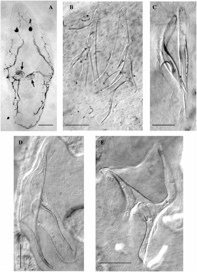
( FIGS 1D View Figure 1 , 5A View Figure 5 , 7A View Figure 7 )
Polycystis hibernica Southern, 1936: 45 View in CoL , 59–61, figs 2–5.
Rogneda hibernica Karling, 1953: 350–352 View in CoL , 354–355, 357–360, 363–364, 366–367, figs 4–8, 15, 17, 23–24, plate 1A, D, E, plate 2A, D, E; Brunet, 1969: 213, 217; Evdonin, 1977: 90, 104, 122, 244–245, figs 54, 112.10; Schockaert, Jouk & Martens, 1989: 24.
Known distribution: Known from several localities in the English Channel, the Irish Sea and the Irish Atlantic coast ( Southern, 1936; Karling, 1953). Also recorded from the sublittoral in the Netherlands Delta area ( Schockaert et al., 1989).
New localities: Ostend ( Belgium), Spuikom, coarse sand from the eulittoral (type locality) .
Material examined: The neotype (a whole mount) ( SMNH, no. 7154). Two whole mounts from Ostend ( HU). Three whole mounts and five serially sectioned specimens from Port Erin ( Isle of Man) ( SMNH, nos. 43554-561) .
Remarks: Karling (1953) described this species adequately. On the newly collected material I observed a bifurcated distal part of plate B1, something not mentioned by Karling (1953), but easily recognizable on the drawings by Southern (1936). Both distal ends are serrated and one end is much broader than the other one. The narrowest one is further referred to as ‘spine of plate B1’, the broader one I consider the distal end of plate B1 proper. As already mentioned by Karling (1953), plate A2 is also distally split. I will refer to the shortest of both these distal ends as ‘spine of plate A2’ (indicated with an arrow in Fig. 5A View Figure 5 ), the longer one considered to be the distal end of plate A2 proper.
Our measurements on the hard parts are as follows [abbreviations in parentheses are from Karling (1953)]. Stylet A: plate A1 (sa + da 1): 127–155 Mm (x¯ = 144; N = 5); plate A2 (da 2): 94–117 Mm (x¯ = 109; N = 5); spine of plate A2 (da 2 partly): 19–30 Mm (x¯ = 24 Mm; N = 5). Stylet B: plate B1 (sb + db 1): 115– 145 Mm (x¯ = 134; N = 3); spine of plate B1: 34–44 Mm (x¯ = 39; N = 2); plate B2 (db 2): 37–42 mm (x¯ = 40; N = 4). These measurements correspond to the measurements given by Karling (1953).
I deem the designation of a neotype necessary for this species, considering the complexity of species recognition within the taxon Rogneda . The type series not longer exists or has never existed, as Southern (1936) based his description on live material only, keeping no slides. Both Karling’s (1953) material (from southern England, the Isle of Man and the northern English Channel), and my material (from Belgium) are a considerable distance from the original locality where Southern (1936) collected his specimens (Valentia Island and Erin, both south-west coast of Ireland). Therefore, I choose a specimen with the most clearly observable hard parts as neotype. Its hard parts are practically identical to those depicted by Southern (1936: fig. 5).
ROGNEDA LICYAE SP. NOV.
( FIGS 5C View Figure 5 , 7C View Figure 7 )
Holotype: A whole-mounted specimen, Corsica ( France), near Pointe Revellata , sand from a sublittoral sample (6 m) off a small sandy beach, 18.ix.1983 ( SMNH, no. 7155).
Other material: Observations on live animals.
Other locality: Corsica ( France), near Ocellutia , sand from 19 m deep, 19.x.1982 .
Etymology: In memory of the late Licy Oeyen, an excellent biologist and even better friend.
Diagnosis: Unpigmented species of Rogneda . Plate A1 proximally rectangular, distally narrowing abruptly to a slender stalk, which ends in an arrowheadshaped point. Plate A2 a simple hook, with a broad and triangular spine. Plate B1 with a slender proximal end and a distal end that consists of a rectangular plate with a serrated distal rim and a broad spine. Plate B2 broad, distal rim serrated.
Description: The whole-mounted specimen is 0.7 mm long and is unpigmented.
Plate A1 is rather long and broad. Distally it abruptly narrows to a stalk, which ends in an arrowhead-shaped point (indicated with a dashed arrow in Fig. 5C View Figure 5 ). Proximally it is rather narrow, ending bluntly. The plate is 121 Mm long. Plate A2 is connected to the proximal half of A1. It is 88 Mm long, and distally reaches as far as the distal tip of plate A1. It evenly tapers towards its distal end, ending rather bluntly. It carries a strong and broad, 37-Mm-long spine somewhat at its midpoint (indicated by an arrow in Fig. 5C View Figure 5 ). The proximal part of plate B1 is rather narrow. Distally plate B1 splits into a narrow, rectangular part, which is serrated distally, and a broad, sharp-ending spine. The plate is 96 Mm long, the spine 29 Mm. Plate B2 has a rather broad distal end with a serrated distal rim. It is 60 Mm long and 25 Mm broad at its broadest. At the place where it is attached to plate B1 it shows a small projection pointing towards the distal tip of the stylet.
ROGNEDA VANGRONSVELDI SP. NOV.
( FIGS 5B View Figure 5 , 7B View Figure 7 )
Holotype: A whole-mounted specimen, Corsica ( France), in the harbour of the marine biological station STARESO, between 4 and 8 m deep, in coarse sand, 19.v.1983 ( SMNH, no. 7156).
Other material: Observations on live animals.
Etymology: Dedicated to Professor J. Vangronsveld, head of the Centre for Environmental Sciences of the Hasselt University.
Diagnosis: Species of Rogneda with a pigmented area at the level of the eyes. Stylet A with a long and slender plate A1, ending in a sharp point, proximally with a short, blunt spine. A2 long and slender, ending in a sharp point, with a relatively long and slender spine that is attached to A2 at about its midpoint and also ends in a sharp point. Stylet B with a plate B1 with a blunt, broad proximal end and a distal end that consists of a rectangular plate with a serrated distal rim and a broad spine.
Description: The specimen studied is 0.9 mm in whole mount. The live animals show an opaque black area around the eyes and a light brown body colour, the latter owing to the presence of parenchymal pigment. In the squashed and damaged holotype, pigment grains are found outside the body, probably the grains responsible for the black area around the eyes observed in the live animal.
Stylet A has a plate A1 that is 125 Mm long and very slender. It tapers distally to a very sharp point. More or less at its midpoint it carries a very short (23 Mm), blunt spine. Plate A2 is connected to the short proximal part of A1. It is very long (158 Mm) and slender, tapering to a sharp distal end. At about its midpoint it carries a very long (79 Mm), slender spine (indicated by an arrow in Fig. 5B View Figure 5 ), which also evenly tapers towards its distal and sharp end. Stylet B is almost identical to that of R. licyae . Plate B1 has a very broad and blunt proximal end. Distally it splits into a rectangular part that is serrated distally, and a broad, sharp-ending spine. The plate is 92 Mm long, the spine 39 Mm. Plate B2, as in R. licyae , is 59 Mm long and 27 Mm broad at its broadest.
ROGNEDA VERVECKENI SP. NOV.
( FIG. 5D View Figure 5 )
Holotype: A whole-mounted specimen, Ferrol (Galicia, Spain), muddy–sandy sediment from the sublittoral near the pump of the marine biological station, 26.viii.2006 ( SMNH, no. 7157).
Etymology: Dedicated to Erwin Vervecken, triple world champion cyclo-cross (2001, 2006, 2007).
Diagnosis: Species of Rogneda without pigment. Stylet A with a long and slender plate A1, distally ending in very narrow, blunt serrated end. Plate A2 long, ending in a sharp point, with a relatively long, triangular spine that is attached to A2 at about its midpoint and also ends in a sharp point. Stylet B with a plate B1 with a long and relatively slender proximal end and a distal end that consists of a rectangular plate with a serrated distal rim and a broad spine. Plate B2 relatively narrow and curved.
Description: The specimen studied is 0.7 mm in whole mount (animal very contracted).
Stylet A has a plate A1 that is slender and 153 Mm long. It tapers distally to a narrow, blunt end. This end is serrated, consisting of four finger-like projections. Plate A2 is connected to plate A1 more or less at about one-third the length of the latter from the proximal tip. It is rather long (107 Mm) and slender, tapering to a sharp distal end. At about its midpoint it carries a long (68 Mm), triangular spine (indicated by an arrow in Fig. 5D View Figure 5 ), which is slightly curved and also evenly tapers towards its distal sharp end. Stylet B has a plate B1 with a relatively long proximal end. Distally it splits into a narrow rectangular part that is serrated distally, and a broad, sharp-ending spine. The plate is 141 Mm long, the spine 56 Mm. Plate B2 is 70 Mm long, somewhat curved, and distally ends in a serrated rim.
POLYRHABDOTA View in CoL -GROUP ROGNEDA POLYRHABDOTA AX, 1959 View in CoL
( FIG. 6A View Figure 6 )
Rogneda polyrhabdota Ax, 1959: 48 View in CoL , 119–123, 145, figs 107–115; Brunet, 1969: 212, 217, 219, 221.
Known distribution: Sea of Marmara ( Turkey), fine sand from the littoral zone in Pendik and Florya ( Ax, 1959).
New locality: Nea Epivates (near Thessalonica, Greece), fine sediment (2.5 m deep), left from the jetty, 6.viii.2002 (coll. HU).
Material examined: Two whole mounts from Nea Epivates.
Remarks: The species was extensively described by Ax (1959). The description of the hard parts by Ax (1959) is accurate, but none of the plates is distally open and funnel-shaped as he describes them. Stylet A consists only of plate A1, and shows a strong transverse band more or less at its midpoint. Distally it is rather delicate, with a serrated distal rim. It is 117–118 Mm long (N = 2) in the specimens from Greece, comparable with the measurements given by Ax (1959). Plate B1 is proximally slightly broader than stylet A. Its distal half is rectangular and also has a serrated distal rim. Plate B2 is attached to plate B1 almost at its midpoint and has a somewhat spatulate distal part. It is not serrated. Its proximal part is very thick and appears very sturdy (pb 2 of Ax, 1959). In the specimens from Greece plate B1 is 119–126 Mm long (N = 2); plate B2 47–51 Mm (N = 2). This is slightly smaller than was mentioned by Ax (1959) (135–163 Mm for the whole stylet).
ROGNEDA SCHAERERI SP. NOV.
( FIGS 1A View Figure 1 , 6B View Figure 6 , 7D, E View Figure 7 )
Holotype: A whole-mounted specimen, Lignano ( Italy), fine sand taken on a very flat beach at low tide, from the water edge down to ± 8 cm deep; oxidized top layer (± upper 2 cm). ( SMNH, no. 7158).
Paratypes: Two whole-mounted specimens, same data as for the holotype (HU, nos. 344–45).
Etymology: Named after Dr Lukas Schärer (University of Basel), who kindly gave me the material of this species.
Diagnosis: Species of Rogneda with four anastomosing, brown, dorsal pigment stripes. Stylet A very simple, consisting of only one plate. Stylet B with two plates. Plate B1 rather long, with a serrated distal rim. Plate B2 attached to plate B1 at about its midpoint, distally ending in a blunt point. Plates B1 and B2 close to each other.
Description: Animals ± 0.9 mm long (measured on whole mounts). Four irregular, anastomosing longitudinal dark brown pigment stripes mark the dorsal side of the body.
Stylet A is very simple, 107–129 Mm long (x¯ = 115; N = 3). It consists of one plate only, which has a thick proximal end. It is somewhat bent, with a linguiform, slightly serrated distal end. Stylet B is of the typical construction, with plate B2 always lying next to plate B1 and attached to B1 at about its midpoint. Plate B1 is 159–207 Mm long (x¯ = 187; N = 3), with a serrated distal end. Plate B2 tapers towards its distal end, ending in a blunt point. It is 83–98 Mm long (x¯ = 92; N = 3). The proximal end of each stylet shows many superficial folds.
STEUERI View in CoL -GROUP ROGNEDA CINCTA BRUNET, 1969 View in CoL
( FIGS 8A View Figure 8 , 9A View Figure 9 )
Rogneda westbladi View in CoL ssp.? Brunet, 1965: 135.
Rogneda cincta Brunet, 1969: 208 View in CoL , 214–221, figs 7–9, 13; Evdonin, 1977: 12, 14, 104, 122, 244–245, figs 2, 112.9; Brunet, 1979: 105.
Known distribution: Bay of Marseilles ( France), Amphioxus View in CoL -sand from the ‘Plateau des Chèvres’ between the island of Jarre and the coast (8–10 m), and ‘Pierre de Joseph’, near the island of Plane, fine sand (17 m) ( Brunet, 1969).
Material examined: The holotype (a whole mount) ( SMNH, no. 3045). The paratype (a whole mount) ( MNHN-P) . Three whole-mounted specimens ( MNHN-P).
Remarks: Brunet (1969) described this species adequately. The brown pigment around the seminal vesicles is typical of this species (arrows in Fig. 9A View Figure 9 ). In the holotype the strong distal spine of plate A1 appears not to be spatulate, as was described by Brunet (1969), but pointed (arrow in Fig. 8A View Figure 8 ). Plate B2, by contrast, is very broad and distally spatulate, not as pointed as was drawn by Brunet (1969). My measurements on the hard parts of this species are as follows [the abbreviations in parentheses are these of Brunet (1969)]. Stylet A: plate A1 (sa + da 1): 69–71 Mm (x¯ = 70; N = 3), spine of plate A (part of da 1): 24–26 Mm (x¯ = 25; N = 3), plate A2 (pa 2 + da 2): 35–39 Mm (x¯ = 38; N = 3). Stylet B: plate B1 (sb + db 1): 60–69 Mm (x¯ = 66; N = 4), plate B2 (pb 2 + dbx): 30–39 Mm (x¯ = 34; N = 4).
ROGNEDA GALLICA AX, 1956 View in CoL
( FIG. 8B View Figure 8 )
Rogneda westbladi gallica Ax, 1956: 5 View in CoL , 146–148, 165, 189, 200, fig. 36; Brunet, 1969: 212; Evdonin, 1977: 243, 245, fig. 112.4.
Rogneda westbladi Brunet (1969) View in CoL : 208, 210–212, figs 2, 14.
Known distribution: The lagoon of Lapalme (Etang de Lapalme), near La Franqui (French Mediterranean coast), in pure fine sand, sometimes with some detritus ( Ax, 1956) (type locality). Bay of Marseilles ( France), near la Mounine (east of the ‘Île de Maire’), fine sand with some detritus (12 m) ( Brunet, 1969).
Material examined: One sectioned specimen from the Etang de Lapalme , designated lectotype ( SMNH, no. 5988). One whole mount from Marseilles ( SMNH, no. 43574) .
Remarks: The whole mount I studied is one of the specimens already studied and discussed by Brunet (1969), who referred to them as R. westbladi . He did not allocate them to one of the two subspecies recognized, but suggested that the Marseilles population might represent a taxonomic entity of its own. He mentioned that stylet A of the specimens from Marseilles is identical to that described by Ax (1956) for R. w. gallica , but indicated some differences in the detailed morphology of stylet B between his specimens and Ax’s (1956) description. These differences are the presence of a short, strong spine at the base of db 1 (distal part of plate B1) and, more importantly, the fact that in the specimens from Marseilles, db 2 (the distal part of plate B2) is somewhat longer than db 1 (plate B1) and has a rounded distal end. The first character is, however, difficult to assess, and was not even visible on the specimen I studied. Ax (1956) did not mention or draw this spine, but it could still be present. The relative lengths of the two parts of stylet B is another doubtful character. Not only is it very difficult to judge from Ax’s (1956) drawings which one is the longest, but even in the material from Brunet himself the relative position of the two distal tips appears not as constant as he indicated. Finally, the distal parts of these stylets are often very difficult to study, and a more or less rounded end is also a doubtful character. These observations suggest that the specimens studied by Brunet (1969) in fact belong to Ax’s (1956) R. w. gallica .
According to Ax (1956), stylet A of R. w. gallica differs from that of the nominal subspecies in the relative length of the two subunits: plates A1 and A2. In R. w. gallica plate A2 is rather long and its distal point projects beyond the distal point of plate A1, whereas in R. w. westbladi plate A2 is much shorter. Moreover, the distal end of plate A1 is serrated in R. w. westbladi , but smooth in the specimen of R. w. gallica I studied. Stylet B is also very different between the two taxa, a feature barely discussed by Ax (1956). In R. w. gallica , plate B1 is somewhat curved, with a blunt distal point. Plate B2 is rather simple, with a somewhat diamond-shaped distal point, and is attached to plate B1 more or less at its midpoint. On Ax’s (1956) drawings this plate is depicted as very narrow, almost threadlike. Possibly he only saw the thick rim of the plate, and not the whole plate. Stylet B of R. w. westbladi is much more complex and indeed not reminiscent of that of R. w. gallica (see further under R. westbladi ). These large and fixed differences between the two taxa indicate that the French population ( R. w. gallica ) belongs to a separate species from the population from the Adriatic Sea ( R. w. westbladi ). Therefore, I consider R. w. gallica a species of its own: R. gallica Ax, 1956 .
Measurements on the hard parts of the whole mount: stylet A: plate A1: 98 Mm; plate A2: 57 Mm. Stylet B: plate B1: 109 Mm, plate B2: 56 Mm.
ROGNEDA MARTENSI SP. NOV.
( FIGS 8C View Figure 8 , 9D, E View Figure 9 )
Holotype: A whole-mounted specimen, south-west Sulawesi ( Indonesia), Kajangan , coral sand from the eulittoral, 22.x.1984 ( SMNH, no. 7159).
Other material: Observations on live material. Etymology: Dedicated to Dr Paul Martens, who collected the material of this and many other kalyptorhynchs present in the collections of the HU.
Diagnosis: Unpigmented species of Rogneda . Plate A1 proximally very broad, square, distally very narrow, ending bluntly. Plate A2 a simple, curved hook, attached more or less at the midpoint of plate A1. Plate B1 curved, ending in a very sharp distal point. Plate B2 attached to the midpoint of plate B1, rectangular, distally ending in a broad and blunt end.
Description: The only specimen available is unpigmented, about 0.9 mm long (measured on the whole mount). It is not in an excellent condition, but enough detail could be seen to mark it undoubtedly as a new species. Interpretation of the construction of plate A1 is difficult. At first sight it appears to be very broad and square proximally, but narrows abruptly at about its midpoint to a rectangular plate that ends bluntly. It is 70 Mm long. Plate A2 is attached to plate A1 more or less at the place where the latter narrows. It is a simple 36-Mm-long, curved hook. However, another interpretation could be that plate A2 partly has come loose from A1 because of the squeezing and is connected to it by a thin, very narrow clasp. If this is the case, plate A1 is very narrow, and the square that can be seen in the holotype is not a plate (as in the first interpretation), but a hole. As such, stylet A, reconstructed, would look very much like that of R. gallica . However, the observations on the live animal (only very slightly squeezed) suggest that the first interpretation is the correct one.
Stylet B is relatively simple and consists of a 116- Mm-long plate B1, which slightly bends at about its midpoint. Proximally it ends bluntly; distally it ends in a sharp point. Plate B2 is attached almost at the midpoint of plate B1, and is a rather broad rectangular plate. It is 68 Mm long.
ROGNEDA PALULA BRUNET, 1969 View in CoL
( FIGS 8D View Figure 8 , 9B View Figure 9 )
Rogneda palula Brunet, 1969: 208 View in CoL , 216–221, figs 10– 11, 15; Evdonin, 1977: 14, 122, 244–245, figs 2, 112.12.
Rogneda patula View in CoL (incorrect subsequent spelling) Watson, 2001: 224, figs 20.23, 20.27, table 20.1.
Known distribution: Bay of Marseilles ( France), Amphioxus View in CoL -sand from between the ‘ Château d’If’ and the island of Ratonneau (14–16 m) ( Brunet, 1969) (type locality) .
New localities: Roscoff ( France), very coarse sand from between rocks in the Fucus serratus zone in the ‘Green Island Channel’ (3.vi.2007). Roscoff ( France), coarse sand from a tide pool in front of the marine station, near to the long jetty (6.vi.2007).
Material examined: One whole mount, designated lectotype (SMNH, no. 3047). One other whole mount the type locality (MNHN-P). Three whole mounts from Roscoff.
Remarks: The species is adequately described by Brunet (1969). Plate B1 is distally split into a bluntending plate and a sharp ending one, the latter further referred to as spine of plate B1. The stylets of the specimens from Roscoff are identical in shape to those of the specimens from the Mediterranean, but are slightly larger. Therefore, measurements on specimens of both areas are given separately. Measurements on the two specimens from Marseilles: plate A1 (sa + da1): 89–90 Mm; plate A2 (da2): 48–51 Mm, plate B1 (sb + part of db1): 80–82 Mm; spine of plate B1 (part of db1): 47–56 Mm; plate B2: 52–56 Mm. Measurements on the three specimens from Roscoff: plate A1: 100–104 Mm (x¯ = 102); plate A2: 57–63 Mm (x¯ = 61), plate B1: 102–107 Mm (x¯ = 104); spine of plate B1: 70–71 Mm (x¯ = 71); plate B2: 71–77 Mm (x¯ = 74).
R. palula is the only species of Rogneda that has both a Mediterranean and an Atlantic population.
ROGNEDA RETICULATA BRUNET, 1969 View in CoL
( FIG. 8E View Figure 8 )
Rogneda reticulata Brunet, 1969: 208 View in CoL , 212–214, 216– 219, 221, figs 4–6, 14; Evdonin, 1977: 14, 104, 122, 244–245, figs 2, 112.11; Brunet, 1979: 105.
Known distribution: Several localities in the Bay of Marseilles, in Amphioxus View in CoL -sand and fine and muddy sands (4–17 m) ( Brunet, 1969).
New locality: Cerbère ( France), Anse de Terrimbou, 10 m deep; coarse clean sand, 12.iv.2002 (coll. HU).
Material: The holotype (a whole mount) ( SMNH, no. 3048). Two whole mounts from Pière de Joseph (bay of Marseilles) ( MNHN-P). Live observations and three whole mounts from Cerbère .
Remarks: Brunet (1969) has described this species adequately. Our measurements (on four specimens) are as follows: plate A1: 77–92 Mm (x¯ = 85); plate A2: 34–51 Mm (x¯ = 45), plate B1: 77–96 Mm (x¯ = 85), plate B2: 36–45 Mm (x¯ = 41). These measurements are completely in accordance with those given by Brunet (1969). The specimens from Cerbère appear to have a larger plate A1 measuring 90, 90 and 92 Mm, respectively (77–86 Mm in the others), but they do not show any difference as to the other measurements.
ROGNEDA STEUERI ( STEINBÖCK, 1933) KARLING, 1953 View in CoL
( FIGS 8F View Figure 8 , 9C View Figure 9 )
Polycystis steueri Steinböck, 1933: 14–15 View in CoL , figs 6–7.
Rogneda steueri Karling, 1953: 350 View in CoL , 356, 358–359, 361, 363, 365–367, figs 2, 13, 19–22, plates 1B–C; Ax, 1959: 121, 123; Brunet, 1969: 217, 221; Evdonin, 1977: 14, 122, 244–245, figs 2, 112.13; Brunet, 1979: 105.
Known distribution: Adriatic Sea, Split ( Croatia), Ciovo, Amphioxus View in CoL -sand ( Karling, 1953) (type locality). Adriatic Sea, Rovinj ( Croatia), Cuvi, in coarse sand and shell-gravel ( Steinböck, 1933).
Material examined: One whole mount from Split, designated neotype ( SMNH, no. 5987). Two other whole mounts from Split, on the same slide as the holotype. One whole mount, on the same slide as the lectotype of R. capulata ( SMNH no. 2677). One other whole mount from Split ( SMNH, no. 43579).
Remarks: Stylet A of this species is very typical, with a long and slender plate A1, distally ending in a sharp point, and a very slender plate A2, which distally projects beyond plate A1. In some of the specimens, plate A2 has a blunt, serrated distal end, in others it ends in a sharp point. We have measured lengths of 104 Mm for plate A1 (N = 2); 62 Mm for plate A2 (N = 1). The length of plate A1 (sa + da1) is comparable with the length mentioned by Karling (1953), whereas the length of plate A2 (pa2 + da2) is ± double the length mentioned by Karling. Judging from his drawings it is clear that the lengths he mentioned for plate A2 are erroneous. Stylet B is also of a very simple construction. Plate B1 is simple, proximally somewhat broader than distally. Attached to it is plate B2, which also is a simple plate, distally ending at the same level as plate B1. In some specimens it seems to end bluntly, with a serrated edge. In other specimens it seems to end in a sharp point (as in Fig. 8F View Figure 8 ). Plate B1 is 73–82 Mm long (N = 2), which is slightly smaller than mentioned by Karling (1953). Plate B2 is 36–47 Mm long (N = 2), which is in complete correspondence with the measurements given by Karling (1953).
TRIPALMATA View in CoL -GROUP ROGNEDA TRIPALMATA ( BEKLEMISCHEV, 1927) KARLING, 1953 View in CoL
( FIG. 10C View Figure 10 )
Polycystis tripalmata Beklemischev, 1927: 195–198 View in CoL , 206–207, plate 1 figs 13–16.
Rogneda tripalmata Karling, 1953: 350 View in CoL , 352, 356, 359–361, 367; Ax, 1959: 119, 121–124, 146, 161; Brunet, 1969: 213, 217, 219, 221; Evdonin, 1977: 8, 101, 104–105, 121, 244–246, figs 62, 112.8.
Known distribution: The Black Sea, the bight of Odessa ( Ukraine), in sand ( Beklemischev, 1927). Sea of Marmara, fine sand from the littoral zone on Heybeli Island ( Turkey) ( Ax, 1959).
Material examined: None.
Remarks: See the discussion by Karling (1953) and Ax (1959). The lengths of the stylets given previously ( Beklemischev, 1927; Ax, 1959) are as follows (my notation used): stylet A: 154–180 Mm; plate B1: 190– 200 Mm; plate B2: 114 Mm. The measurement of B2, however, includes the proximal extension of plate B2 (‘pb2’ of Karling, 1953). If the plate is measured axially, as I did for the other species, it probably will be smaller.
ROGNEDA VALCKEI SP. NOV.
( FIGS 10A, B View Figure 10 , 12A, B View Figure 12 )
Holotype: A whole-mounted specimen, Corsica ( France), near Ocellutia , sand from 10 m deep, 25.x.1982 ( SMNH, no. 7160).
Paratypes: Two whole-mounted specimens, same data as for the holotype (HU, nos. 346–47).
Other material: Observations on live animals.
Other localities: Several localities near Calvi ( Corsica, France): off Pointe Revellata, sand from 6 m deep, 25.xi.1984; sublittoral sample from the bay of Calvi, sand from 20 m deep, 30.i.1984.
Etymology: Dedicated to Professor R. Valcke, head of the research group Molecular and Physical Plant Physiology of the UHasselt.
Diagnosis: Species of Rogneda with two dorsal, longitudinal pigment stripes. Stylet A represented by a single long, narrow plate with a small distal spine. Stylet B consisting of two plates. Plate B1 distally ending in a sharp point, with a small hook not far from the distal point. Rim between hook and distal point serrated. Plate B2 connected to plate B1 above the midpoint of B1, distally ending in a sharp point. A small hook present at about its midpoint.
Description: Animals 0.7– 1 mm long, measured on whole mounts. Dorsally, at both sides of the body, two stripes of brown pigment stretch from the level of the eyes towards the caudal end of the body.
Stylet A is very simply built. It consists of a relatively slender plate that distally carries a small spine. It is 135–175 Mm long (x¯ = 158; N = 3), with a relatively broad and blunt proximal end and a more pointed and much narrower distal end. The spine is connected to this main plate between the second and the distal third. It is 39–42 Mm long (x¯ = 41; N = 3), and can end in a sharp or blunt point, depending on the specimen. Plate B1 is 150–192 Mm long. Its distal end is serrated at one side, and ends in a sharp point. Just proximal from the serrated rim, the plate shows a 18–19-Mm-long hook (N = 2). Plate B2 is attached to plate B1 somewhat above the midpoint of B1 (in one of the paratypes it has come loose: see Fig. 10B View Figure 10 ). It has a straight distal end, and tapers towards a sharp point. At about its midpoint it shows a relatively broad hook. Plate B2 is 100–117 Mm long (x¯ = 108; N = 3); the hook is 23–30 Mm long (N = 2).
WESTBLADI View in CoL -GROUP ROGNEDA ANGLICA KARLING, 1953 View in CoL
( FIGS 11C View Figure 11 , 12C View Figure 12 )
Rogneda anglica Karling, 1953: 350 View in CoL , 354–356, 358– 361, 363–366, figs 1, 14, 18, 31–32, plates 1F, 2B, C, F; Brunet, 1965: 153; Brunet, 1969: 217, 219–220; Evdonin, 1977: 14, 101, 110, 122, 243, 245, figs 2, 112.2.
Known distribution: The English Channel , several localities near Plymouth (type locality, no more details available) and Millport ( UK), mostly in loamy bottoms (10–36 m) ( Karling, 1953) .
New localities: Kristineberg ( Sweden), Gullmarfjord, Essvik , muddy sediment, 20 m deep (coll. HU). Ferrol (Galicia, Spain), muddy–sandy sediment from the sublittoral near the pump of the marine biological station (26.viii.2006) .
Material examined: Observations on live animals from Kristineberg. The lectotype (a whole mount) ( SMNH, no. 2676). Four whole mounts (on the same slide) and three serially sectioned specimens from Plymouth ( SMNH, nos. 43575–78). Two whole mounts and three serially sectioned specimens from Kristineberg (coll. HU). Two whole mounts from Galicia (coll. HU). Three of the nine whole-mounted specimens do not allow accurate measuring .
Remarks: Stylet A is relatively simple, and the basic construction can still be recognized. Plate A1 is more or less S-shaped. It corresponds to Karling’s (1953) sa + the broad ending part of da 2. It is 105–140 Mm long (x¯ = 118; N = 6). At about its midpoint, plate A shows a rather broad, sharp-ending hook (da1 of Karling, 1953), which is 21–38 Mm long (x¯ = 26; N = 6). Plate A2 is connected to plate A just proximally from this hook. It is curved and lies adjacent to the distal part of plate A1. It ends in a sharp point (part of da2 of Karling, 1953). It is 46–63 Mm long (x¯ = 56; N = 6). Stylet B is much more complex than is depicted by Karling (1953), and apparently the distal parts of both constituting plates have grown together at some places, making it rather difficult to distinguish them. Plate B1 [sb + part of db1 of Karling (1953)] is rather long, somewhat curved, and distally ends in a rather narrow, oblong part with a serrated distal rim. Plate B2 [other part of Karling’s (1953) db1] is connected to plate B1 more or less at its midpoint, and proximally protrudes extensively at both sides of B1; the projections are called pb2 (z in Figs 11C View Figure 11 , 12C View Figure 12 ) and db3 (indicated with an arrow in Figs 11C View Figure 11 , 12C View Figure 12 ), respectively, by Karling (1953). Distally it is very broad, and has a relatively long serrated part. At the opposite part of this serrated rim, a broad distal extension of plate B1 (x? in Figs 11C View Figure 11 , 12C View Figure 12 ) seems to be connected to plate B2. The lengths of the different parts are as follows: plate B1: 123–157 Mm (x¯ = 136; N = 6); plate B2: 44–88 Mm (x¯ = 69; N = 6).
ROGNEDA COLPAERTI SP. NOV.
( FIGS 1B View Figure 1 , 11B View Figure 11 , 12D, E View Figure 12 )
Holotype: A whole-mounted specimen, Corsica ( France), bay of Calvi , off Pointe Revellata, sand from a sublittoral sample (± 40 m), 26.xi.1982 ( SMNH, no. 7161).
Paratypes: Two whole-mounted specimens, same data as for the holotype (HU, nos. 348–49).
Other material: Observations on live material by Dr P. Martens.
Etymology: Dedicated to Professor J. Colpaert, mycologist at the HU.
Diagnosis: Unpigmented species of Rogneda . Stylet A simple. Plate A1 narrow, rounded proximally, serrated distally, with a strong hook almost at its midpoint. Plate A2 a simple curved plate, ending in a sharp distal point. Stylet B complex. Plate B1 with a broad and short proximal part, distally splitting into a short pointed part and a rectangular part with a serrated distal rim. Small triangular tooth present at base of rectangular part. Plate B2 S-shaped, with a serrated distal rim and one strong, thick wall. Narrow pointed projection present at base of plate B2.
Description: Animals unpigmented, rather small, 0.4–0.7 mm long in whole mounts.
Stylet A is rather simple. Plate A1 is narrow, with a rounded proximal and a serrated distal end. It is S-shaped, 94–98 Mm long (x¯ = 96; N = 3). At about its midpoint, near to the place where plate A2 is attached, it forms a relatively broad hook, about 20 Mm long in the holotype and in one of the paratypes (not visible in the other paratype, probably because of the poor quality of the slide). Plate A2 is very simple, with a broad proximal end, tapering towards its distal end, which is a very sharp point. It is 44–62 Mm long (x¯ = 51; N = 3). Stylet B is very complex, and can be best described on the holotype. Plate B1 is curved and has a short and broad proximal part, which distally splits into a short and pointed projection (x in Figs 11B View Figure 11 , 12E View Figure 12 ) and a longer, rectangular part with a serrated distal rim. At the base of the rectangular plate, at the opposite side of the projection, there is a small triangular tooth (y in Figs 11B View Figure 11 , 12E View Figure 12 ). Measured from the proximal point of the plate to the serrated rim of the rectangular plate the stylet is 113 Mm long, with the projection 25 Mm long and the tooth 12 Mm. Plate B2 makes an S-shaped curve. Distally it ends in a serrated rim, which can only be followed for a short distance. At the opposite side of the pointed projection of plate B1, plate B2 shows a narrow and very pointed projection (indicated with a dashed arrow in Figs 11B View Figure 11 , 12E View Figure 12 ). The proximal part of plate B2 extends beyond the curve of plate B1 (z in Figs 11B View Figure 11 , 12E View Figure 12 ) and has a smaller projection at the opposite side of this (indicated with an arrow in Figs 11B View Figure 11 , 12E View Figure 12 ). Plate B2 is 73 Mm long. In the paratypes, the overall shape of stylet B is easily recognizable but some details are somewhat obscured and difficult to find. The measurements in these specimens are as follows (some of the measurements were impossible to take on one or both of the specimens): plate B1: 93 and 111 Mm; projection of B1: 25 Mm; tooth of B1: 11 Mm; length B2: 56 Mm.
ROGNEDA WESTBLADI KARLING, 1953 View in CoL
( FIG. 11A View Figure 11 )
Rogneda westbladi Karling, 1953: 350 View in CoL , 354, 358–359, 361, 363–367, figs 16, 25–28; Ax, 1956: 147, 189; Brunet, 1969: 217, 220.
Rogneda westbladi westbladi Ax, 1956: 148 View in CoL , 189; Brunet, 1965: 153; Brunet, 1969: 212, 221.
Known distribution: The Adriatic Sea, Isola Lunga, Canal di Leme (type locality) and Baia Lone (Rovinj, Croatia), in sand or loam (4–35 m) ( Karling, 1953) .
Material examined: The lectotype (a whole mount) ( SMNH, no. 2678). Four serially sectioned specimens ( SMNH, nos. 53000–003).
Remarks: See the original description by Karling (1953) and the remarks on R. gallica . Plate A1 is 98 Mm long, plate A2 only 34 Mm. Stylet B of R. westbladi is rather complex, in fact much more complex than that of R. gallica , and it is rather difficult to distinguish the different parts. It apparently consists of a plate B1 (Karling’s sb + db1) that is curved and has a narrow rectangular distal part, ending in a serrated rim. It is 116 Mm long. It carries two extensions at the place where the plate becomes rectangular, a blunt-ending one (x in Fig. 11A View Figure 11 ), 17 Mm long, and a sharp-ending one at the opposite side, 17 Mm long (y in Fig. 11A View Figure 11 ). Plate B2 (db2) is attached to plate B1 at about its midpoint and has a very large proximal part (Karling’s pb2; z in Fig. 11A View Figure 11 ) that extends far (± 32 Mm) beyond the curve of plate A1. It is very broad, 42 Mm long and has a serrated distal rim.


| SMNH |
Department of Paleozoology, Swedish Museum of Natural History |
| HU |
University of Zhejiang |
| R |
Departamento de Geologia, Universidad de Chile |
No known copyright restrictions apply. See Agosti, D., Egloff, W., 2009. Taxonomic information exchange and copyright: the Plazi approach. BMC Research Notes 2009, 2:53 for further explanation.
|
Kingdom |
|
|
Phylum |
|
|
Order |
|
|
Family |
Rogneda
| Artois, Tom J. 2008 |
Rogneda patula
| Watson NA 2001: 224 |
Rogneda exilis Brunet, 1979: 108
| Brunet M 1979: 108 |
Rogneda acuta
| Brunet M 1979: 107 |
Rogneda cincta Brunet, 1969: 208
| Brunet M 1979: 105 |
| Evdonin L 1977: 12 |
| Brunet M 1969: 208 |
Rogneda westbladi
| Brunet M 1969: 208 |
Rogneda palula Brunet, 1969: 208
| Evdonin L 1977: 14 |
| Brunet M 1969: 208 |
Rogneda reticulata Brunet, 1969: 208
| Brunet M 1979: 105 |
| Evdonin L 1977: 14 |
| Brunet M 1969: 208 |
Rogneda falcata Brunet, 1965: 153–158
| Brunet M 1979: 107 |
| Evdonin L 1977: 122 |
| Brunet M 1969: 208 |
| Brunet M 1965: 158 |
Rogneda westbladi
| Brunet M 1965: 135 |
Rogneda polyrhabdota Ax, 1959: 48
| Brunet M 1969: 212 |
| Ax P 1959: 48 |
Rogneda westbladi gallica
| Evdonin L 1977: 243 |
| Brunet M 1969: 212 |
| Ax P 1956: 5 |
Rogneda westbladi westbladi
| Brunet M 1969: 212 |
| Brunet M 1965: 153 |
| Ax P 1956: 148 |
Rogneda capulata Karling, 1953: 350
| Brunet M 1979: 105 |
| Evdonin L 1977: 14 |
| Brunet M 1969: 208 |
| Ax P 1959: 121 |
| Karling T 1953: 350 |
Rogneda hibernica
| Schockaert E & Jouk P & Martens P 1989: 24 |
| Evdonin L 1977: 90 |
| Brunet M 1969: 213 |
| Karling T 1953: 352 |
Rogneda steueri
| Brunet M 1979: 105 |
| Evdonin L 1977: 14 |
| Brunet M 1969: 217 |
| Ax P 1959: 121 |
| Karling T 1953: 350 |
Rogneda tripalmata
| Evdonin L 1977: 8 |
| Brunet M 1969: 213 |
| Ax P 1959: 119 |
| Karling T 1953: 350 |
Rogneda anglica Karling, 1953: 350
| Evdonin L 1977: 14 |
| Brunet M 1969: 217 |
| Brunet M 1965: 153 |
| Karling T 1953: 350 |
Rogneda westbladi Karling, 1953: 350
| Brunet M 1969: 217 |
| Ax P 1956: 147 |
| Karling T 1953: 350 |
Polycystis hibernica
| Southern R 1936: 45 |
Polycystis steueri Steinböck, 1933: 14–15
| Steinbock O 1933: 15 |
Polycystis tripalmata
| Beklemischev W 1927: 198 |
Polycystis minuta
| Steinbock O 1933: 14 |
| von Graff L 1913: 335 |
| von Graff L 1905: 136 |
Rogneda minuta
| Brunet M 1979: 103 |
| Evdonin L 1977: 11 |
| Brunet M 1969: 217 |
| Ax P 1959: 119 |
| Karling T 1953: 349 |
| Pereyaslawsewa S 1892: 282 |
| von Graff L 1882: 328 |
| Uljanin W 1870: 23 |