
Proterochersis porebensis, Szczygielski & Sulej, 2016
|
publication ID |
https://doi.org/ 10.1111/zoj.12374 |
|
publication LSID |
lsid:zoobank.org:pub:A208DBD2-7C7E-4779-B0AB-1782371E7053 |
|
persistent identifier |
https://treatment.plazi.org/id/6C090C43-DCCA-4D1C-98B7-37C413FA9517 |
|
taxon LSID |
lsid:zoobank.org:act:6C090C43-DCCA-4D1C-98B7-37C413FA9517 |
|
treatment provided by |
Marcus |
|
scientific name |
Proterochersis porebensis |
| status |
sp. nov. |
PROTEROCHERSIS POREBENSIS SP. NOV.
( FIGS 2 View Figure 2 , 3A – I View Figure 3 , 4 – 8 View Figure 4 View Figure 5 View Figure 6 View Figure 7 View Figure 8 , 11B View Figure 11 )
cf. Proterochersis Fraas, 1913 : Sulej et al.,
2012: 1034, fig. 4. cf. Proterochersis robusta Fraas, 1913 :
Niedzwiedzki et al., 2014: 1123.
Occurrence and distribution: Zbaszynek Beds (subzone IVb of the Corollina meyeriana zone, middle – upper Norian) in Poreba, Silesian Voivodeship, Poland.
Etymology: From Poreba, the place of discovery.
Diagnosis: Articular surface of femoral head triangular in dorsal view. Differing from Proterochersis robusta in caudal notch triangular, anterior margin of the carapace nearly straight, anterior edge of the third marginal only slightly rounded, and lower shell. Differing from Keuperotesta limendorsa gen. et sp. nov. in acromion and coracoid forming an angle of ~ 120 °.
Holotype: ZPAL V.39/48.
Paratypes: ZPAL V.39/34, ZPAL V.39/49, ZPAL V.39/72, ZPAL V.39/370 .
There are two shell morphotypes known to date from Poreba ( Fig. 2 View Figure 2 ). One of them, represented by ZPAL V.39/34, was preliminarily described and reconstructed by Sulej et al. (2012). Despite its overall similarity to Proterochersis robusta , and because of its somewhat different morphology (especially when it comes to peripheral elements of the carapace and plastron), the exact taxonomical status of this specimen was left undecided. The second morphotype, represented by previously undescribed specimens (among others ZPAL V.39/48, the holotype of Proterochersis porebensis sp. nov., ZPAL V.39/49, and ZPAL V.39/72, all described herein), is much more similar to Proterochersis robusta .
Carapace and vertebrae
The composition of the shell is almost identical to that of Proterochersis robusta (SMNS 17561 and others). There are five vertebrals, four pairs of pleurals, and three pairs of supramarginals; the marginal count is 14 for ZPAL V.39/49 (the same as in SMNS 17561) and 15 for ZPAL V.39/48 ( Fig. 2A, B View Figure 2 ). The anterior marginals have nearly straight outer borders (with the exception of the third one, which is gently rounded), so the anterior rim of the carapace is smooth, in contrast with the slightly serrated condition found in Proterochersis robusta . Sulci are usually sinuous and radial ridges are present. There is some variability between ZPAL V.39/48 and ZPAL V.39/49 (as well as the other specimens from Poland and Germany) in the shape and position of some scutes, but this will be discussed in future papers.
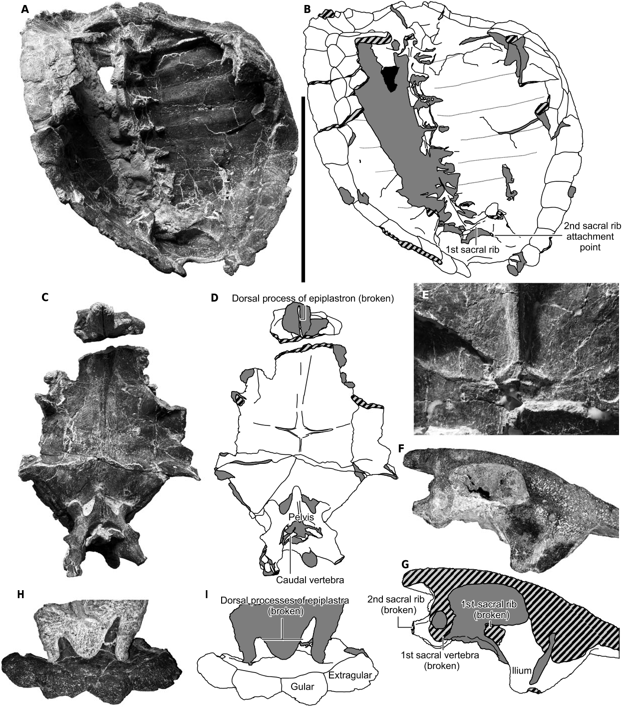
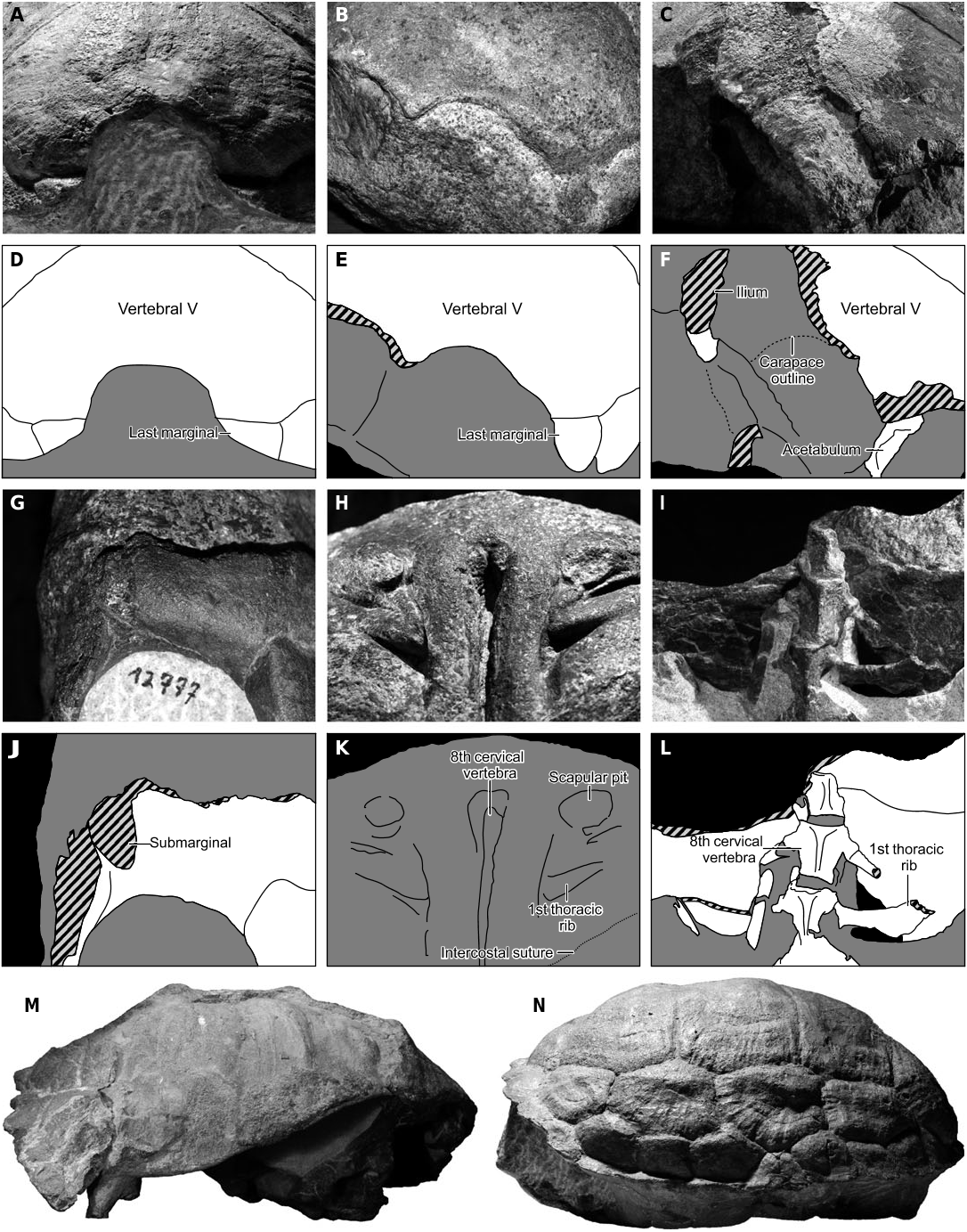
The last cervical vertebra is fused with (or sutured to) the first thoracic vertebra, and with the carapace ( Fig. 3E, F View Figure 3 ). There are ten pairs of thoracic ribs, with each apparently forming ten fully developed costals ( Figs 2F View Figure 2 , 4A, B View Figure 4 ). The first pair of ribs is different from the others because it forms a distinct ridge on the visceral side of the costal (similar to that of Proganochelys quenstedti , but less pronounced; its prominence differs between specimens), but (unlike Proganochelys quenstedti ) it is roughly parallel with the second pair, and there is what appears to be a clear suture between the corresponding costals. The intercostal sutures, similarly as in Proganochelys quenstedti , form long, shallow, and narrow grooves; Gaffney (1990) interpreted them instead as being vascular or nerve imprints, because of their lack of interdigitation. Numerous isolated, disarticulated costals found in Poreba indicate, however, that the intercostal sutures in most cases are indeed straight and longitudinal, and not interdigitated. On some costals there is a second groove, lying along the suture, possibly where the nerves or vessels were. In any case, even if the grooves seen on the articulated carapaces are not sutures themselves, both of these structures are close to each other and, as concluded by Gaffney (1990), the grooves are at least a good approximation of the sutural positions. The distal tip of the first thoracic rib is visible, yet it is not entirely free, but articulates the carapace with its anterior surface, and is probably sutured. The sutures between bones forming the posterior part of the carapace are not visible, but each of the three last ribs independently reaches the carapace and they do not meet with each other, so, similarly as in other Late Triassic turtles, each one of them probably forms its own costal as well.
Plastron
The plastron composition does not differ from that of Proterochersis robusta , with two pairs of abdominal scutes and an additional three scutes at the caudal end ( Figs 2D, E View Figure 2 , 3C, D View Figure 3 ). The dorsal processes of epiplastra ( Fig. 4C, D, H, I View Figure 4 ) are relatively large (the preserved broken process of ZPAL V.39/48 is at least 6 cm long), very thin dorsally (dorsal end of the preserved part has a circumference of 2 mm and is circular in cross section), and lacks a sutural contact with the carapace. No sutures or any signs of discreteness are visible at the base of the processes in any of the specimens, which favours their interpretation as parts of the epiplastra ( Gaffney, 1990; Lyson et al., 2013b) and not the cleithra ( Jaekel, 1918; Joyce, Jenkins & Rowe, 2006). The posterior process of the entoplastron is large, reaching as far as the middle portion of the bridge ( Figs 2G View Figure 2 , 4C, D View Figure 4 ). Its point of contact with the mesoplastra is peculiar, as it splits into two lateral projections (forming a bow, with ends turning anteriorly and nearly reaching the level of the inframarginals, most likely along the suture) and one posterior projection, disappearing halfway towards the point of contact with the epipubic process. In the middle part of this split and along the bases of the lateral arms there is a slight depression with rough surface ( Fig. 4E View Figure 4 ). This structure is visible as an imprint in Proterochersis robusta (SMNS 12777 and SMNS 16603) too, but is absent in Proganochelys quenstedti (the posterior process of entoplastron ends blindly instead), possibly because only one pair of mesoplastra is present in that taxon. The hypoplastron – mesoplastron suture in Proganochelys quenstedti is most probably bowed also, however, as seen in SMNS 17203.
Scapulocoracoid
The scapular process ( Fig. 5 View Figure 5 ) is rod-like, almost straight, and set at around 100 ° to the acromion. In its dorsal part it is tear-shaped in cross section, with a rounded ridge turned lateroposteriorly, and gradually changes to oval in the middle part. At approximately one-third of its height a ridge spans from the scapular process to the acromion. The acromion is relatively long and triangular at its base, projecting three distinct ridges: one towards the scapular process, one towards the glenoid, and one linking it with the medial edge of the coracoid. The glenoid is N – shaped. The coracoid is bee wing-shaped (with the lateral edge straight or slightly concave, the posterior tip rounded, and the medial edge markedly convex), plate-like, thicker near the glenoid, and forms an angle with the acromion of around 120 °. Its dorsal surface is wavy, unlike any other Triassic turtle, possibly being imprinted by some soft tissues. A small part of its posteromedial rim is broken, but a comparison with SMNS 17757 and IVPP V15653 View Materials as well as the waves on the dorsal surface of the coracoid itself (seemingly parallel with the edge) show that less than 0.5 cm is missing. The coracoid foramen is oval. The dorsal end of the dorsal process of scapula bears a circular pit similar to that in Proganochelys quenstedti specimen SMNS 16980. We agree with the statement of Gaffney (1990) that in vivo it might have been filled with cartilage, possibly indicating a subadult age of the specimen.
Sacrum and pelvic girdle
There are two sacral vertebra and two pairs of sacral ribs. Unlike in other turtles, the neural spines of the sacral vertebrae are sutured or fused to the visceral surface of the carapace ( Fig. 4F, G View Figure 4 ). This osseous contact is visible in lateral view in ZPAL V.39/49, and in cross section in ZPAL V.39/370. The first sacral rib is strong, triangular in cross section (with flat dorsal surface, apex turned ventrally, and posterior surface slightly concave), and the second pair is weaker and compressed dorsoventrally. At the level of their contact with the ischium a thin medial lamella of bone is present, linking both articular sites and projecting caudally beyond the second sacral rib. Above the lamella there is a noticeable depression (the structure is more pronounced in ZPAL V.39/49 than in ZPAL V.39/48). The pelvis ( Figs 6 View Figure 6 , 7 View Figure 7 ) is fused with the carapace (via ilia) and plastron (in three spots, with lateral pubic processes and with ischium, and possibly also involving posterior part of pubis). Its overall shape is similar to that in Palaeochersis talampayensis (as illustrated by Sterli et al., 2007) and Proganochelys quenstedti (as illustrated by Gaffney, 1990). Anteriorly it projects a long, triangular epipubic process, which in ZPAL V.39/49 turns downwards at about two-thirds of its length, and contacts a pit on the visceral surface of the plastron. In ZPAL V.39/48 the epipubic process is shorter and does not reach the plastron. There are paired small, ovoid thyroid foramina located laterally just in front of the anterior flange of triangular acetabulum, similar to those in Odontochelys semitestacea (as shown by Li et al., 2008). No sutures are visible, but it appears that the ischia have grown together at the midline, and anteriorly they contact the posterior part of the pubis, forming a rounded depression of the posteromedial part of the pelvis, similarly as in Proganochelys quenstedti and probably as in Palaeochersis talampayensis , but deeper. The ilium is broadened at the point of contact with the carapace, and it is L – shaped in cross section at that point, where it projects two processes – laterally and posteriorly (or slightly lateroposteriorly). The posterior process of ilium is similar to that in Odontochelys semitestacea ( Li et al., 2008) , Palaeochersis talampayensis (Sterli et al., 2007) , and Proganochelys quenstedti ; however, its caudal tip is not free, but instead it contacts the ventral surface of the carapace in its entirety. An identical composition seems to be present in Proterochersis robusta .
Femur
The femur ( Fig. 8 View Figure 8 ) is very similar to that of Proganochelys quenstedti and Palaeochersis talampayensis , but in its slenderness is more similar to the femur of Odontochelys semitestacea . It is 12.5 cm long, broken, and distorted in a few places along the shaft (causing an unnatural angle between its ends), and its distal end is slightly damaged (probably as an effect of the post-mortem transport of the specimen before burial). The trochanter minor is finger-like, roughly hexagonal in cross section, seems to be less angled than in Palaeochersis talampayensis , and its tip is rounded and slightly slanted ventrally. At its base there are two small tubercles or condyles turned anteriorly and separated by a very shallow fossa. Posterodorsally the trochanter minor is linked to the femoral head by a strong, rounded ridge, being about two-thirds of its height, almost half its width at its top, and gradually broadening towards its base. The intertrochanteric fossa is similar to that of Palaeochersis talampayensis ; however, its ventral end is not well rounded, but it is rimmed by a low ridge connecting bases of both trochanters instead. The trochanter major is weaker than the trochanter minor, being only about half its width. It is more angled than in Proganochelys quenstedti or Palaeochersis talampayensis . It forms a slight protrusion along its anteroventral surface (apparently weaker than in Palaeochersis talampayensis ), and anterodorsally it projects a strong ridge (as high and almost as broad as itself) towards the femoral head, merging with it fluently. The femoral head is the highest and largest structure on the proximal end of the femur, and although in proximal view it is similar as in other Triassic turtles, in dorsal view the articular surface is triangular, not rectangular ( Fig. 3I – K View Figure 3 ). This is caused by a lack of a distal posterior apex of this surface. A similar condition is present in Odontochelys semitestacea (T. Sulej, pers. observ.), but the distribution of this character may have low phylogenetic value, as a similar shape (albeit with less acute anterior apex) seems to be present in Cretaceous Kallokibotion ( Gaffney & Meylan, 1992), as well as in recent trionychids and emydids ( Zug, 1971). The shaft is slender, slightly S – shaped, circular in cross section near the proximal end, and gradually broadening towards the distal end. The distal end is roughly triangular and has three ridges projecting along its ventral surface, terminating in three epicondyles (the anterior and middle ridges are particularly distinct). Even further anteriorly, another small epicondyle is visible. There are some grooves and pits on the articular surface (most notably a distinct pit on the surface contacting tibia), but this area is damaged, so any visible structures may be artificial or distorted.
Caudal notch
Unlike Proterochersis robusta , the caudal notch in turtles from Poreba is an inverted V – shape, not an inverted U-shape ( Figs 3G, H View Figure 3 , 10A – F View Figure 10 ). The exact geometry of the carapace, however, is difficult to restore because of compaction and breakage. The right side of ZPAL V.39/48 is geometrically similar to that of Proterochersis robusta (SMNS 17561), but paradoxically it is more broken than the flatter left side and appears to be compacted laterally, resulting in its more vertical arrangement. A lower carapace profile (closer to that of Proganochelys quenstedti ) appears to be consistent with other specimens from Poreba, but again an unambiguous determination of the degree of shape distortion is currently impossible.
Second morphotype
After a detailed study of ZPAL V.39/34 it turns out that there are some errors in the reconstruction from Sulej et al. (2012; Fig. 2C, E View Figure 2 ). There are at least 12 marginals (not 11), the pattern of scutes at the posterior end of the specimen is somewhat different, and the anterior end of the plastron is not as featureless as was illustrated. Paired extragulars and gulars are clearly present, although they are flat and do not form the tubercles seen in other specimens. This, however, is congruent with the condition seen in SMNS 16603 (see figs 4A – C of plate I in de Broin, 1984). Both shells are roughly the same size (around 31 cm), and notably smaller than the other specimens (SMNS 17561 is around 35 cm, ZPAL V.39/48 is 43.5 cm, and ZPAL V.39/49 is 48 cm). Thus the differences most likely arise from their young age, and we include this specimen in Proterochersis porebensis sp. nov.


| ZPAL |
Zoological Institute of Paleobiology, Polish Academy of Sciences |
No known copyright restrictions apply. See Agosti, D., Egloff, W., 2009. Taxonomic information exchange and copyright: the Plazi approach. BMC Research Notes 2009, 2:53 for further explanation.