
Feron Kinsey, 1937
|
publication ID |
https://doi.org/ 10.11646/zootaxa.5366.1.1 |
|
publication LSID |
lsid:zoobank.org:pub:D5CD7765-C984-48E6-83E9-05C79C92F2E7 |
|
persistent identifier |
https://treatment.plazi.org/id/1662613E-FFC9-FFE8-FF8A-A1FCFA31F950 |
|
treatment provided by |
Plazi |
|
scientific name |
Feron Kinsey, 1937 |
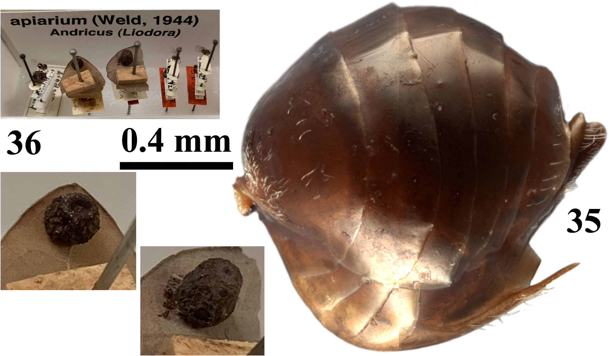
| status |
|
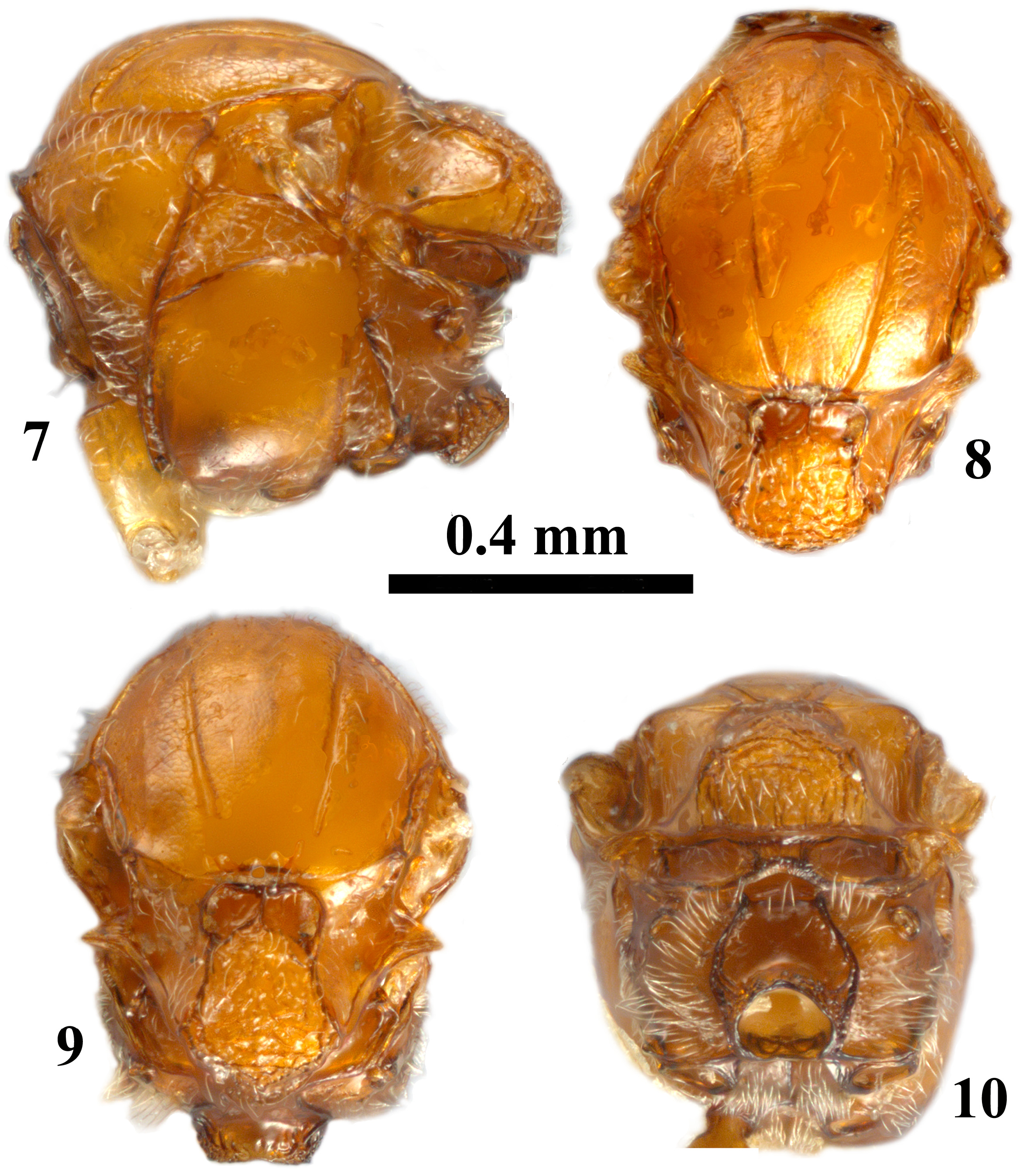
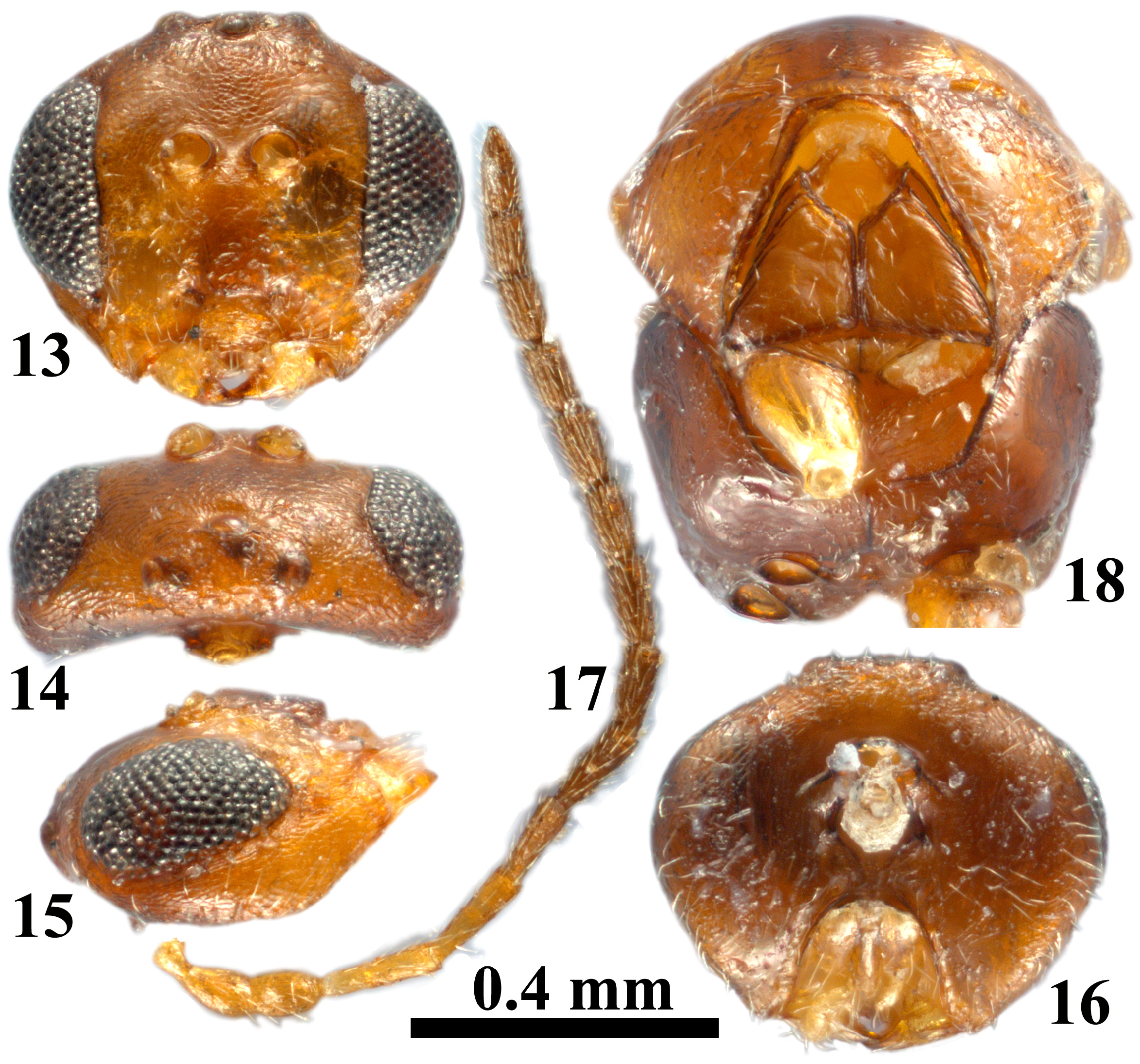
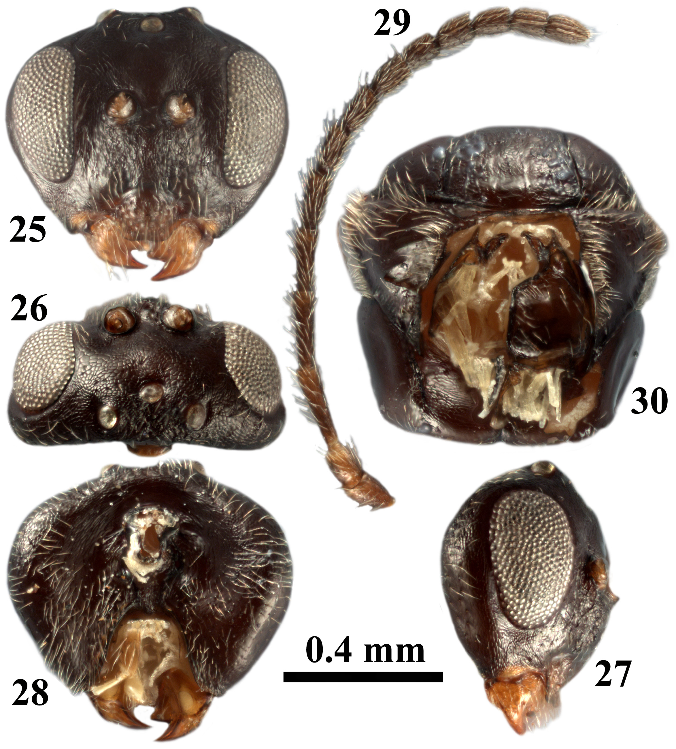
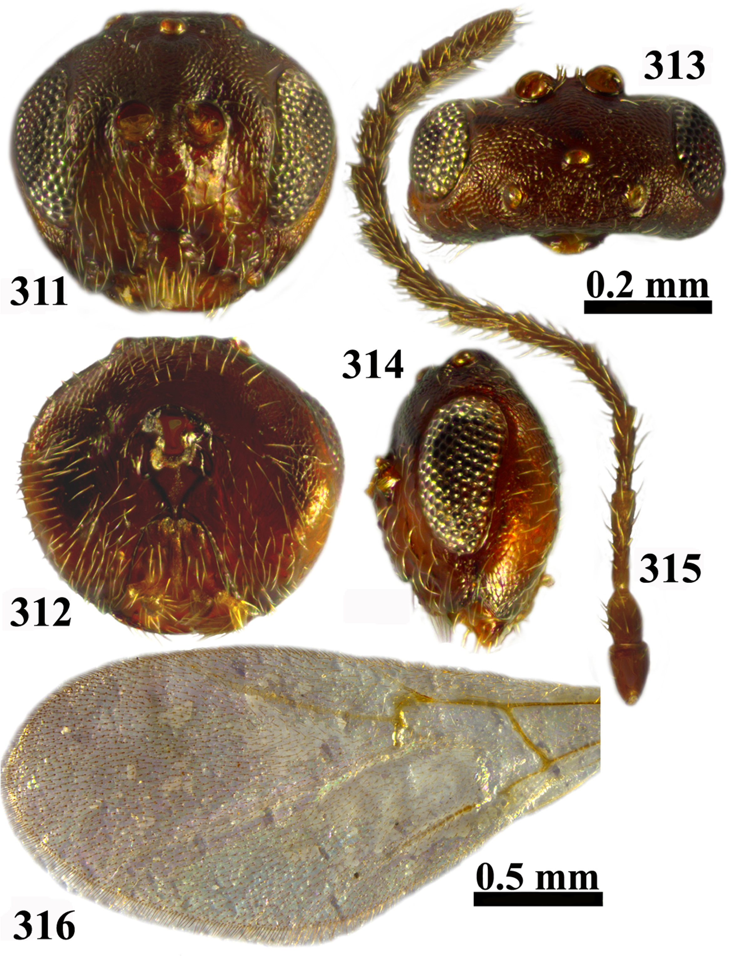
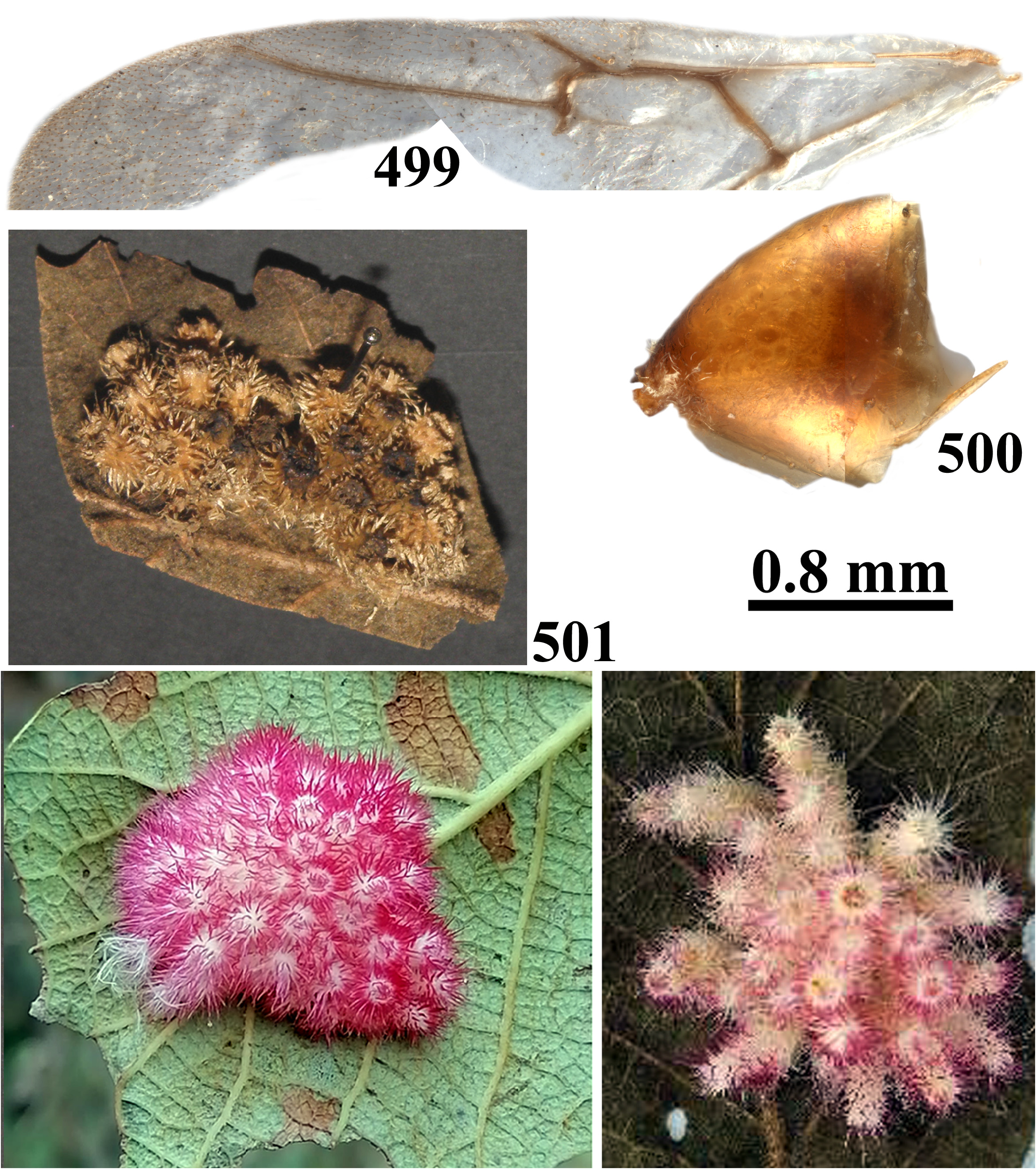
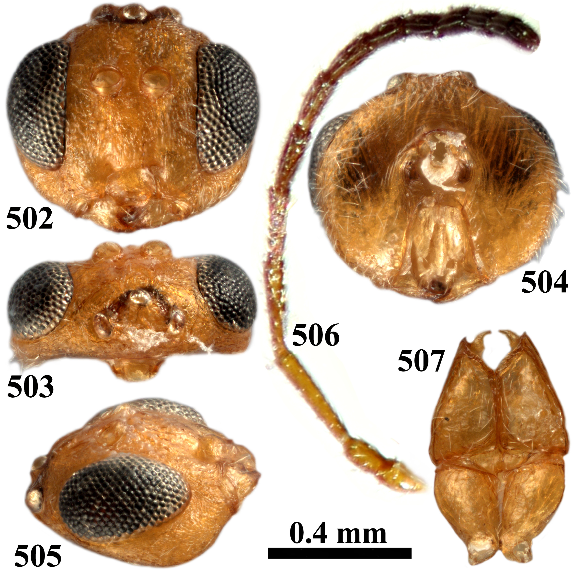
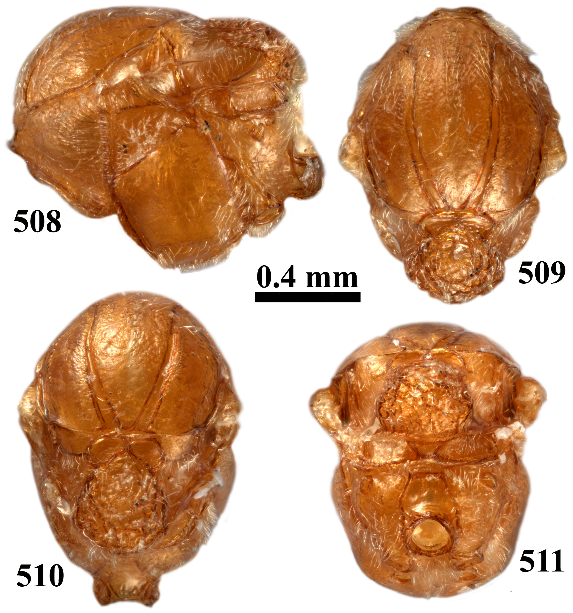
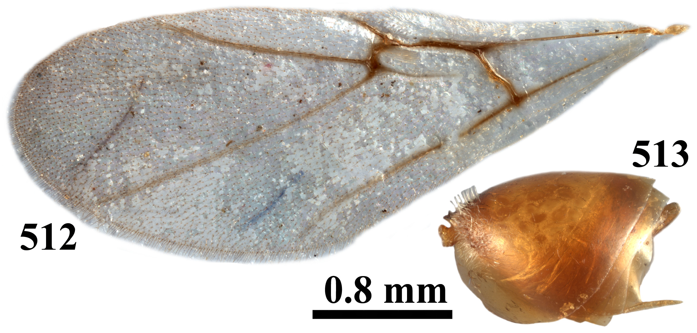
Figs 1–513 View FIGURES 1–6 View FIGURES 7–10 View FIGURES 11–12 View FIGURES 13–18 View FIGURES 19–22 View FIGURES 23–24 View FIGURES 25–30 View FIGURES 31–34 View FIGURES 35–36 View FIGURES 37–42 View FIGURES 43–46 View FIGURES 47–50 View FIGURES 51–56 View FIGURES 57–60 View FIGURES 61–67 View FIGURES 68–71 View FIGURES 72–77 View FIGURES 78–81 View FIGURES 82–83 View FIGURES 84–89 View FIGURES 90–93 View FIGURES 94–96 View FIGURES 97–102 View FIGURES 103–106 View FIGURES 107–109 View FIGURES 110–115 View FIGURES 116–120 View FIGURES 121–123 View FIGURES 124–129 View FIGURES 130–133 View FIGURES 134–135 View FIGURES 136–141 View FIGURES 142–145 View FIGURES 146–147 View FIGURES 148–149 View FIGURES 150–154 View FIGURES 155–158 View FIGURES 159–161 View FIGURES 162–167 View FIGURES 168–171 View FIGURES 172–173 View FIGURES 174–178 View FIGURES 179–183 View FIGURES 184–188 View FIGURES 189–194 View FIGURES 195–198 View FIGURES 199–202 View FIGURES 203–208 View FIGURES 209–212 View FIGURES 213–215 View FIGURES 216–221 View FIGURES 222–225 View FIGURES 226–227 View FIGURES 228–233 View FIGURES 234–237 View FIGURES 238–240 View FIGURES 241–249 View FIGURES 250–255 View FIGURES 256–261 View FIGURES 262–265 View FIGURES 266–268 View FIGURES 269–273 View FIGURES 274–278 View FIGURES 279–283 View FIGURES 284–292 View FIGURES 293–297 View FIGURES 298–299 View FIGURES 300–303 View FIGURES 304–307 View FIGURES 308–310 View FIGURES 311–316 View FIGURES 317–320 View FIGURES 321–323 View FIGURES 324–327 View FIGURES 328 View FIGURES 329–334 View FIGURES 335–338 View FIGURES 339–341 View FIGURES 342–347 View FIGURES 348–351 View FIGURES 352–353 View FIGURES 354–359 View FIGURES 360–363 View FIGURES 364–366 View FIGURES 367–369 View FIGURES 370–371 View FIGURES 372–376 View FIGURES 377–380 View FIGURES 381–383 View FIGURES 384–388 View FIGURES 389–392 View FIGURES 393–394 View FIGURES 395–400 View FIGURES 401–404 View FIGURES 405–408 View FIGURES 409–418 View FIGURES 419–425 View FIGURES 426–428 View FIGURES 429–434 View FIGURES 435–438 View FIGURES 439–440 View FIGURES 441–446 View FIGURES 447–450 View FIGURES 451–452 View FIGURES 453–457 View FIGURES 458–461 View FIGURES 462–463 View FIGURES 464–468 View FIGURES 469–472 View FIGURES 473–475 View FIGURES 476–481 View FIGURES 482–485 View FIGURES 486–488 View FIGURES 489–494 View FIGURES 495–498 View FIGURES 499–501 View FIGURES 502–507 View FIGURES 508–511 View FIGURES 512–513
Etymology. Kinsey (1937) did not indicate the etymology of this genus name in the original description. The name Feron could come from either the Greek φέρω (phérō) or Latin fero, both of which are verbs meaning “to carry” in which case its gender would be neuter; it could alternatively come from Φηρῶν which is a mythological name of a centaur, in which case it would be masculine. The suffixes in the epithets of the original species described under the name Feron , correspond to a neuter word, thus it must refer to a verb and not to a noun. Here, all species names’ terminals have been changed to neuter when applicable.
Type species: Feron verutum Kinsey, 1937 (according to the original description).
Kinsey (1937) described this genus with six new Mexican species, Feron tibiale , F. tostum , F. uterinum , F. validum , F. verutum and F. vitreum , which induce crystalline galls on leaves of white oaks in the subgenus Quercus View in CoL (section Quercus View in CoL , subsection Leucomexicana; Denk et al. 2017, Manos & Hipp 2021). Kinsey (1937: 70) commented that Feron should include numerous species that had previously been assigned to Andricus View in CoL and other Western Palearctic genera; in addition, two other genera, Dros and Druon , were described in the same paper and Kinsey mentioned that the limits of all three genera would be determined in a monographic revision of the entire group. However, this revision was never published. Moreover, Kinsey constantly compared his new Feron species with Andricus tecturnarum but never transferred that species to Feron ; our phylogenetic study shows that A. tecturnarum falls within the genus Feron . According to Weld (1951; 1952a), morphologically Feron closely resembles Andricus View in CoL , and Weld (1951: 632) synonymized both genera. However, the original description of Feron does not mention several important characters that differentiate it from Andricus View in CoL so for this reason, Feron is re-described below.
Diagnosis. Feron has the lower face with two smooth or almost smooth areas between the clypeus and eye, the malar sulcus absent; gena broadened or not behind the eye in frontal view, toruli usually located in the upper half of the head, the antenna always with 11–12 flagellomeres, the notaulus is usually complete, the mesopleuron is smooth or with a few vertical carinae on the most posterodorsal part of the speculum or with weak longitudinal carinae anteriorly; the transscutal articulation is present; lateral propodeal carinae bent outwards, sometimes strongly; the prominent part of the ventral spine of the hypopygium relatively long, usually around 4.0–6.0× as long as broad. According to these characters, Feron is similar to some sexual Andricus species. Andricus do not exhibit smooth areas on the lower face, toruli are always located at mid-height of the head, and the prominent part of the ventral spine of the hypopygium is usually shorter (around 2.0–3.5× as long as broad). Exceptionally, Andricus with longer ventral spines of the hypopygium have lateral propodeal carinae subparallel or bent slightly outwards and the mesoscutum is completely smooth or partially delicately alutaceous; even in some sexual female Andricus the antenna has 13 flagellomeres. Biologically, an important character to differentiate Feron from Andricus is the galls: one group of Feron species induces pubescent, crystalline or conical galls with rigid extensions on leaves, while the galls of Andricus never form gregarious pubescent masses on leaves; also, other lineages of Feron species induce disc-shaped, often star-like, spangle galls on leaves, a morphology that never occurs in Andricus . A few sexual-generation Andricus species, for example, the Western Palearctic A. quercusramuli ( Linnaeus, 1761) , induce Feron -like pubescent galls but only on catkins; in addition, those sexual females differ from Feron in the absence of smooth areas on the lower face, the mesopleuron is uniformly and very delicately transversally striate and the prominent part of the ventral spine of the hypopygium is usually short. The galls of some Feron species are similar to those induced by Druon species, but in Druon galls are covered in softer, more wool-like pubescent compared to the more crystalline hairs on Feron galls. The adults of Druon differ from Feron by the mesopleuron striate, and the lower face is entirely sculptured without smooth areas. Finally, some species of Feron can resemble Dros , but in Dros the frons is completely smooth (always sculptured in Feron ) and the galls are significantly different.
Re-description. Females. Body yellowish to light brown, chestnut brown or dark brown, legs always lighter; antenna light brown to dark brown, with scape, pedicel and flagellomeres from F1 until F4 lighter than subsequent flagellomeres.
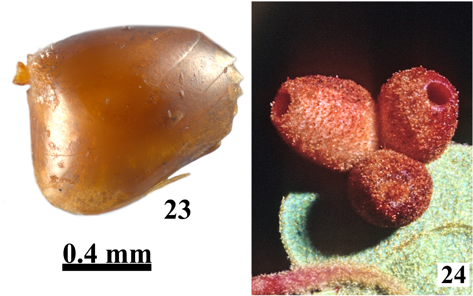
Head trapezoid, rounded, slightly ovate or semiquadrangular, with sparse white setae, denser on lower face, 1.1–1.2× as broad as high, slightly broader than mesosoma in small specimens in frontal view; 1.6–2.3× as broad as long in dorsal view. Gena alutaceous-reticulate, not broadened or very slightly broadened behind eye in frontal view, narrower than the transverse diameter of the eye in lateral view. Malar space alutaceous-reticulate, with striae radiating from clypeus sometimes reaching the eye, malar sulcus absent; eye 1.7–6.1× as high as the length of malar space. Inner margins of eyes parallel, converging, or slightly divergent. POL 1.3–3.0× as long as OOL (longer in males), OOL 1.0–3.3× as long as the diameter of lateral ocellus and slightly shorter to 2.0× as long as LOL, all ocelli ovate, of the same size. Transfacial distance longer or slightly shorter than the height of eye (shorter in some sexual forms); toruli usually located in the upper half of head and then frons shorter than lower face, the diameter of antennal torulus 1.0-2.0× as long as the distance between them, distance between torulus and eye around 1.0–1.5× as long as diameter of torulus; lower face smooth or slightly alutaceous between clypeus and eye, glabrous or with sparse white setae; slightly elevated median area alutaceous to smooth, with a few setae. Clypeus trapezoid, rectangular or quadrangular, smooth, or delicately coriaceous or striate, glabrous or with a few long setae scattered all over; ventrally rounded, emarginate, with or without median incision; anterior tentorial pit small, rounded, indistinct, epistomal sulcus distinct, clypeo-pleurostomal line well impressed. Frons alutaceous-reticulate or imbricate; interocellar area and vertex alutaceous-reticulate, without striae or setae; area under central ocellus impressed, alutaceous; occiput, postocciput and postgena alutaceous-reticulate, with few setae; posterior tentorial pit large, elongated, area below impressed; occipital foramen as high as height of postgenal bridge; hypostomal carina emarginate, continuing into strong postgenal sulci which diverge strongly toward occipital foramen, postgenal bridge anteriorly slightly broader than occipital foramen. Antenna longer than head+mesosoma, with 11–12 flagellomeres (13 in males); pedicel longer than broad; F1 1.5–2.6× as long as pedicel and from equal with to 1.4× as long as F2; F2 equal to or longer than F3; all subsequent flagellomeres shorter, sometimes thicker; placodeal sensilla from F2 to F 5 in females (in all flagellomeres in males); F 1 in males with a variable shape, sometimes 1.6× as long as F2.
Mesosoma as long as high, with a few white setae along propleura. Pronotum smooth, glabrous or more or less pubescent laterally, foveolate or not along propleuron, laterally smooth or with delicate parallel striae in anteroposterior part; propleuron smooth, glabrous or setose. Mesoscutum as long as broad or slightly longer than broad (greatest width measured across mesoscutum level with base of tegulae), uniformly delicately coriaceous or alutaceous to rugose-reticulate, sometimes with smooth areas and punctures (smooth in some sexual forms). Notaulus complete or not, deep or shallow, posteriorly converging and usually broader than anteriorly, bottom smooth, glabrous; in most posterior section the distance between notauli shorter than distance between notaulus and side of mesoscutum. Anterior parallel line indistinct or distinct, impressed, smooth, glabrous, reaching at most 1/2 length of mesoscutum; parapsidal line indistinct or marked with broad smooth, glabrous stripe; median mesoscutal line absent or in the form of a short triangle; parascutal carina broad, reaching notaulus. Mesoscutellum ovate or trapezoid, longer than broad, broader at posterior end; disc of mesoscutellum uniformly rugoso-coriaceous, reticulate or smooth in central part, rugose in most posterior section and along sides, with some longitudinal rugae, overhanging metanotum. Mesoscutellar foveae present, divided by a rugose elevated central area or fused in the form of an anterior transverse impression, with smooth, glabrous bottom. Mesopleuron entirely smooth or with a few vertical carinae in most posterodorsal section, glabrous or with setae only along ventral edge; mesopleural triangle delicately coriaceous, glabrous or with white setae; dorsal and lateral axillar areas smooth, with or without dense setae; axillula with delicate parallel longitudinal striae; subaxillular bar smooth, glabrous, triangular, posteriorly as high as height of metanotal trough; metapleural sulcus reaching mesopleuron at half of its height, in some species slightly higher or lower, lower part of sulcus delimiting broad triangular coriaceous area; upper part of sulcus either distinct, separating smooth, glabrous area or indistinct. Metascutellum coriaceous or smooth, glabrous, as high as height of smooth, glabrous ventral impressed area; metanotal trough smooth, glabrous or with short setae; central propodeal area lyre-shaped, smooth, without or with only a few strong short longitudinal rugae; lateral propodeal carinae strong, broad and high, bent outwards in posterior 1/3; lateral propodeal area smooth, rarely alutaceous, glabrous or with long white setae, each seta with piliferous point at the base. Nucha with parallel longitudinal sulci dorsally and laterally or with a net of short irregular rugae. Tarsal claws with basal lobe.
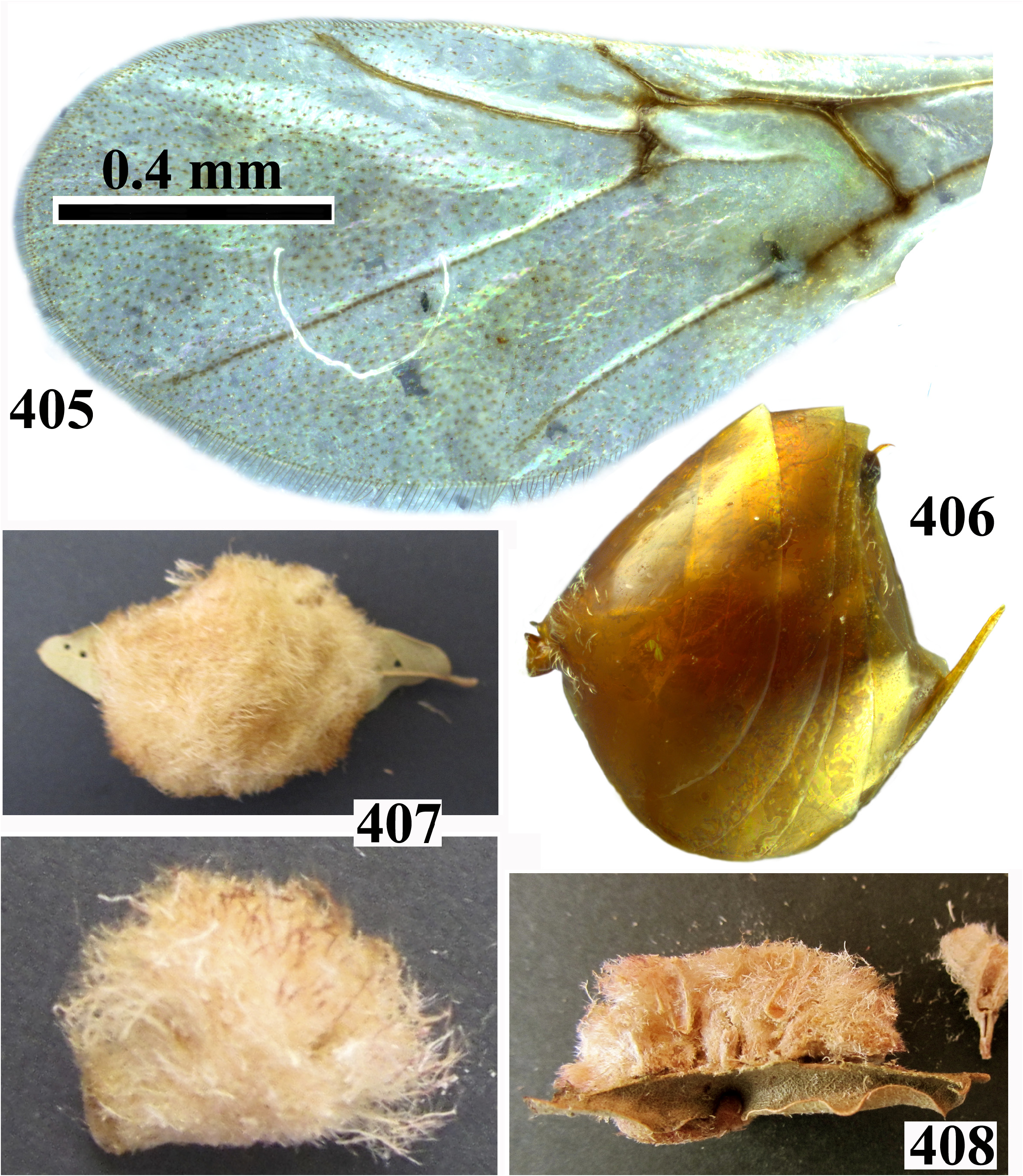
Fore wing longer than body, except in brachypterous forms, hyaline, with dense cilia on margin, veins brown, radial cell open, 3.5–5.2× as long as broad; Rs and R1 not or nearly reaching wing margin; areolet triangular, enclosed by distinct veins. Rs+M distinct along entire length or along 1/3 to 2/3 of distance between areolet and basalis, its projection reaching basalis at half of its height.
Metasoma longer than head+mesosoma; 2nd metasomal tergum extending to 1/3–5/6 length of metasoma in dorsal view, with patch of dense white setae anterolaterally, with or without micropunctures; all subsequent terga and hypopygium with or without micropunctures; prominent part of ventral spine of hypopygium variable, 2.0–8.0× as long as broad in ventral view, with or without a few short white setae ventrally.
Body length 0.9–3.1 mm.
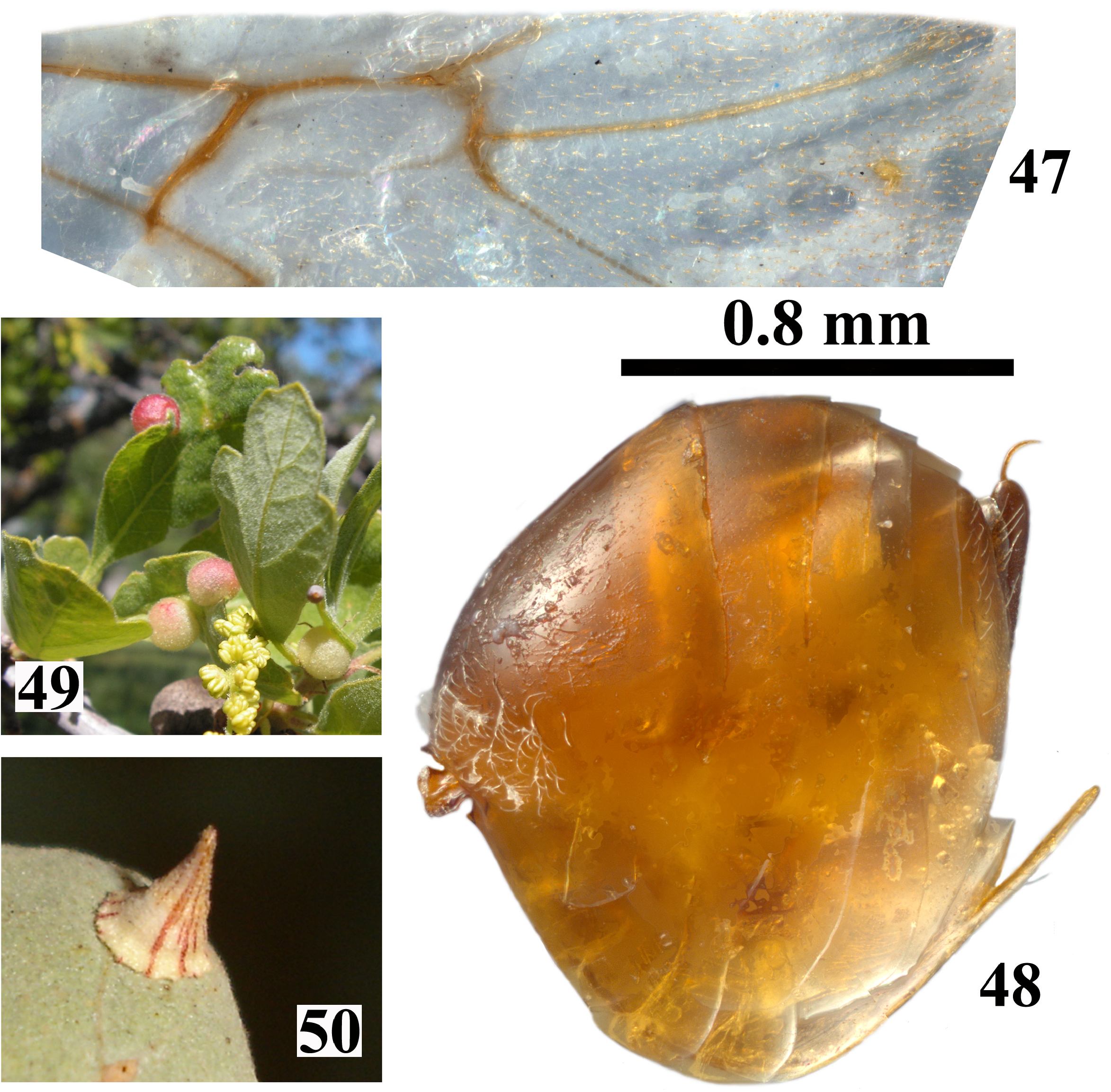
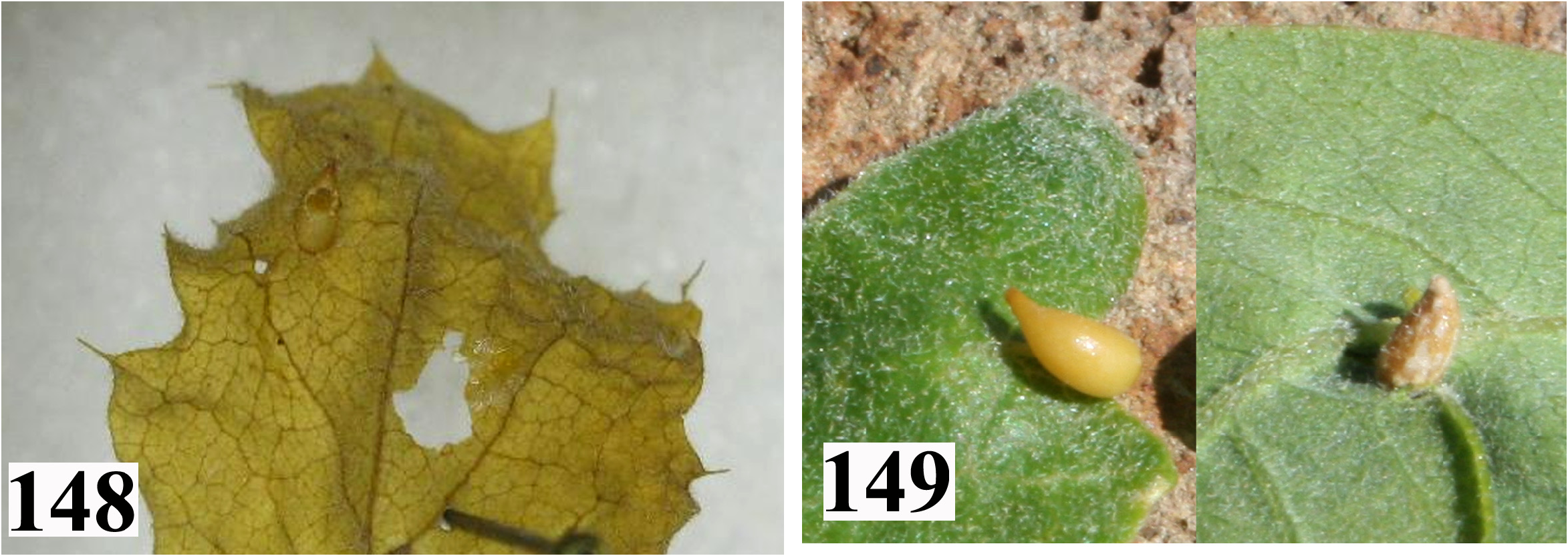
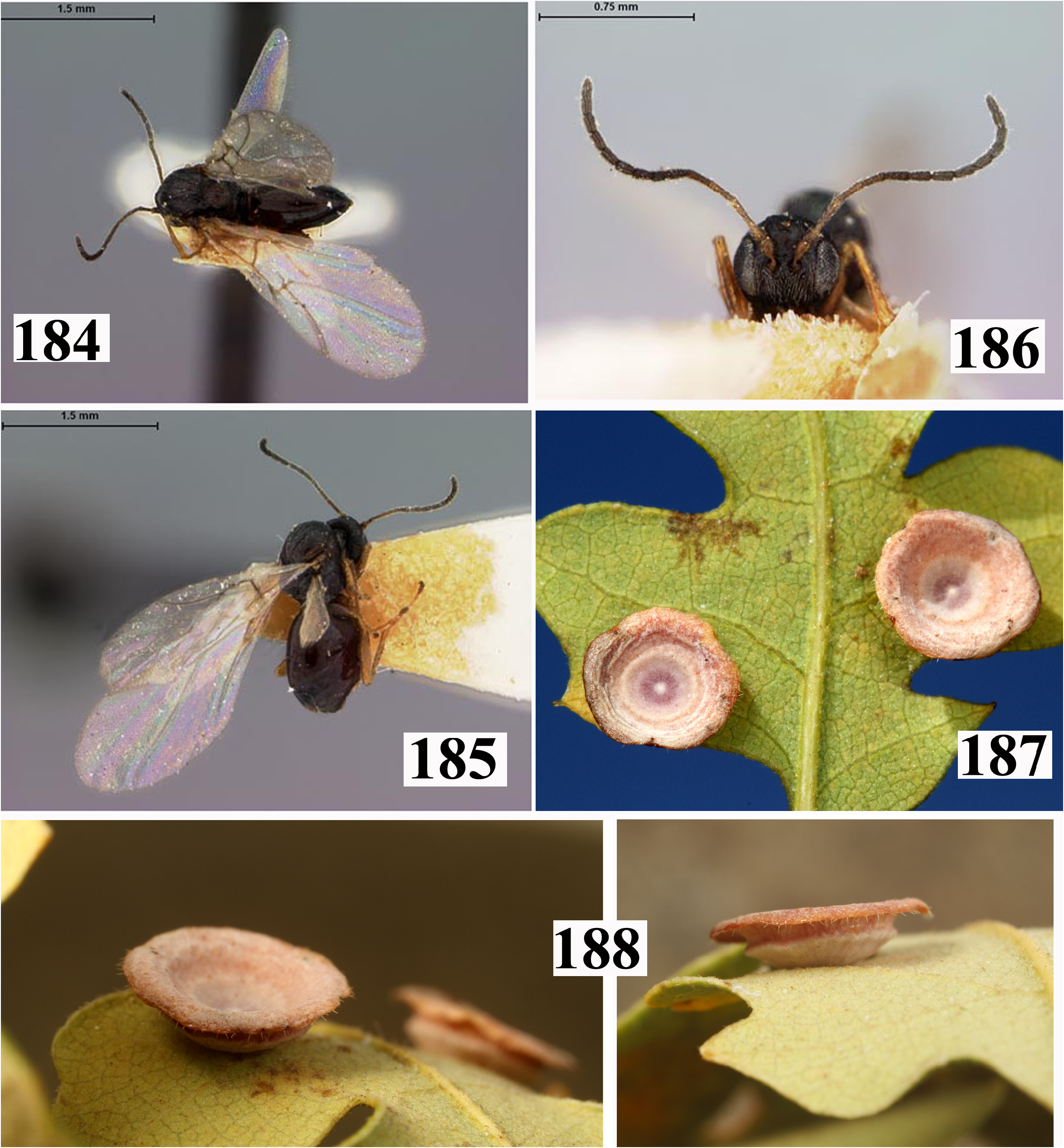
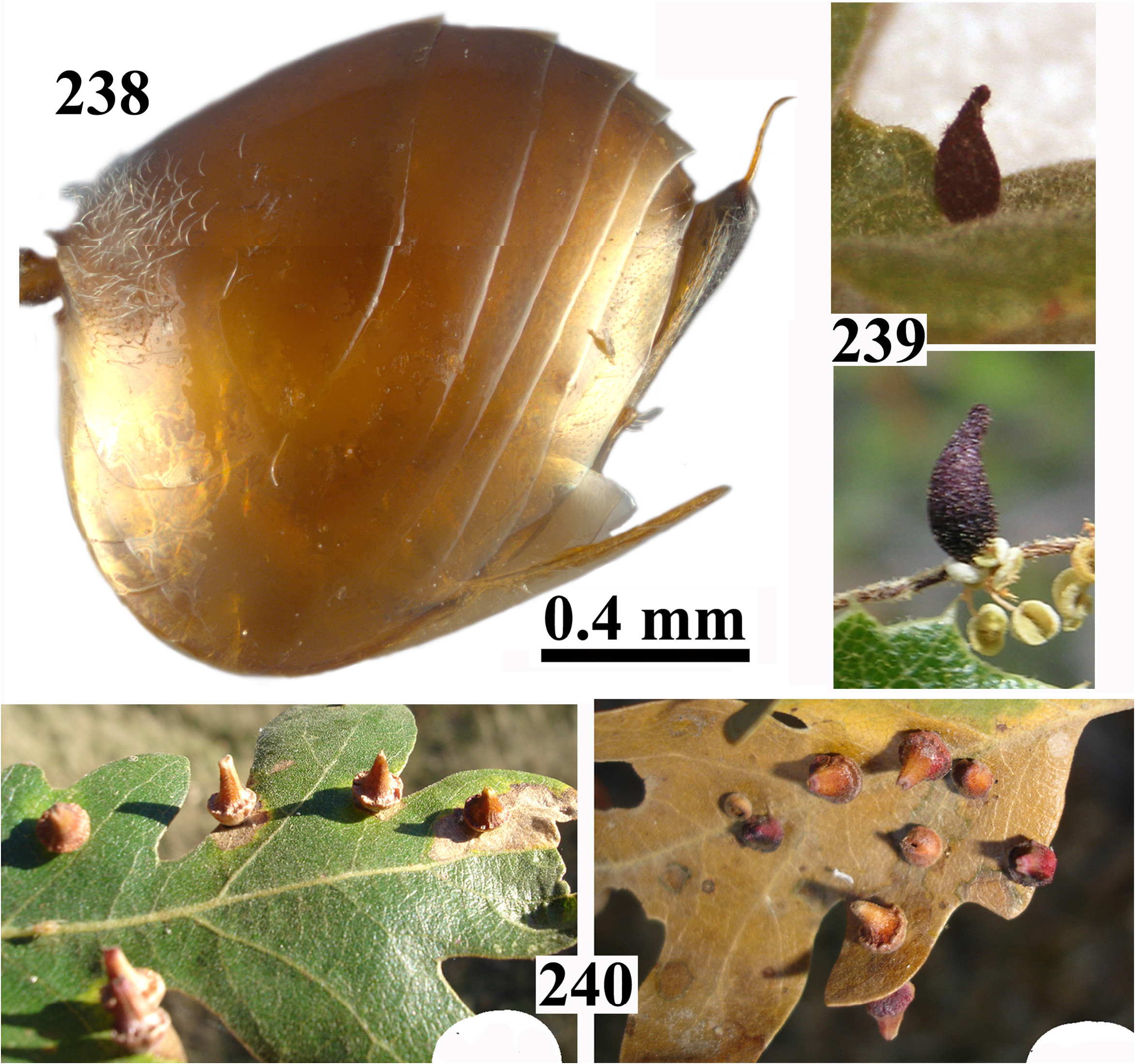
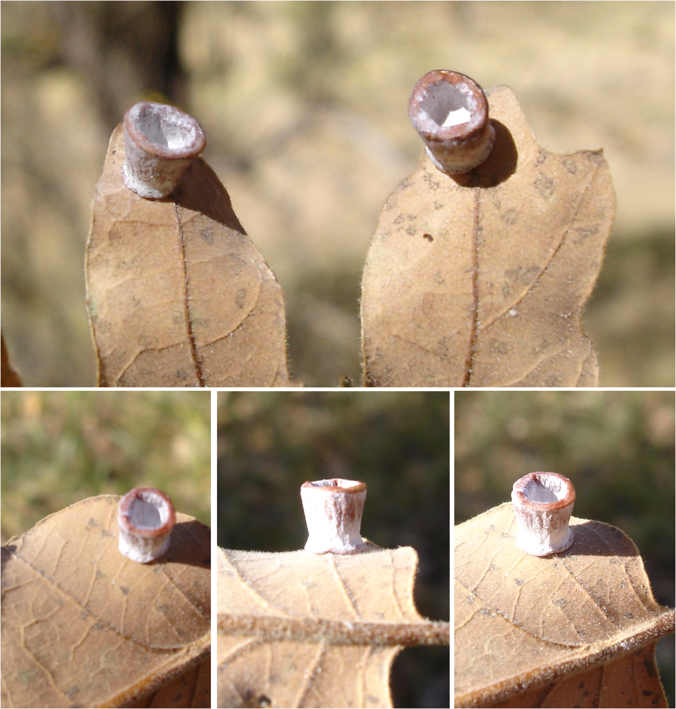
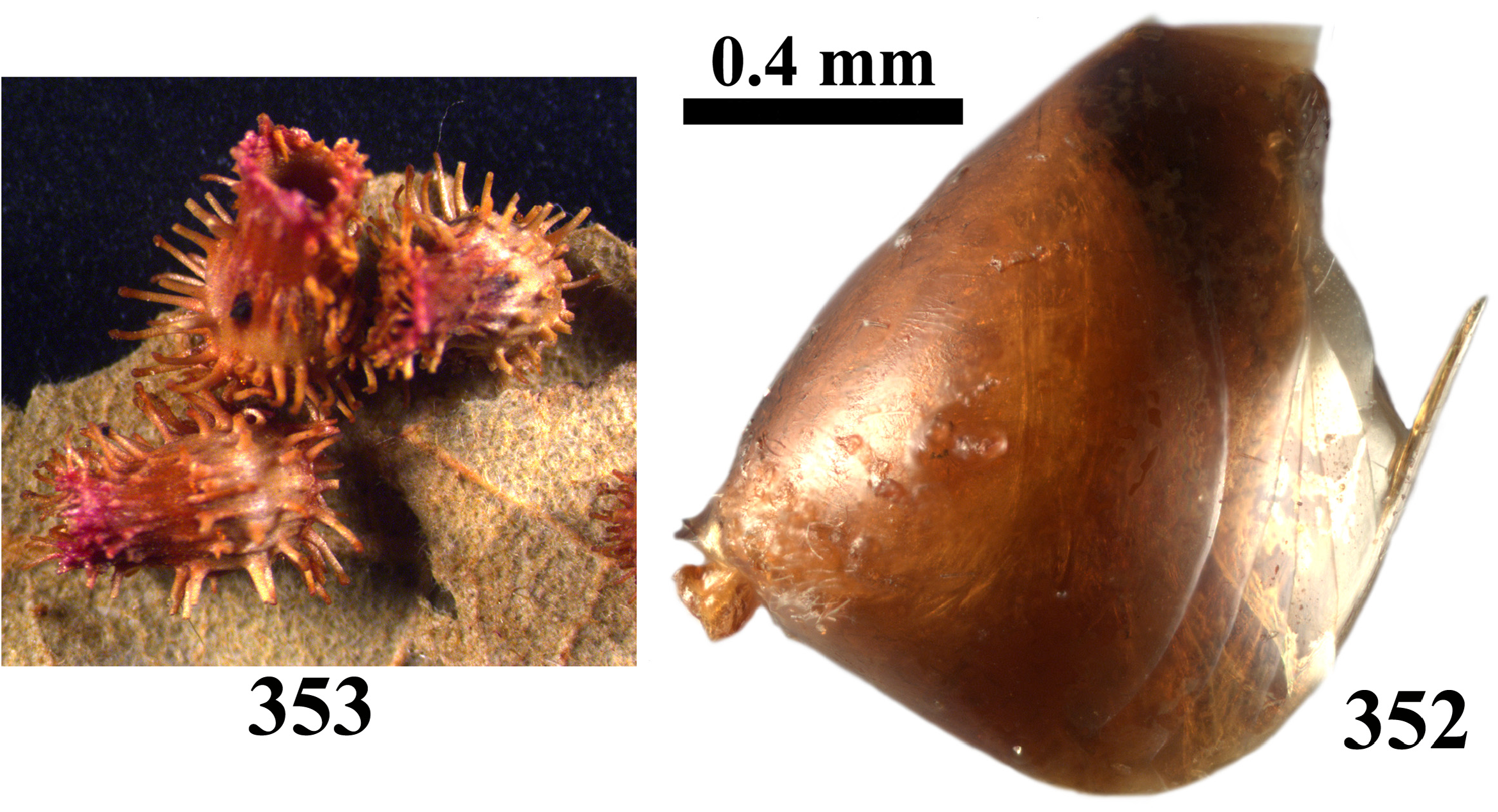
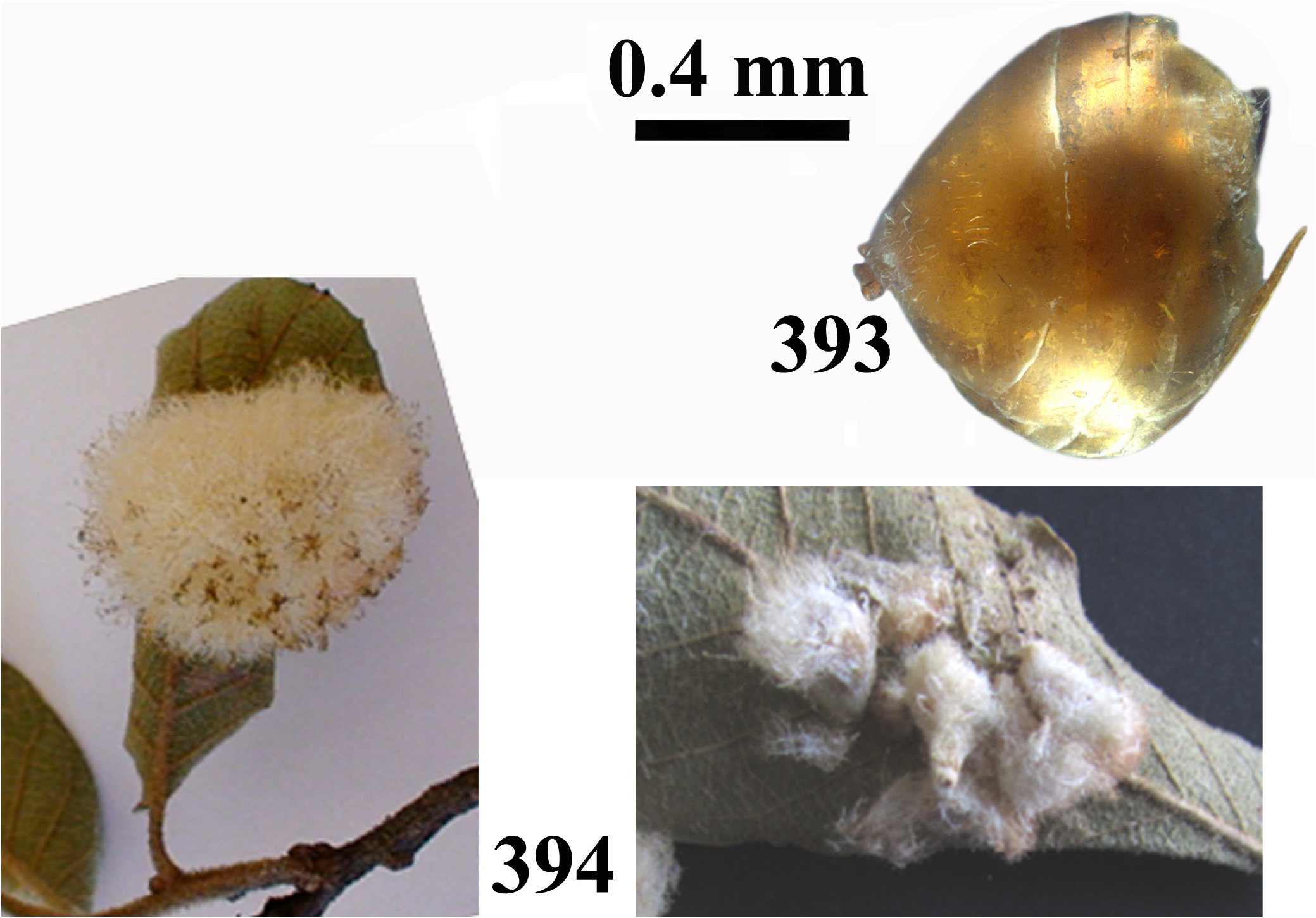
Gall. The asexual generation’s galls are always found on leaves but have a wide range of morphologies that can mainly be divided into two subgroups: One with pubescent masses, crystalline (resembling mineral crystalline structures, usually erected tubular structures with short rigid and brittle filaments radiating from it) or conical galls with rigid extensions on leaves; and a second with disc-shaped, often star-like, spangle galls (disc-shaped with a central round protrusion). The sexual generation’s gall
Biology. Adults galling on oaks from Section Quercus . The galls of the asexual generation mature during autumn, and adults overwinter in the galls and emerge between late January and May. The sexual generation occurs in late spring. The galls maduration and emergence of adults spans between April and May.
Distribution. Southern Canada, USA, and Mexico.
Descriptions of the sexual females and males are given in the species section below.
Thirty-four species are currently recognized within Feron :
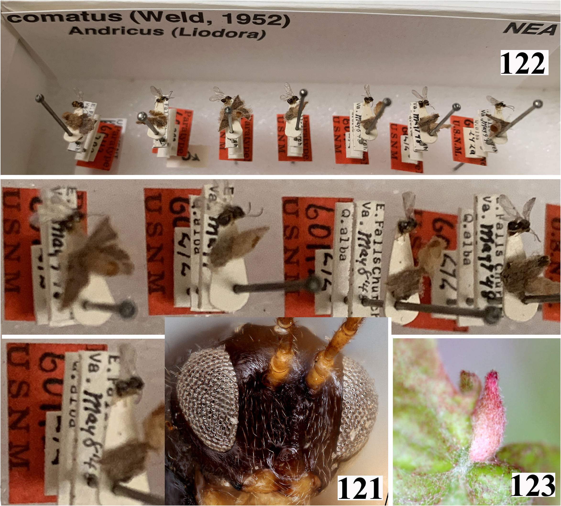
F. albicomus (Weld, 1952) , comb. nov., F. amphorus ( Weld, 1926) , comb. nov., F. apiarium ( Weld, 1944) , comb. nov., F. atrimentum ( Kinsey, 1922) , comb. nov., F. bakkeri ( Lyon, 1984) , comb. nov., F. caepula ( Weld, 1926) , comb. nov., F. californicum ( Beutenmueller, 1911) , comb. nov., F. clarkei ( Bassett, 1890) , comb. nov., F. comatum (Weld, 1952) , comb. nov., F. crystallinum ( Bassett, 1900) , comb. nov., F. cylindratum ( Kinsey, 1937) , comb. nov., F. discale ( Weld, 1926) , comb. nov., F. discularis ( Weld, 1926) , comb. nov., F. dumosae ( Weld, 1957) , comb. nov., F. gigas ( Kinsey, 1922) , comb. nov., F. izabellae Melika, Nicholls & Stone , sp. nov., F. kingi ( Bassett, 1900) , comb. nov., F. parmula ( Bassett, 1900) , comb. nov., F. pattersonae ( Fullaway, 1911) , comb. nov. (= Andricus pedicellatus ( Kinsey, 1922) , syn. nov.), F. roberti Melika, Nicholls & Stone , sp. nov., F. rucklei Melika, Nicholls & Stone , sp. nov., F. scutellum ( Weld, 1930) , comb. nov., F. serranoae Pujade-Villar & Cuesta-Porta , sp. nov., F. splendens ( Weld, 1919) , comb. nov., F. stellare ( Weld, 1926) , comb. nov., F. stellulum ( Burnett, 1974) , comb. nov., F. sulfureum ( Weld, 1926) , comb. nov., F. syndicorum Pujade-Villar & Cuesta-Porta , sp. nov., F. tecturnarum ( Kinsey, 1920) , comb. nov., F. tetyanae Melika , sp. nov., F. tibiale Kinsey, 1937 , comb. rev. (= F. tostum Kinsey, 1937 , syn. nov., Feron uterinum Kinsey, 1937 , syn. nov.), F. tubifaciens ( Weld, 1926) , comb. nov., F. verutum Kinsey, 1937 , comb. rev. and F. vitreum Kinsey, 1937 , comb. rev. (= F. validum Kinsey, 1937 , syn. nov.).
Key to Feron species
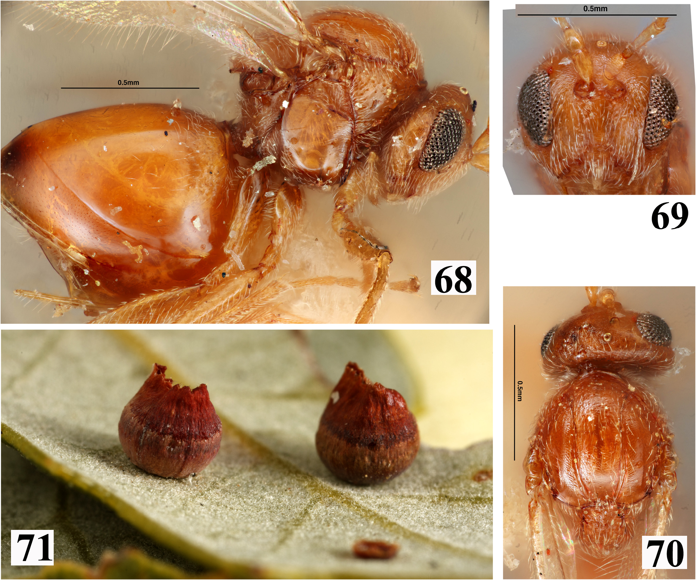
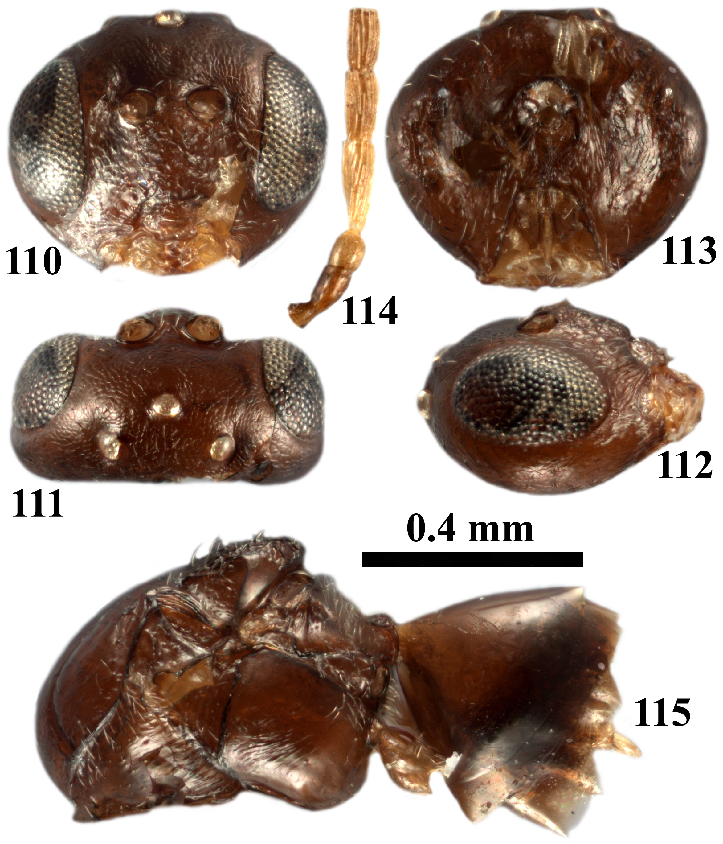
1. Males ( Figs 62–67 View FIGURES 61–67 , 110–115 View FIGURES 110–115 , 146–147 View FIGURES 146–147 , 245–249 View FIGURES 241–249 , 288–292 View FIGURES 284–292 ).................................................... 2
- Females ( Figs 1–11 View FIGURES 1–6 View FIGURES 7–10 View FIGURES 11–12 , 51–61 View FIGURES 51–56 View FIGURES 57–60 View FIGURES 61–67 , 68 View FIGURES 68–71 , 116–120 View FIGURES 116–120 ).................................................................. 9
2. Ocelli strongly elevated above frons in frontal view ( Fig. 62 View FIGURES 61–67 ), OOL shorter than diameter of lateral ocellus and inner margins of eyes subparallel ( Figs 62–63 View FIGURES 61–67 ); mesoscutum uniformly reticulate between notauli in anterior half and laterad to notauli, smooth between notauli in posterior half....................................................... atrimentum comb. nov.
- Ocelli moderately or not elevated above frons; if not then inner margins of eyes strongly converging laterally and/or OOL longer ( Figs 110–111 View FIGURES 110–115 , 146 View FIGURES 146–147 , 245, 247 View FIGURES 241–249 , 288–289 View FIGURES 284–292 ); mesoscutum smooth or partially alutaceous only anteriorly.............. 3
3. Notaulus narrow, distinct only posteriorly, fragmented or absent anteriorly where mesoscutum delicately coriaceous.............................................................................................. gigas comb. nov.
- Notaulus distinct, complete, reaching pronotum............................................................. 4
4. Inner margins of eyes slightly diverging ventrally ( Fig. 146 View FIGURES 146–147 ); mesoscutellar foveae absent, present in the form of a transverse smooth anterior impression, continuing into smooth mesoscutellar disc ( Fig. 146 View FIGURES 146–147 ); body yellowish to amber ( Figs 146–147 View FIGURES 146–147 )................................................................................. crystallinum comb. nov.
- Inner margins of eyes parallel or slightly converging ventrally ( Figs 110 View FIGURES 110–115 , 245 View FIGURES 241–249 , 288 View FIGURES 284–292 ); mesoscutellar foveae present, delimited posteriorly and delimited or not medially; mesoscutellar disc sometimes smooth; body brown to black ( Figs 110–115 View FIGURES 110–115 , 245–248 View FIGURES 241–249 , 288–292 View FIGURES 284–292 )............................................................................................ 5
5. Mesoscutellum with irregular rugae at least in lateral and posterior part; sometimes dorsocentral part smooth, shining, without piliferous points...................................................................................... 6
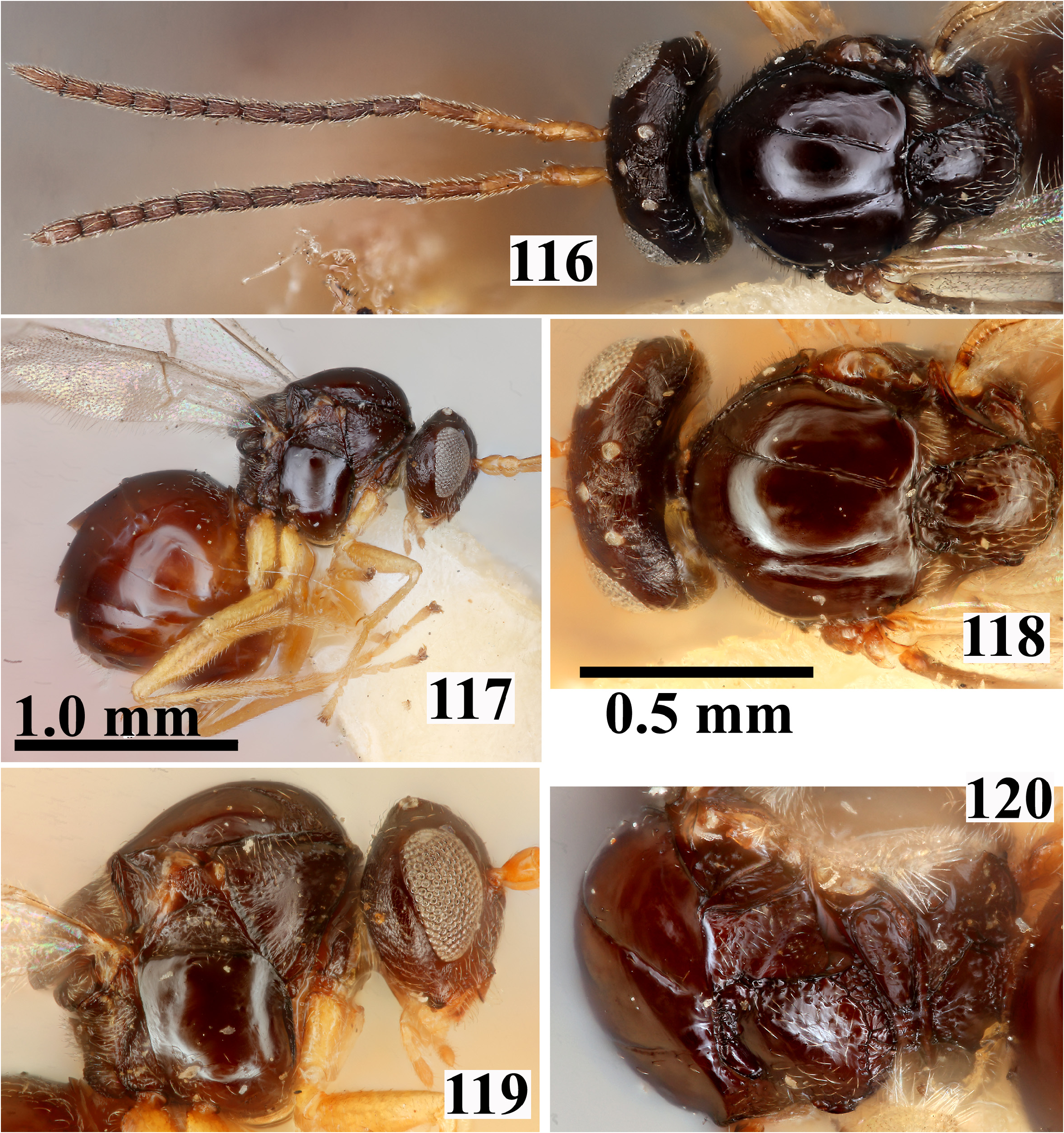
- mesoscutellum uniformly alutaceous, with numerous setae on piliferous points; body brown or chestnut ( Fig. 115 View FIGURES 110–115 ); body black ( Fig. 116 View FIGURES 116–120 )........................................................................................... 7
6. F1 1.6× as long as F2; transfacial distance as long as or slightly shorter than height of eye............ dumosae comb. nov.
- F1 1.3× as long as F2; transfacial distance longer than height of eye............................... clarkei comb. nov.
7. F1 equal in length to scape+pedicel, 1.45× as long as F2, slightly broadened and curved, flagellomeres lighter than scape and pedicel.............................................................................. comatum comb. nov.
- F1 longer than scape+pedicel, 1.3× as long as F2, straight, not broadened and curved, flagellomeres, scape and pedicel uniformly coloured............................................................................................ 8
8. Head ovate in frontal view; internal margin of eyes parallel; space between central elevated area of lower space to lateral margin of eye sculptured; OOL shorter than diameter of lateral ocellus..................................... kingi comb nov.
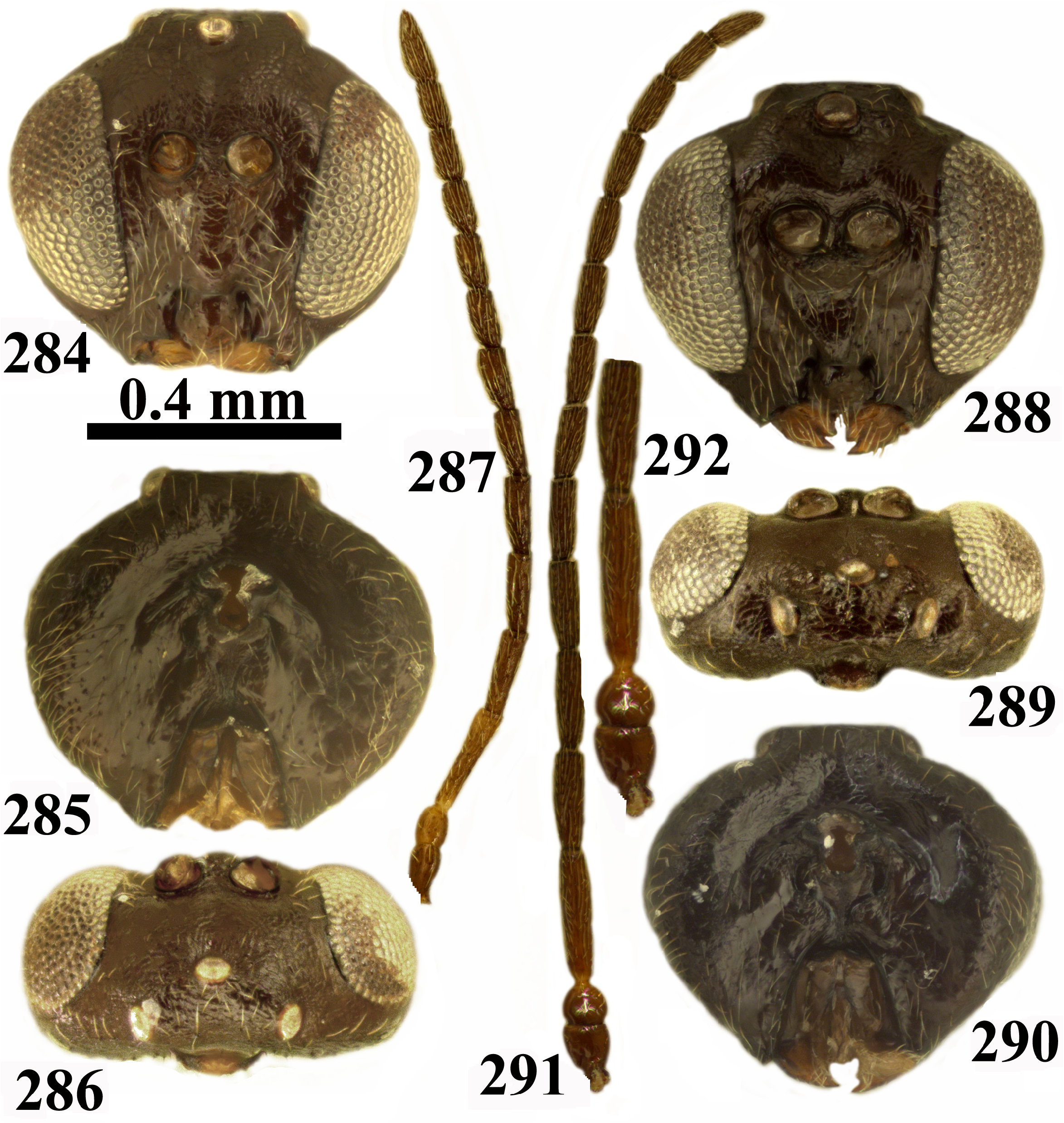
- Head triangular in frontal view ( Fig. 288 View FIGURES 284–292 ); inner margin of eyes converging ventrally; space between central elevated area of lower face to lateral margin of eye smooth ( Fig. 288 View FIGURES 284–292 ); OOL as long as diameter of lateral ocellus ( Fig. 289 View FIGURES 284–292 )................................................................................................ pattersonae comb. nov.
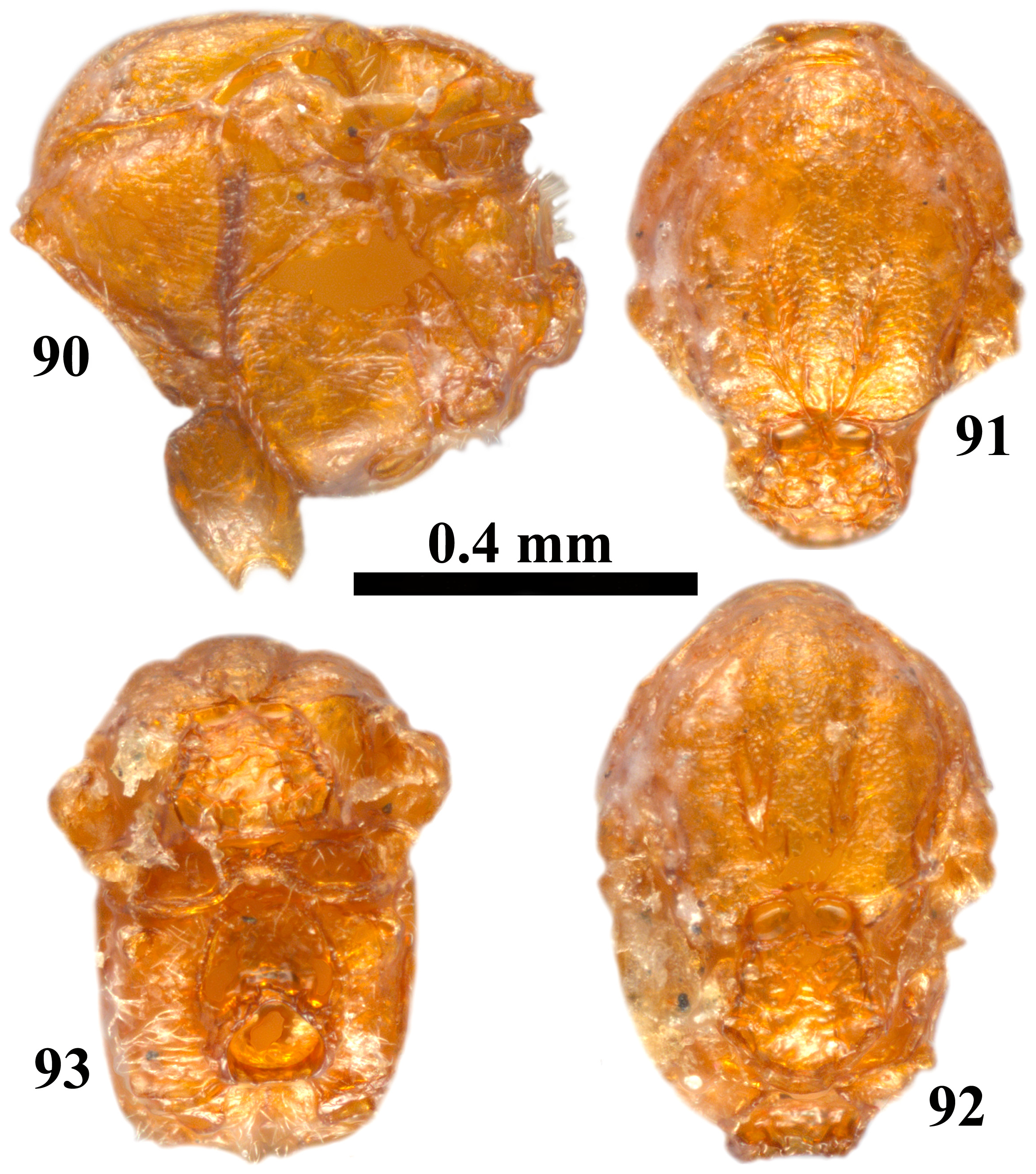
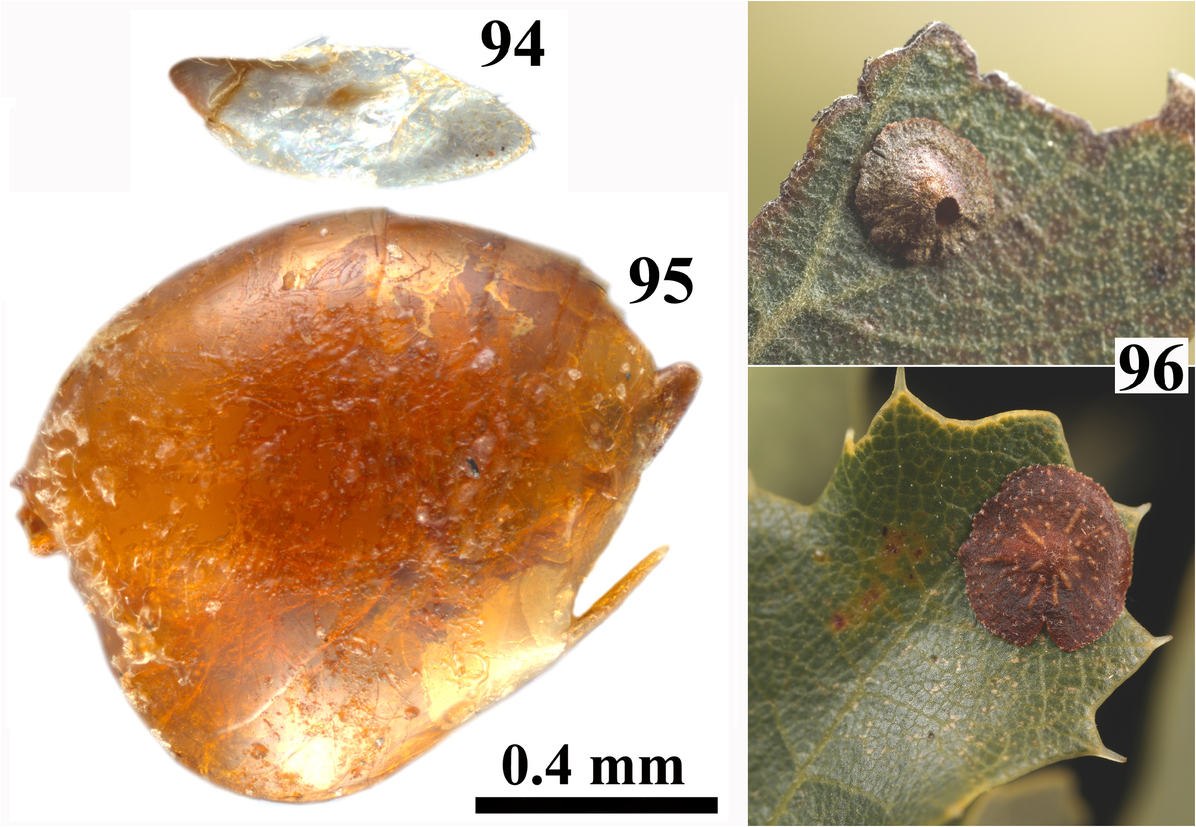
9. Fore wing rudimentary, as long as head+mesosoma but not reaching the mid-length of metasoma, with indistinct veins ( Fig. 94 View FIGURES 94–96 )........................................................................ californicum comb. nov. (asex.)
- Fore wing longer than length of body with distinct veins ( Figs 184–185 View FIGURES 184–188 )........................................ 10
10. Mesoscutum smooth or partially finely alutaceous anteriorly; always glabrous ( Figs 20–21 View FIGURES 19–22 , 32–33 View FIGURES 31–34 , 104–105 View FIGURES 103–106 , 118, 120 View FIGURES 116–120 , 143– 144 View FIGURES 142–145 , 156–157 View FIGURES 155–158 , 169–170 View FIGURES 168–171 , 196–197 View FIGURES 195–198 , 251–252 View FIGURES 250–255 , 295–296 View FIGURES 293–297 )....................................................... 11
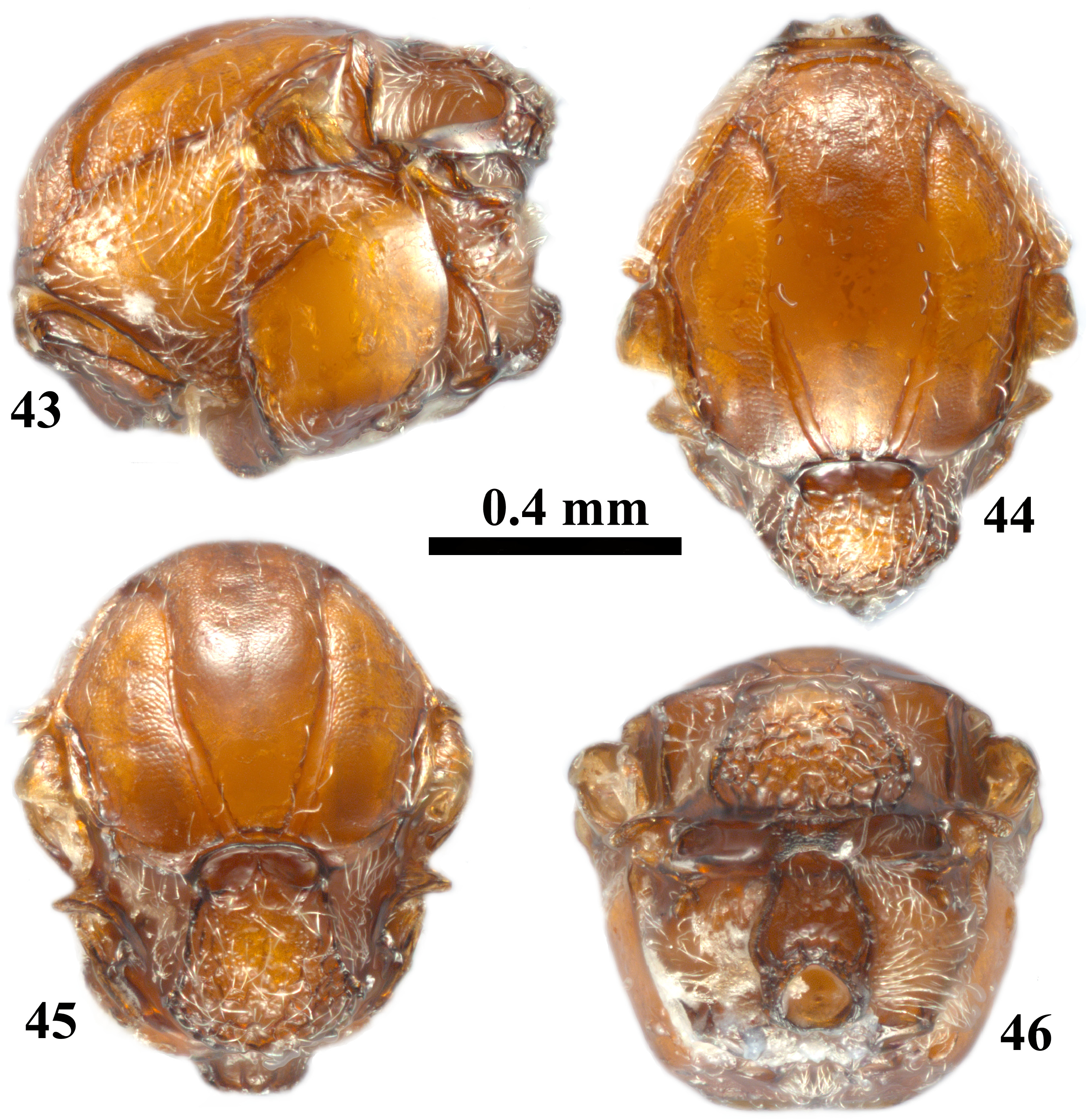
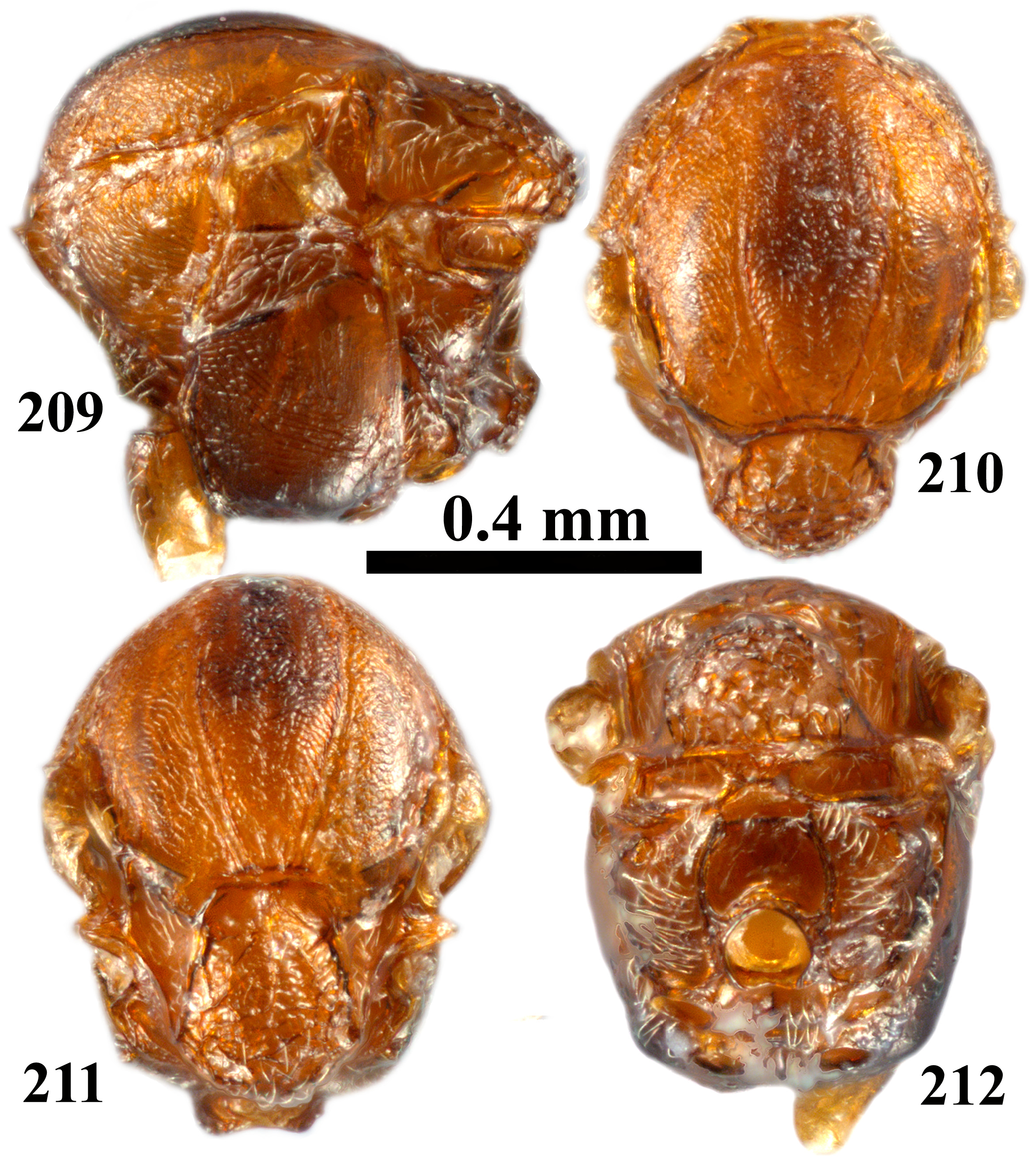
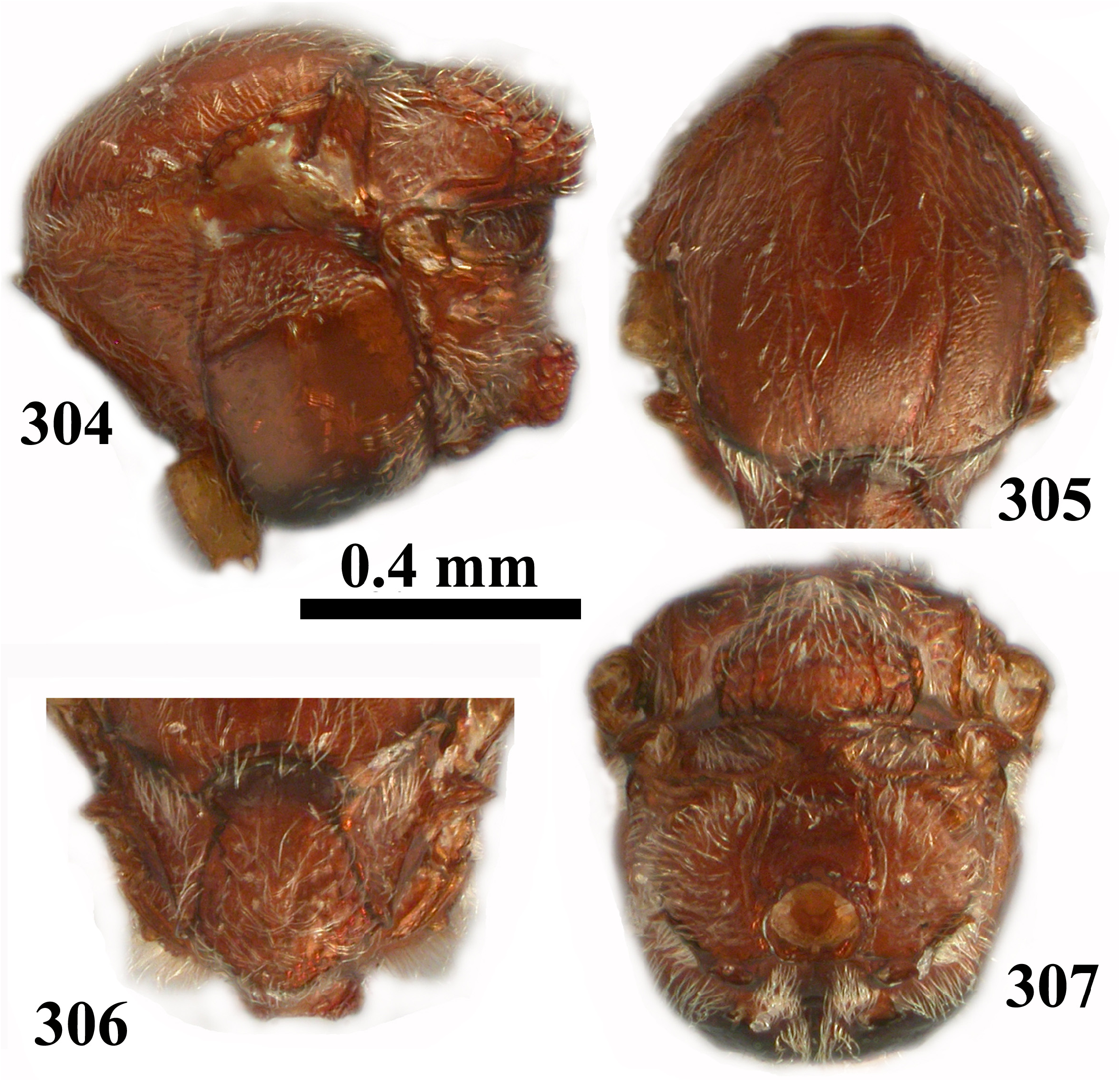
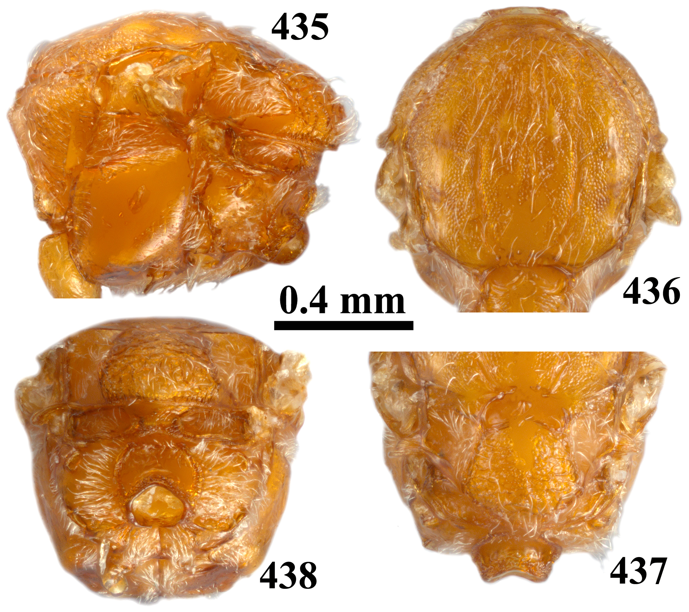
- Mesoscutum alutaceous to coriaceous, rugose-reticulate, reticulate, sometimes with small smooth areas and piliferous points; glabrous or pubescent ( Figs 8–9 View FIGURES 7–10 , 44–45 View FIGURES 43–46 , 70 View FIGURES 68–71 , 131 View FIGURES 130–133 , 210–211 View FIGURES 209–212 , 305 View FIGURES 304–307 , 436 View FIGURES 435–438 )........................................... 21
11. Mesoscutellum uniformly reticulate ( Fig. 157 View FIGURES 155–158 )...................................... cylindratum comb. nov. (asex)
- Mesoscutellum with different sculpture ( Figs 21 View FIGURES 19–22 , 105 View FIGURES 103–106 , 118, 120 View FIGURES 116–120 , 170 View FIGURES 168–171 , 277 View FIGURES 274–278 )....................................... 12
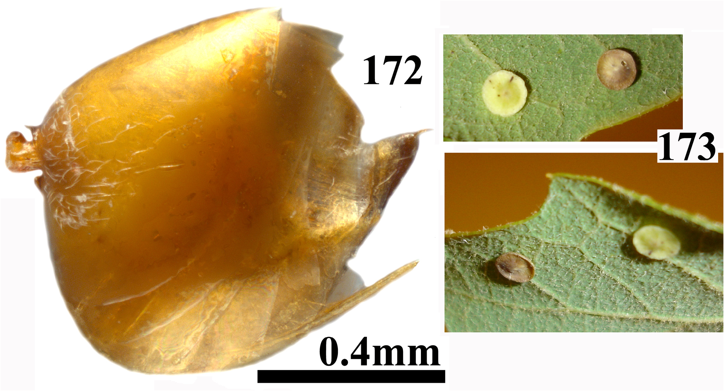
12. Frons bulging in frontal view ( Fig. 162 View FIGURES 162–167 ); inner margins of eyes strongly converging ventrally ( Fig. 162 View FIGURES 162–167 ); mesoscutellum in central part smooth, glabrous, posteriorly and laterally rugose ( Figs 170–171 View FIGURES 168–171 ); body yellowish to reddish brown ( Figs 162– 172 View FIGURES 162–167 View FIGURES 168–171 View FIGURES 172–173 )............................................................................ discale comb. nov. (asex)
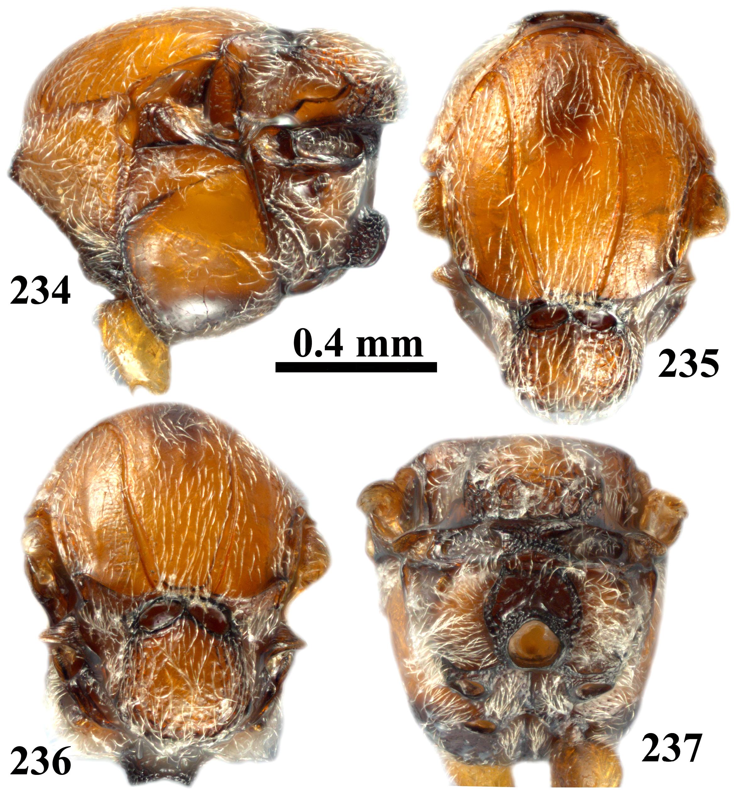
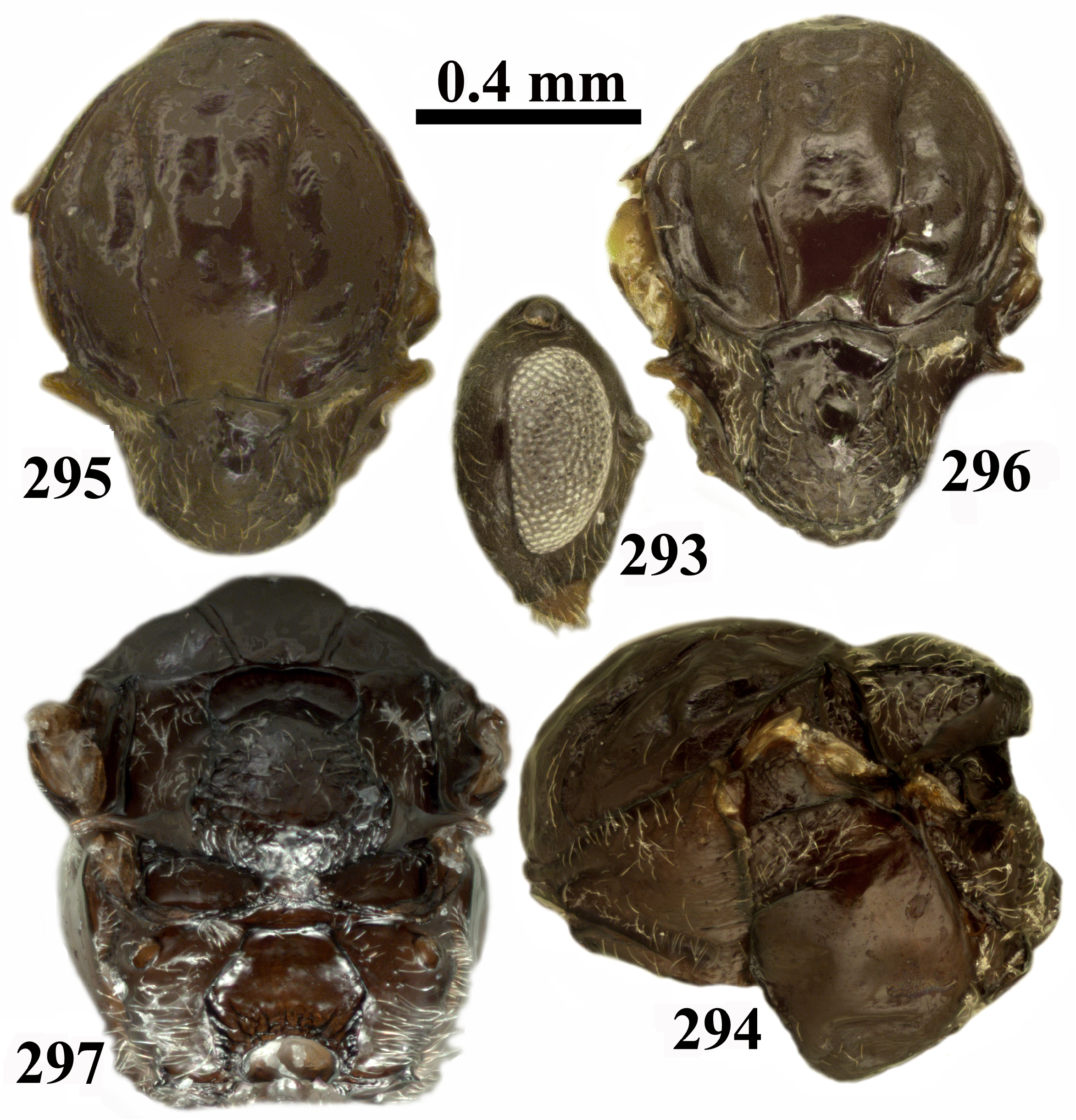
- Frons flat, not or only slightly bulging in frontal view ( Fig. 121 View FIGURES 121–123 ); inner margins of eyes parallel or only slightly converging ventrally; mesoscutellum rugose and/or with piliferous points ( Figs 116, 118, 120 View FIGURES 116–120 ); darker specimens ( Figs 13–23 View FIGURES 13–18 View FIGURES 19–22 View FIGURES 23–24 , 25–35 View FIGURES 25–30 View FIGURES 31–34 View FIGURES 35–36 , 97–106 View FIGURES 97–102 View FIGURES 103–106 , 116–121 View FIGURES 116–120 View FIGURES 121–123 , 241–243 View FIGURES 241–249 , 250–253, 254 View FIGURES 250–255 , 293–299 View FIGURES 293–297 View FIGURES 298–299 )........................................................ 13
13. mesoscutellum uniformly alutaceous with numerous setae on piliferous points ( Figs 33 View FIGURES 31–34 , 120 View FIGURES 116–120 , 252 View FIGURES 250–255 , 296 View FIGURES 293–297 ); body black ( Figs 25–35 View FIGURES 25–30 View FIGURES 31–34 View FIGURES 35–36 , 116–121 View FIGURES 116–120 View FIGURES 121–123 , 241–243 View FIGURES 241–249 , 250–253, 254 View FIGURES 250–255 , 293–299 View FIGURES 293–297 View FIGURES 298–299 )......................................................... 14
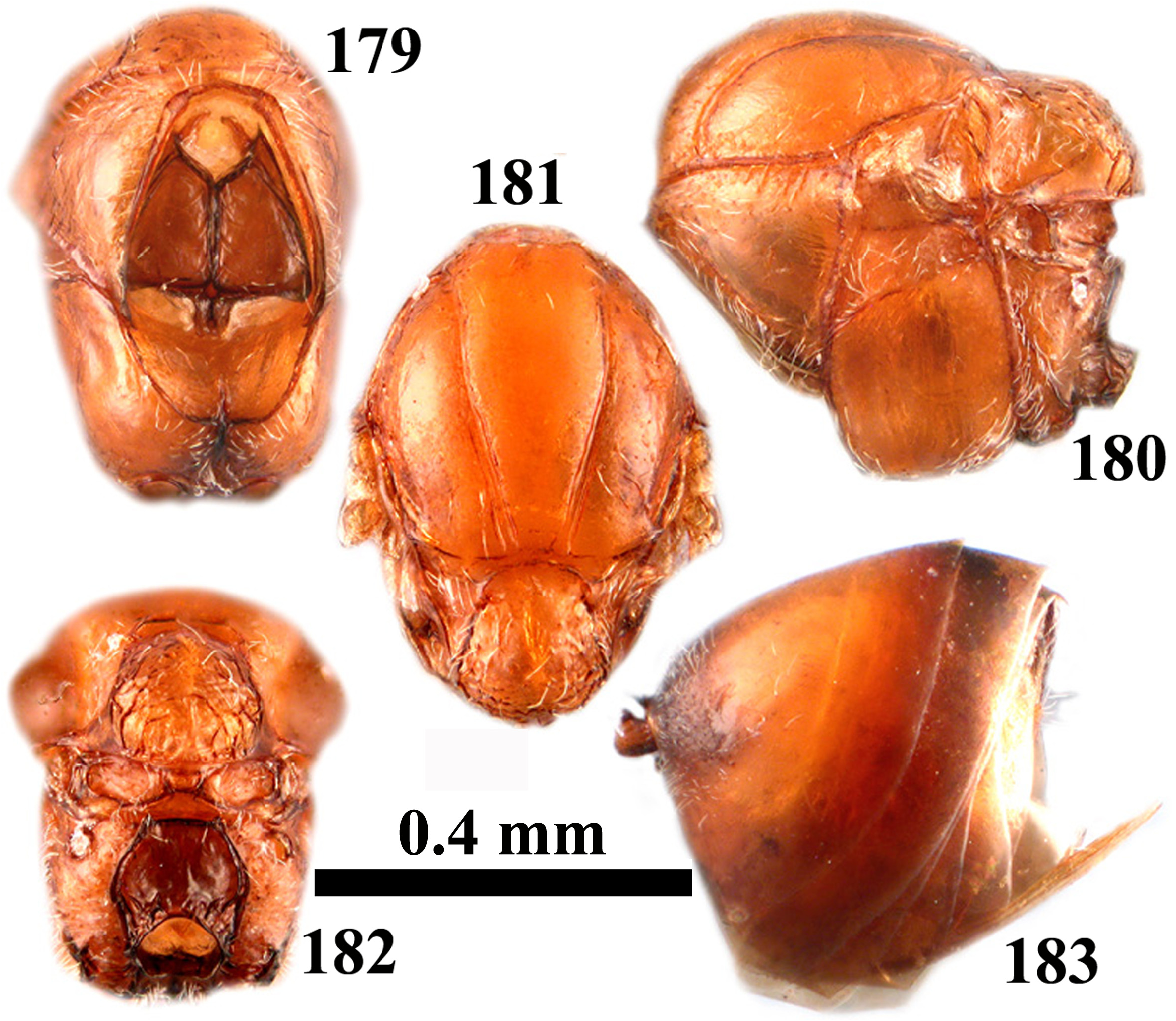
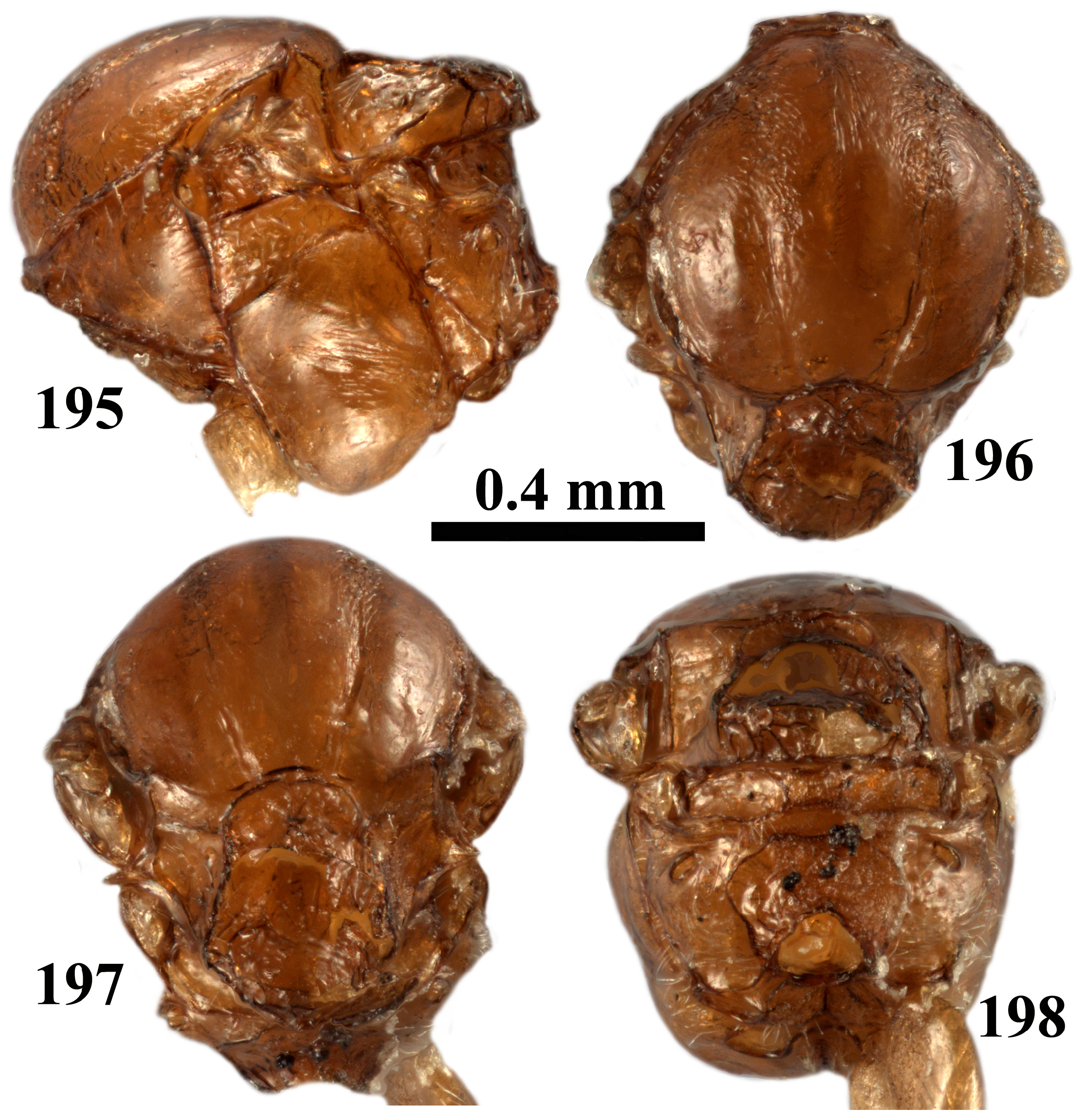
- Mesoscutellum with irregular rugae at least in lateral and posterior parts, sometimes central part smooth, shining, without piliferous points ( Figs 21 View FIGURES 19–22 , 105 View FIGURES 103–106 , 144 View FIGURES 142–145 , 197 View FIGURES 195–198 ); body light brown to chestnut brown, rarely darker ( Figs 13–23 View FIGURES 13–18 View FIGURES 19–22 View FIGURES 23–24 , 97–106 View FIGURES 97–102 View FIGURES 103–106 , 136–145 View FIGURES 136–141 View FIGURES 142–145 , 189–198 View FIGURES 189–194 View FIGURES 195–198 )........................................................................................... 18
14. Notaulus narrow, distinct only posteriorly, discontinuous or absent and finely coriaceous in anterior part; mesopleuron smooth, with transverse striae in central part...................................................... gigas comb. nov. (sex)
- Notaulus distinct, complete, reaching pronotum ( Figs 32 View FIGURES 31–34 , 116, 118 View FIGURES 116–120 , 251 View FIGURES 250–255 , 295 View FIGURES 293–297 ); mesopleuron entirely smooth, without transverse striae in central part ( Figs 31 View FIGURES 31–34 , 117–119 View FIGURES 116–120 , 250 View FIGURES 250–255 , 294 View FIGURES 293–297 )........................................................... 15
15. Toruli at half the height of head ( Fig. 121 View FIGURES 121–123 ); scape, pedicel, F1–F2 yellowish ( Fig. 116 View FIGURES 116–120 ); legs including hind coxa yellowish ( Fig. 117 View FIGURES 116–120 ); prominent part of ventral spine of hypopygium without setae ventrally.................. comatum comb. nov. (sex)
- Toruli in the upper half of head ( Figs 25 View FIGURES 25–30 , 241 View FIGURES 241–249 , 284 View FIGURES 284–292 ; scape, pedicel, F1–F2 dark brown, sometimes light brown but never yellowish ( Figs 29 View FIGURES 25–30 , 244 View FIGURES 241–249 , 287 View FIGURES 284–292 ); legs reddish brown, at least hind coxa darker; prominent part of ventral spine of hypopygium with setae ventrally................................................................................... 16
16. Eyes parallel; eye 1.7× as high as length of malar space ( Fig. 25 View FIGURES 25–30 ); antennomeres with long and erect setae ( Fig. 29 View FIGURES 25–30 ); OOL only slightly longer than diameter of lateral ocellus ( Fig. 26 View FIGURES 25–30 ); notaulus complete, deep ( Fig. 32 View FIGURES 31–34 ); prominent part of ventral spine of hypopygium with long dense white setae ventrally ( Fig. 35 View FIGURES 35–36 ).............................. apiarium comb. nov. (asex)
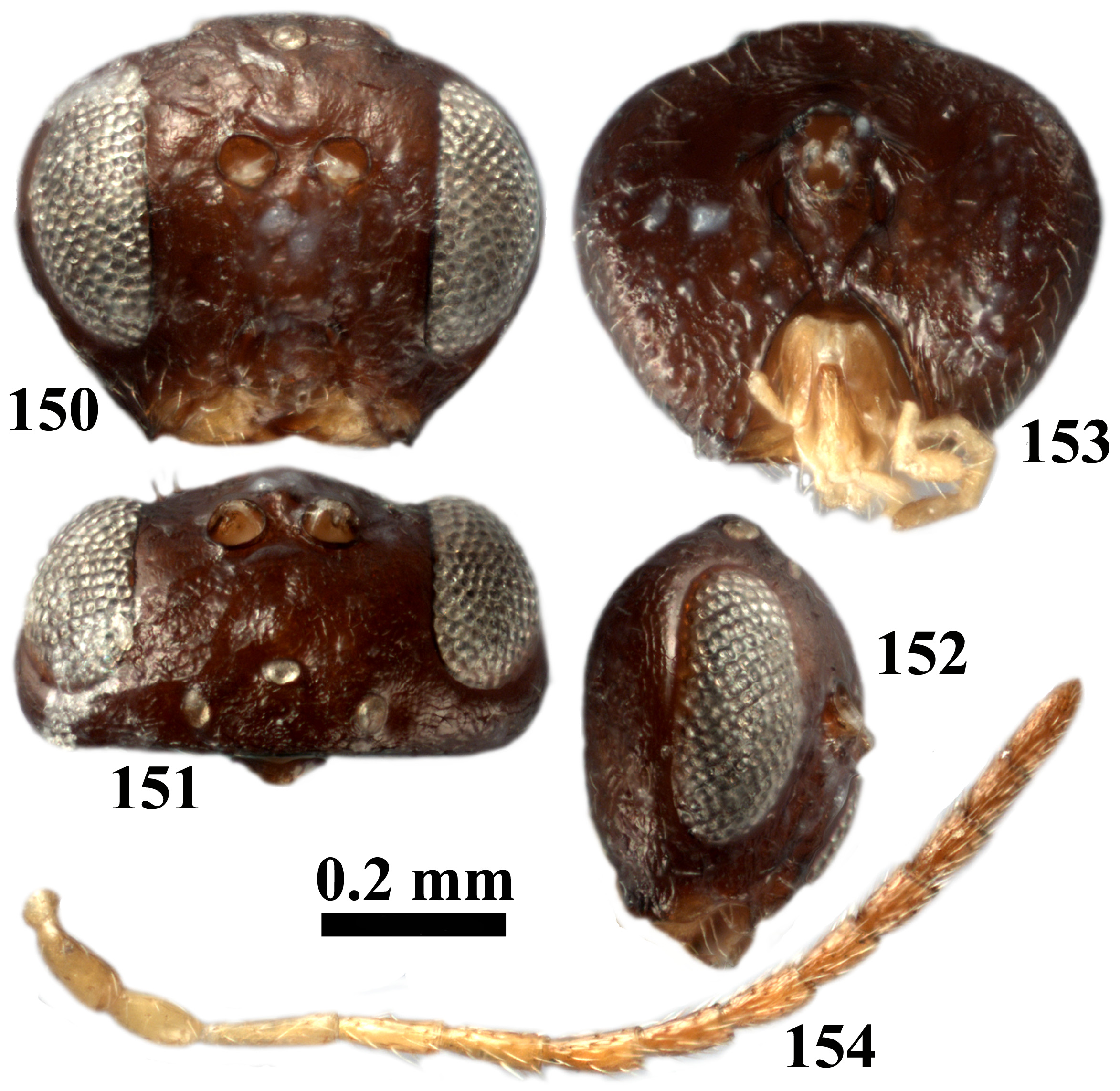
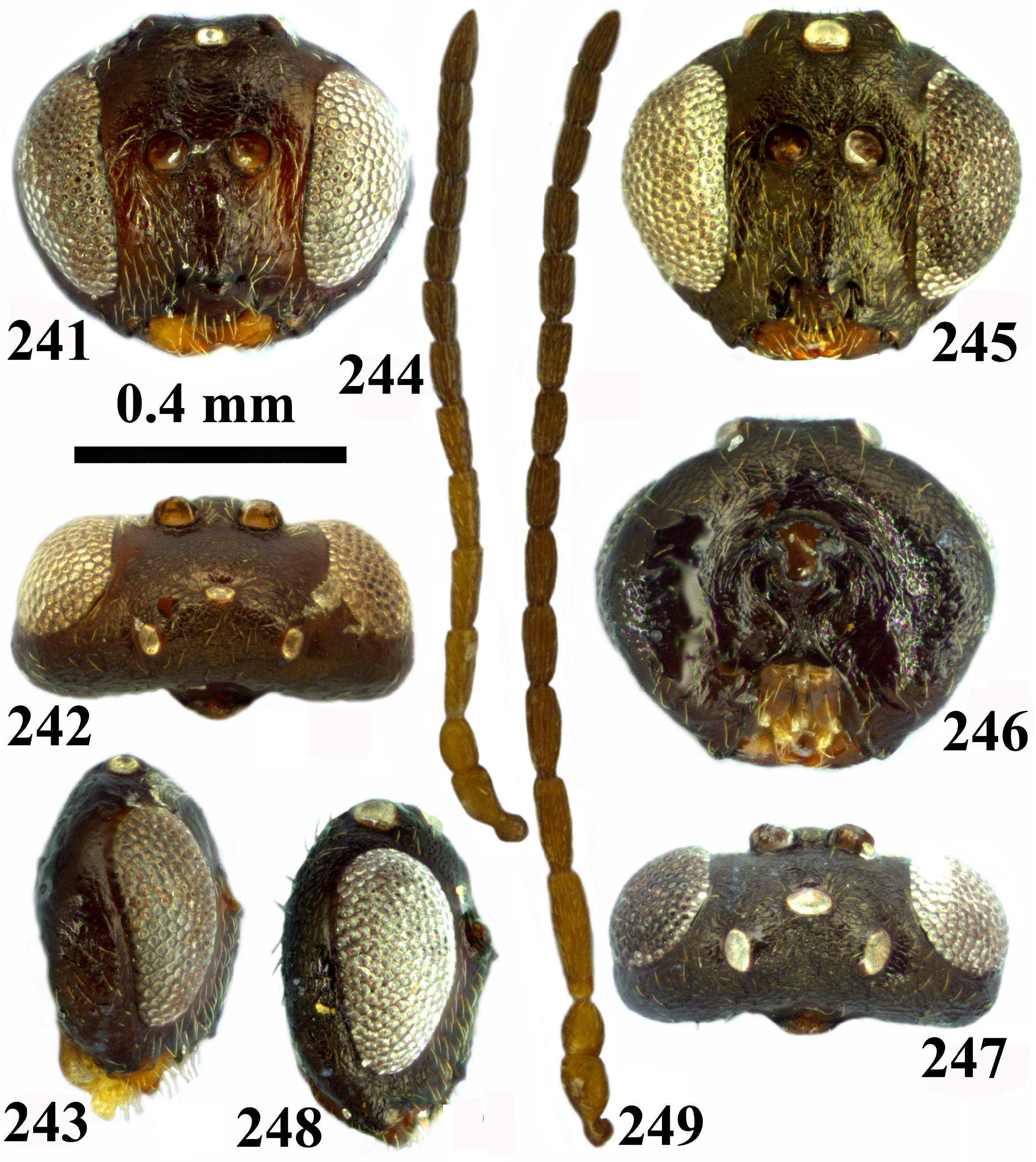
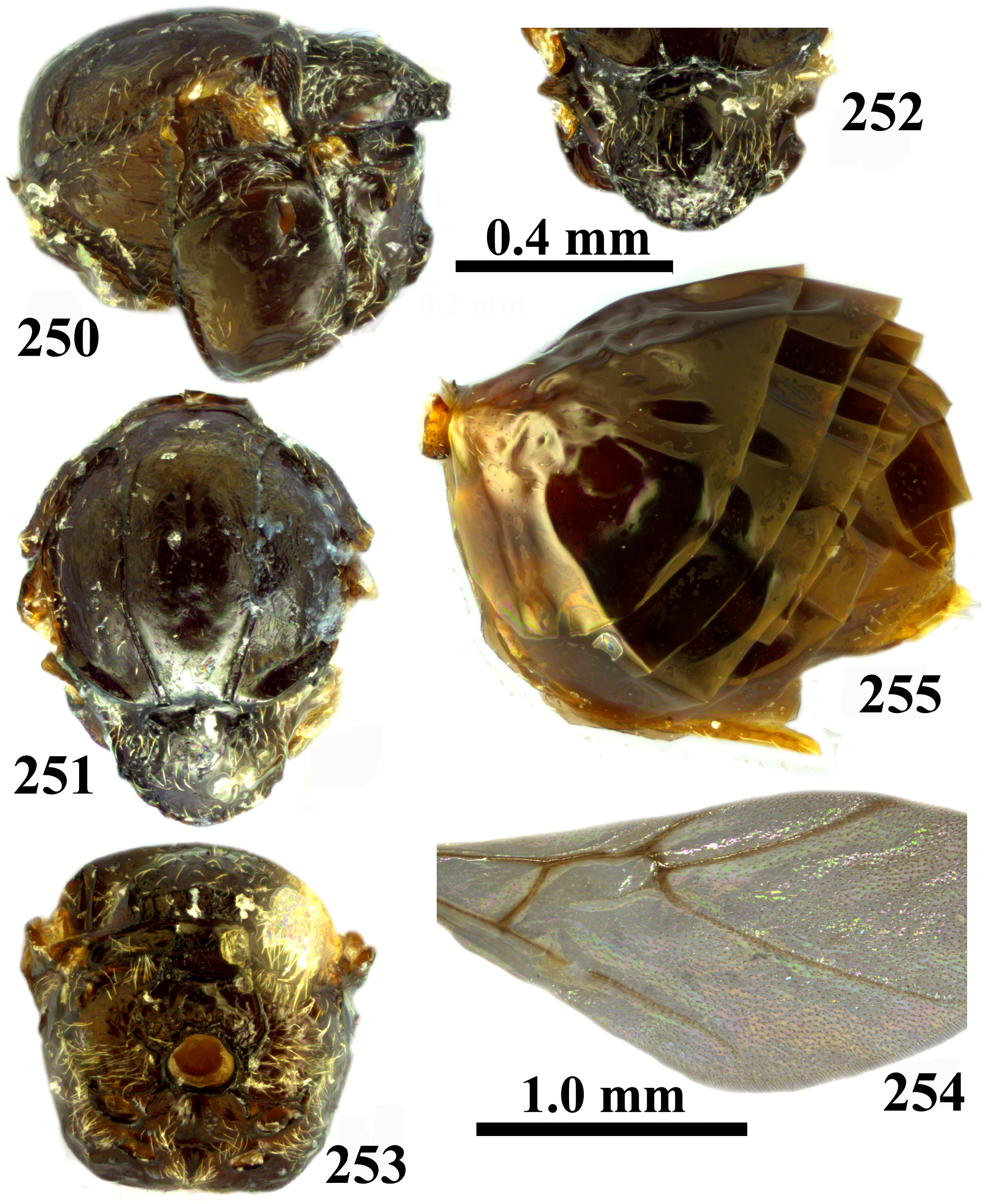
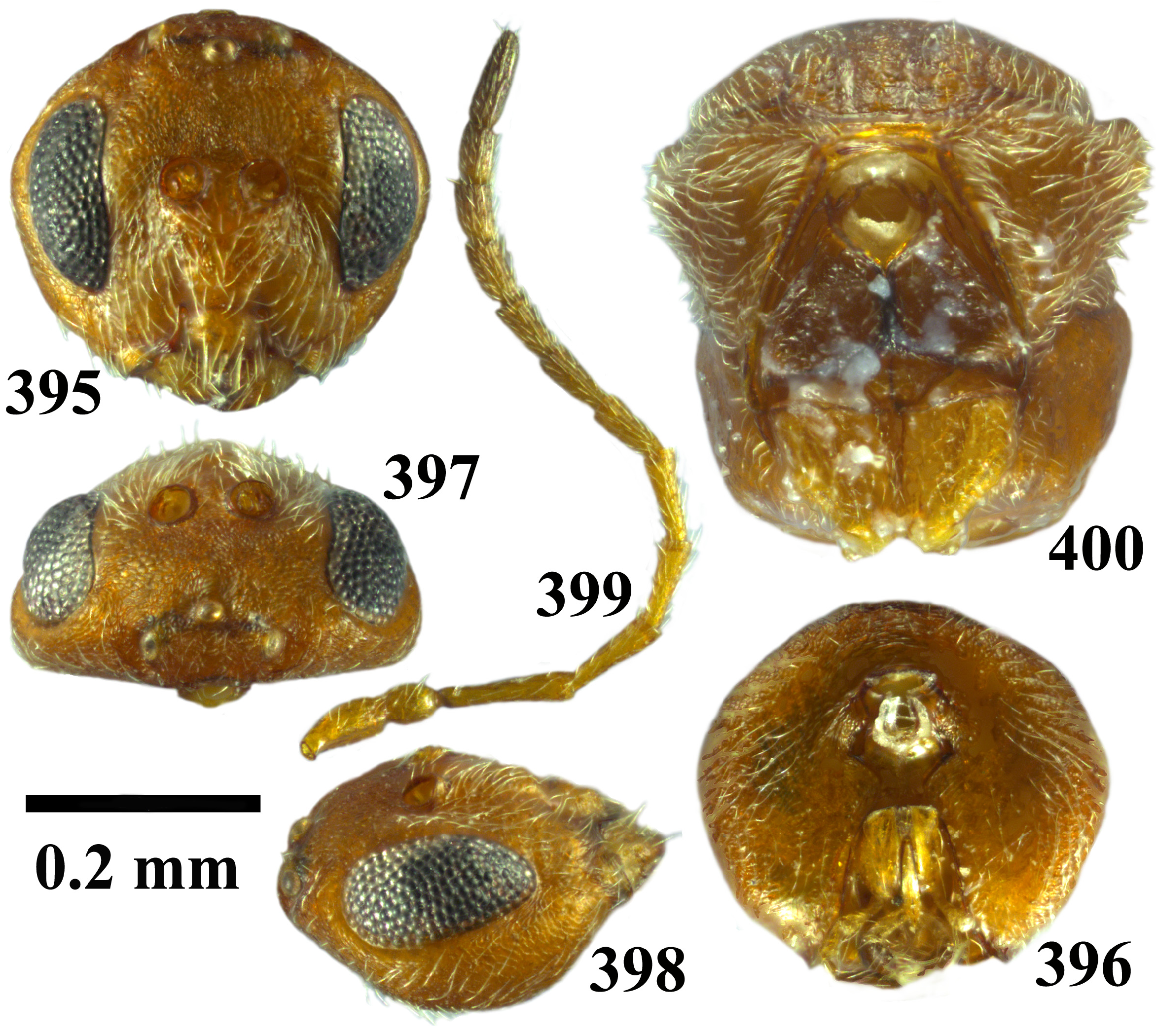
- Eyes converging ventrally; eye at least 4.0× as high as length of malar space ( Figs 241 View FIGURES 241–249 , 284 View FIGURES 284–292 ); antennomeres with short setae ( Figs 244 View FIGURES 241–249 , 287 View FIGURES 284–292 ); OOL 1.5× as long as diameter of lateral ocellus ( Figs 242 View FIGURES 241–249 , 286 View FIGURES 284–292 ); notaulus complete, sometimes weakly impressed anteriorly ( Figs 251 View FIGURES 250–255 , 295 View FIGURES 293–297 ); prominent part of ventral spine of hypopygium with few short setae ventrally ( Figs 255 View FIGURES 250–255 , 299 View FIGURES 298–299 )............................................................................................... 17
17. Antenna with 11 flagellomeres; F1 subequal to F2 and at most 2.0× as long as pedicel ( Fig. 244 View FIGURES 241–249 ); head distinctly narrower than mesosoma and genae broadened in frontal view ( Fig. 241 View FIGURES 241–249 ); mesoscutellar foveae separated by narrow elevated coriaceous central carina; areolet inconspicuous ( Figs 251–252 View FIGURES 250–255 )........................................ kingi comb. nov. (sex)
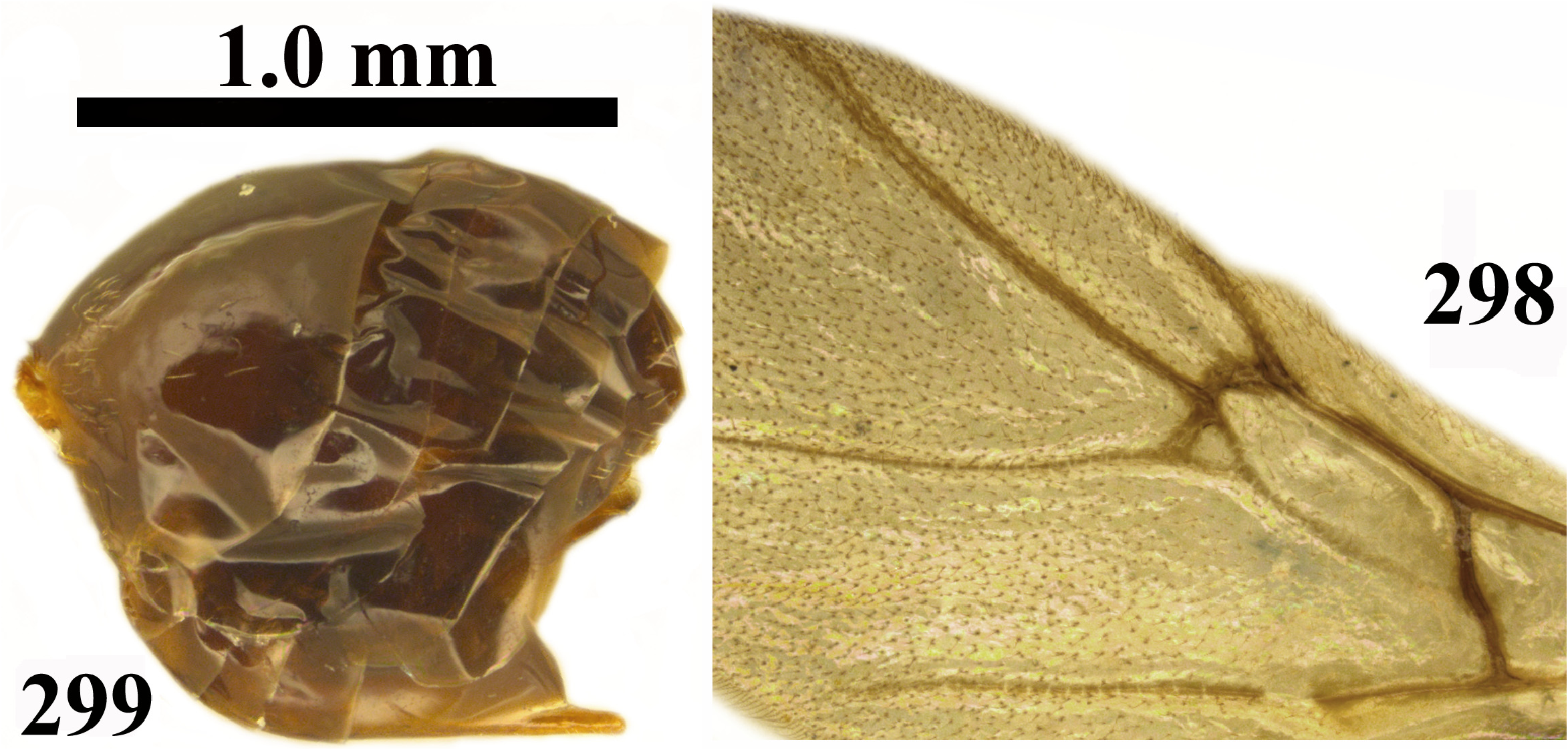
- Antenna with 12 flagellomeres; F1 1.2× as long as F2 and more than 2.0× as long as pedicel ( Fig. 287 View FIGURES 284–292 ); head only slightly narrower than mesosoma and genae not broadened ( Fig. 284 View FIGURES 284–292 ); mesoscutellar foveae fused in the form of a narrow, semi-lunar depression ( Figs 296–297 View FIGURES 293–297 ); areolet triangular ( Fig. 298 View FIGURES 298–299 )................................ pattersonae comb. nov. (sex)
18. Transfacial distance as long as or slightly shorter than height of eye; eye more than 3.6× as high as length of malar space ( Figs 136 View FIGURES 136–141 , 189 View FIGURES 189–194 ); pronotum laterally with short carinae only along posterior margin ( Figs 142 View FIGURES 142–145 , 195 View FIGURES 195–198 )........................ 19
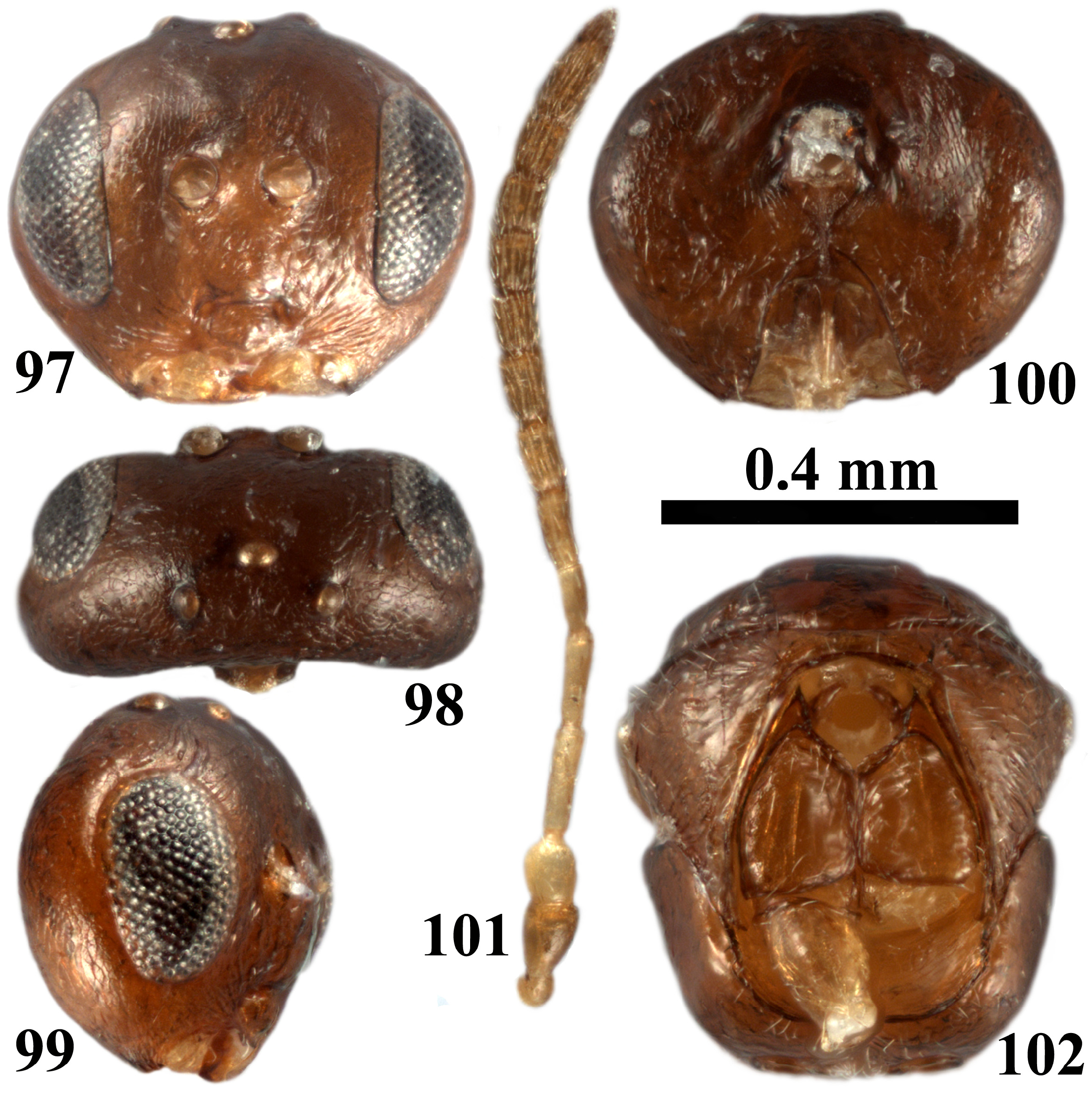
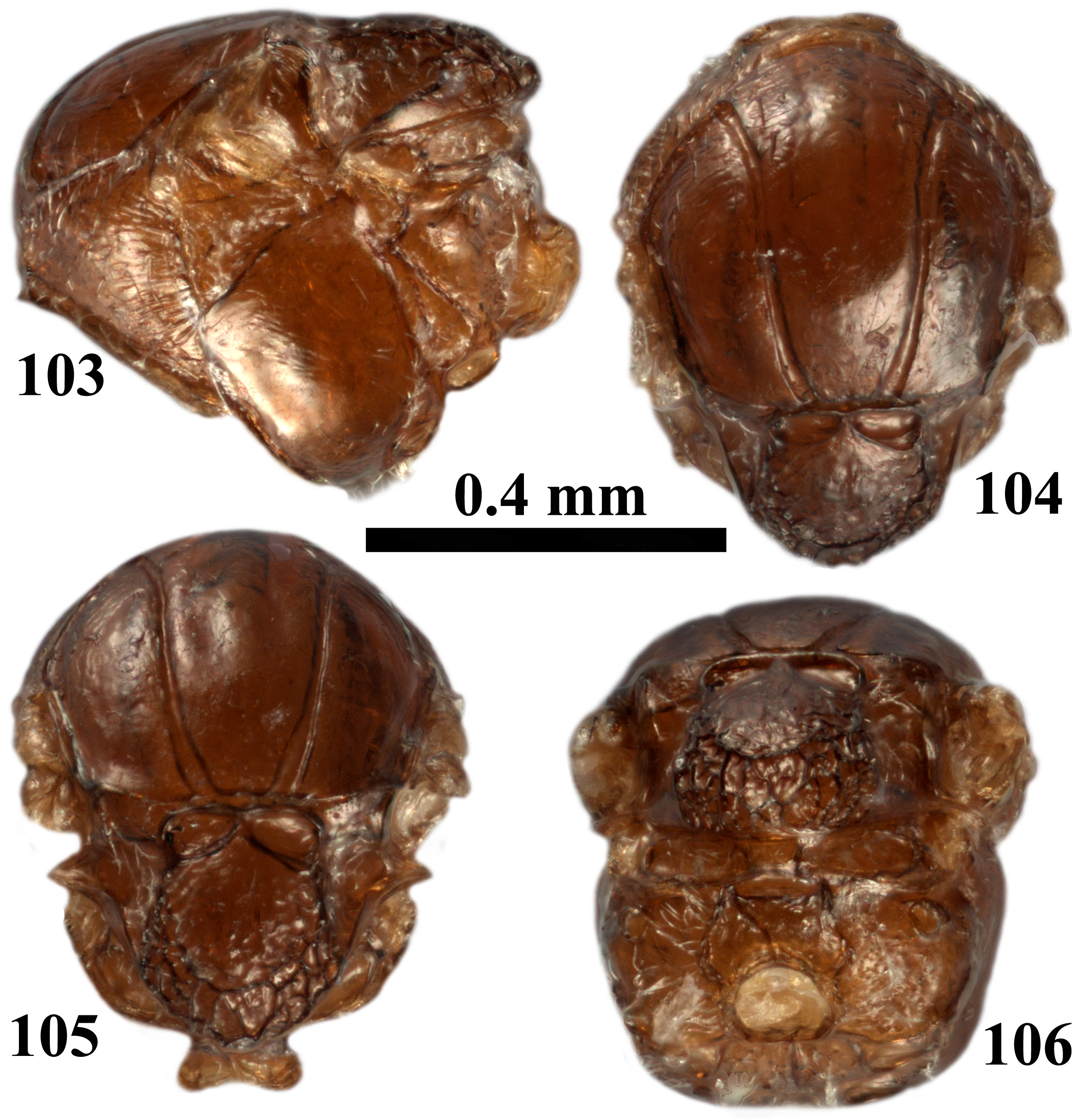
- Transfacial distance longer than height of eye; eye less than 3.0× as high as length of malar space ( Figs 13 View FIGURES 13–18 , 97 View FIGURES 97–102 ); pronotum with carinae going across entire lateral surface ( Figs 19 View FIGURES 19–22 , 103 View FIGURES 103–106 )..................................................... 20
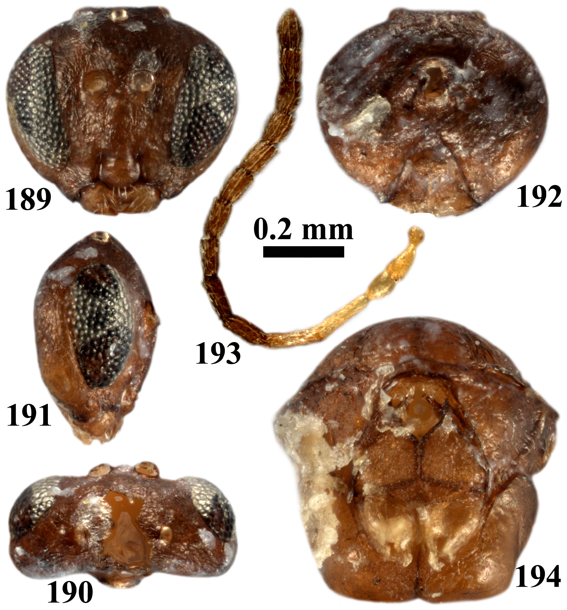
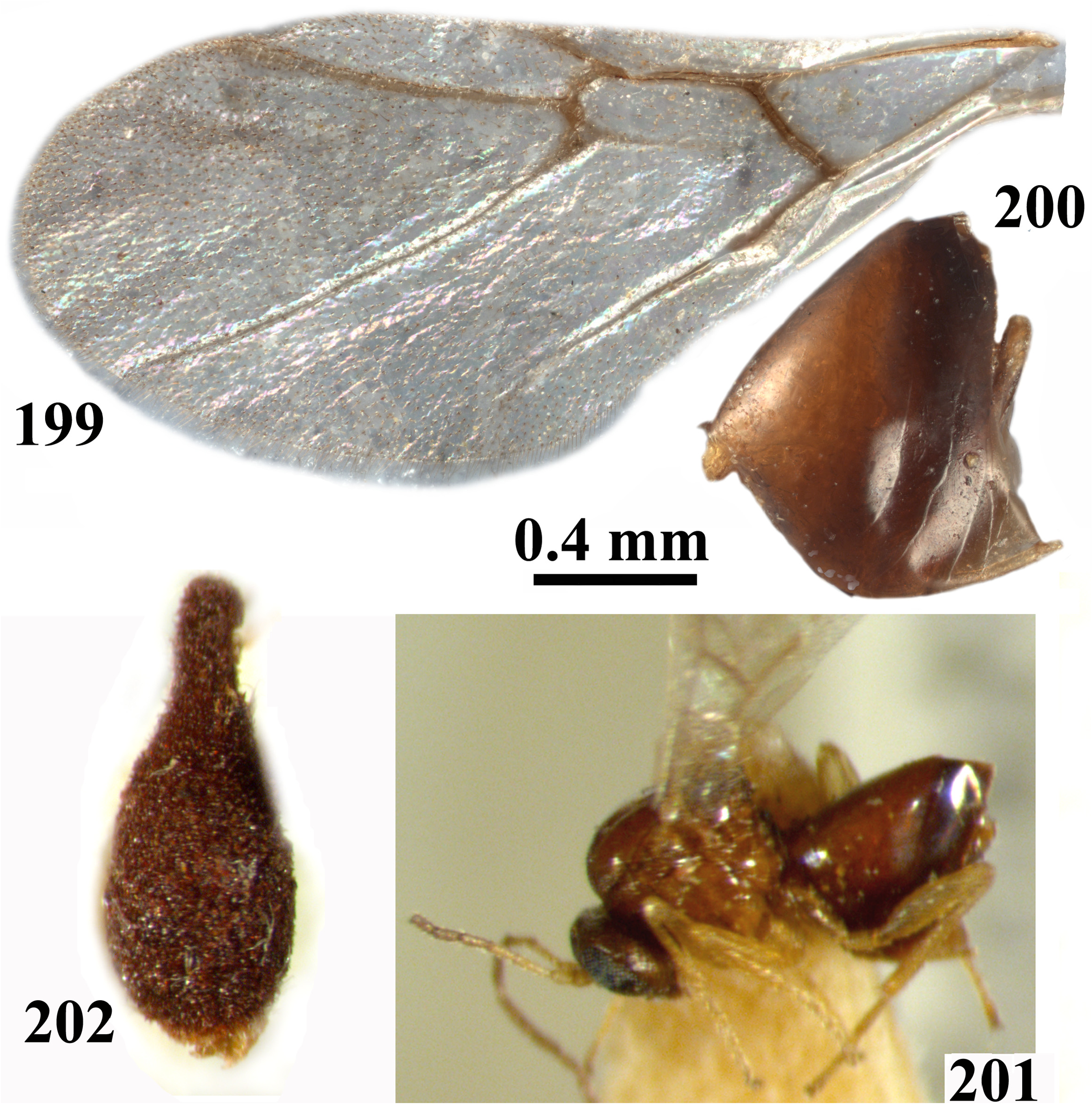
19. Eyes converging ventrally ( Fig. 189 View FIGURES 189–194 ); F1 longer than F2 ( Fig. 193 View FIGURES 189–194 ); mesopleuron smooth, shining, with delicate transverse subparallel striae in central part ( Fig. 195 View FIGURES 195–198 ); prominent part of ventral spine of hypopygium slightly longer than broad, without setae ( Fig. 200 View FIGURES 199–202 ); brown coloured ( Figs 189–198 View FIGURES 189–194 View FIGURES 195–198 , 200 View FIGURES 199–202 ).................................... dumosae comb. nov. (sex)
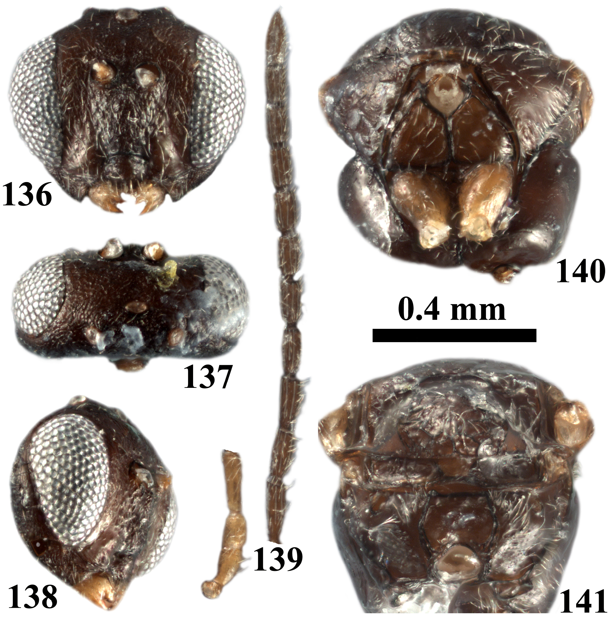
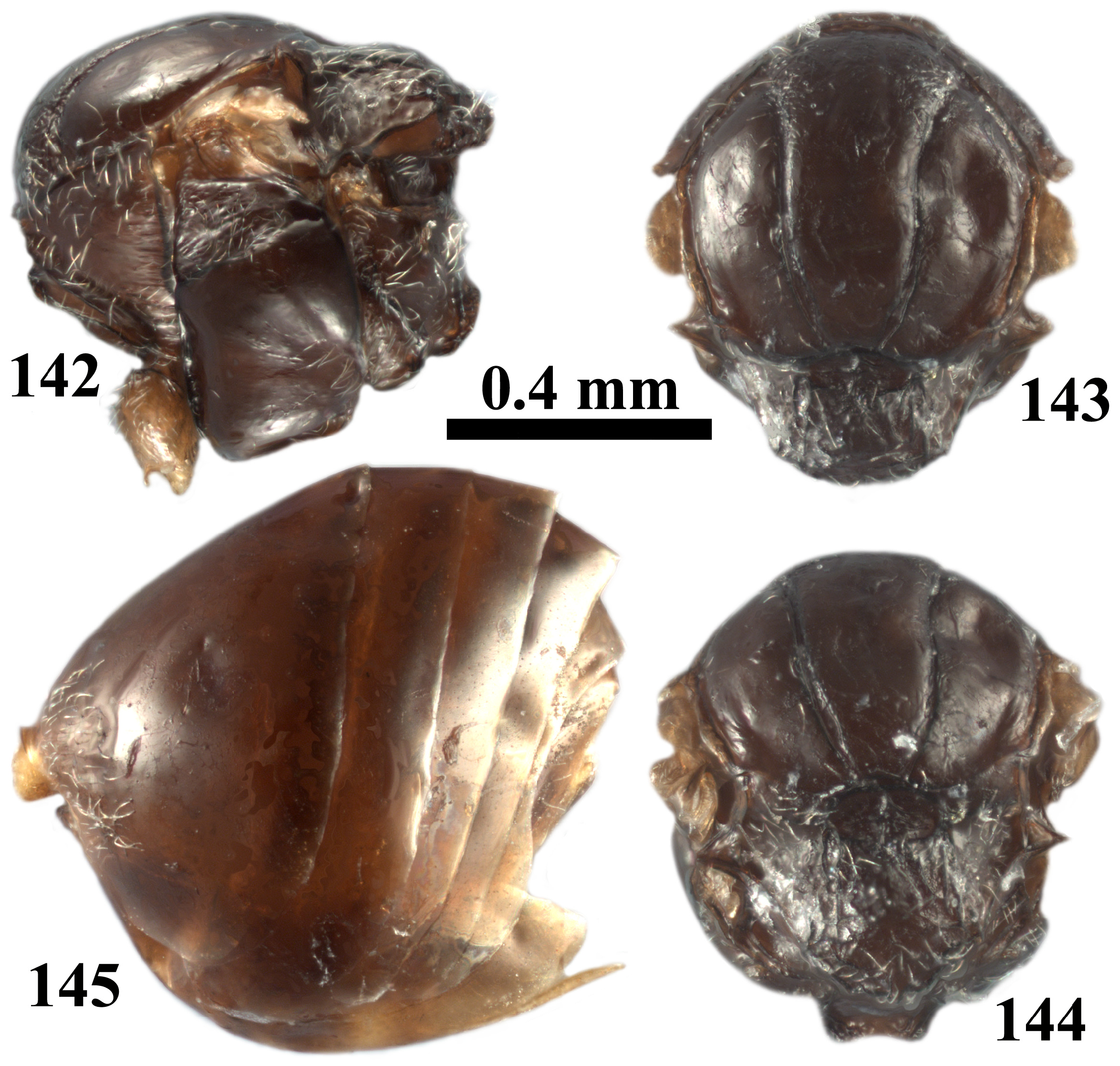
- Eyes parallel or only slightly converging posteriorly ( Fig. 136 View FIGURES 136–141 ); F1= F2; mesopleuron completely smooth, without striae ( Fig. 139 View FIGURES 136–141 ); ventral spine of hypopygium 3.7× as long as broad, with short white setae ventrally ( Fig. 145 View FIGURES 142–145 ; dark-coloured body( Figs 136–138 View FIGURES 136–141 , 142–145 View FIGURES 142–145 )............................................................. crystallinum comb. nov. (sex)
20. Gena not broadened behind eye in frontal view ( Fig. 13 View FIGURES 13–18 ); mesoscutellar foveae in the form of an anterior transverse impression, posteriorly not defined, continuing into disc of mesoscutellum ( Fig. 21 View FIGURES 19–22 ); mesopleuron completely smooth; metapleural sulcus reaching mesopleuron at half of its height ( Fig. 19 View FIGURES 19–22 ); 2nd metasomal tergum with setae anteriorly ( Fig. 23 View FIGURES 23–24 ); body light brown ( Figs 13–23 View FIGURES 13–18 View FIGURES 19–22 View FIGURES 23–24 )................................................................... amphorus comb. nov. (asex)
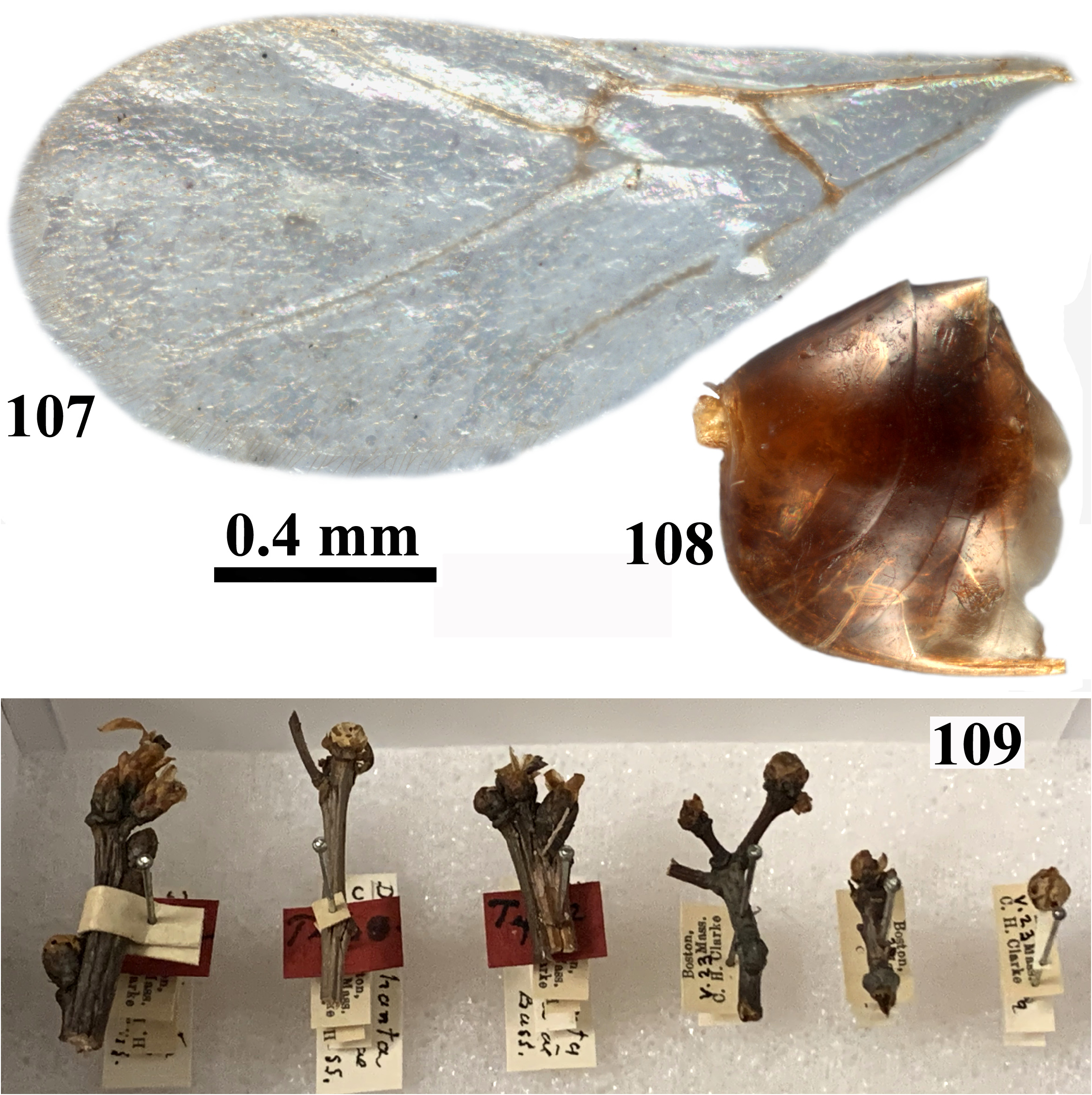
- Gena slightly broadened behind eye in frontal view ( Fig. 97 View FIGURES 97–102 ); mesoscutellar foveae well-delimited by a carina posteriorly, separated medially ( Fig. 105 View FIGURES 103–106 ); mesopleuron with delicate indistinct transverse subparallel striae in anterodorsal part at mid height; metapleural sulcus reaching mesopleuron above half of its height ( Fig. 103 View FIGURES 103–106 ); 2nd metasomal tergum without setae ( Fig. 108 View FIGURES 107–109 ); body dark brown to chestnut brown ( Figs 97–106 View FIGURES 97–102 View FIGURES 103–106 , 108 View FIGURES 107–109 )............................... clarkei comb. nov. (sex)
21. Pronotum laterally smooth to coriaceous, without carinae ( Figs 7 View FIGURES 7–10 , 43 View FIGURES 43–46 , 130 View FIGURES 130–133 , 222 View FIGURES 222–225 , 275 View FIGURES 274–278 , 378 View FIGURES 377–380 )........................... 22
- Pronotum with longitudinal carinae laterally ( Figs 57 View FIGURES 57–60 , 68 View FIGURES 68–71 , 78 View FIGURES 78–81 , 209 View FIGURES 209–212 , 304 View FIGURES 304–307 , 360 View FIGURES 360–363 , 401 View FIGURES 401–404 , 470 View FIGURES 469–472 )............................. 27
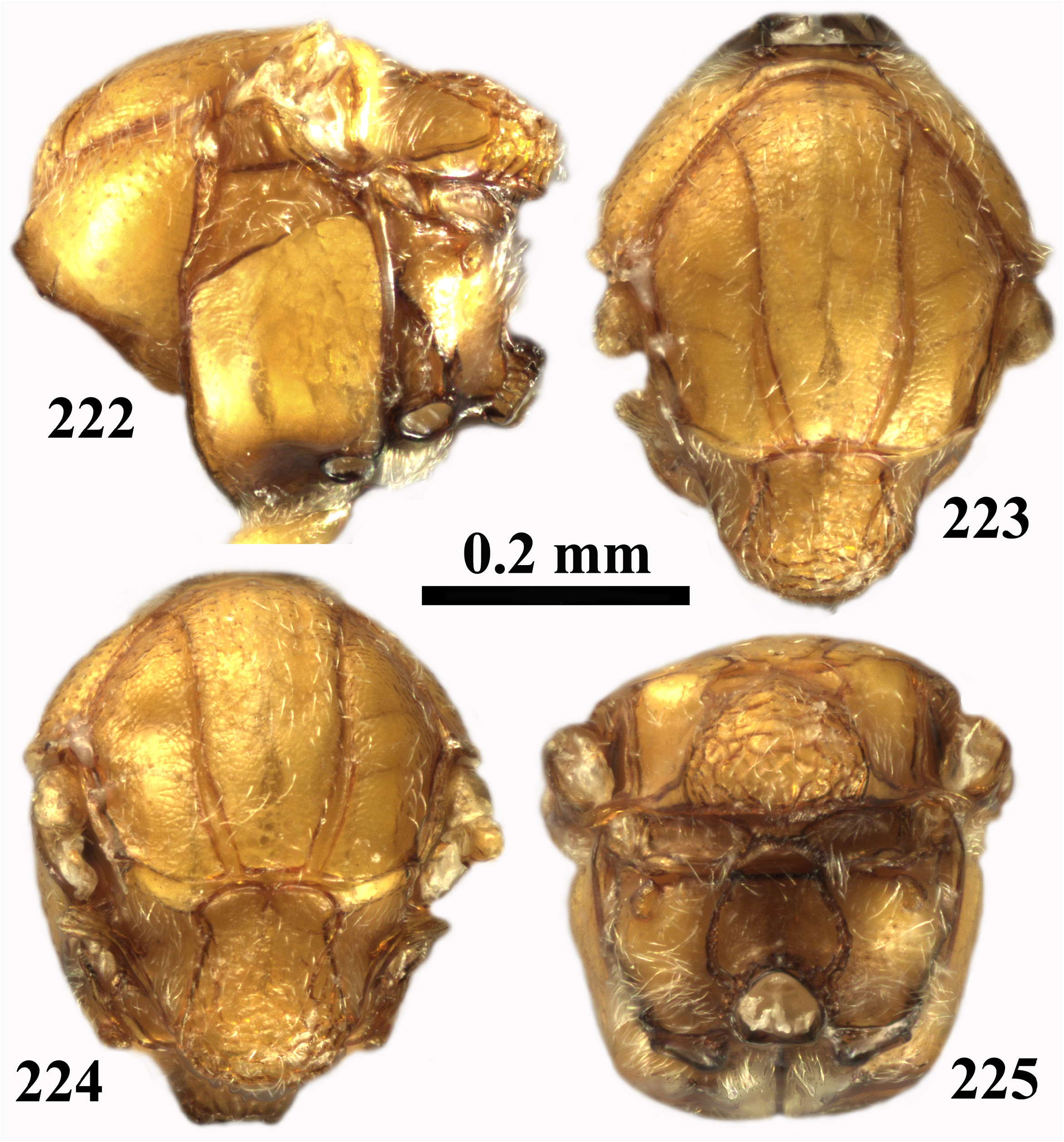
22. Frons bulging in frontal view ( Figs 1 View FIGURES 1–6 , 216 View FIGURES 216–221 ); ocelli not elevated above head; transfacial distance 1.4× longer than height of eye ( Figs 1 View FIGURES 1–6 , 216 View FIGURES 216–221 ); eyes slightly converging ventrally, metapleural sulcus reaching mesopleuron on upper 2/3 of its height ( Figs 7 View FIGURES 7–10 , 222 View FIGURES 222–225 )............................................................................................... 23
- Frons not bulging in frontal view, ocelli sometimes elevated above head; transfacial distance subequal or 1.2× longer than height of eye ( Figs 37 View FIGURES 37–42 , 124 View FIGURES 124–129 , 269 View FIGURES 269–273 , 372 View FIGURES 372–376 ); metapleural sulcus reaching mesopleuron at half of its height ( Figs 43 View FIGURES 43–46 , 130 View FIGURES 130–133 , 275 View FIGURES 274–278 , 378 View FIGURES 377–380 ). .................................................................................................. 24
23. Median mesoscutal line in the form of a short smooth, shining triangle ( Fig. 8 View FIGURES 7–10 ); mesoscutellar foveae divided by a thin carina ( Fig. 9 View FIGURES 7–10 ); ventral spine of hypopygium 11.0× longer than broad in ventral view ( Fig. 11 View FIGURES 11–12 ); head and mesosoma uniformly reddish brown ( Figs 1–10 View FIGURES 1–6 View FIGURES 7–10 ).............................................................. albicomus comb. nov. (asex)
- Median mesoscutal line absent ( Figs 223–224 View FIGURES 222–225 ); mesoscutellar foveae divided by an elevated coriaceous triangle ( Fig. 224 View FIGURES 222–225 ); ventral spine of hypopygium 8.0× as long as broad in ventral view ( Fig. 226 View FIGURES 226–227 ); head and mesosoma amber ( Figs 216–225 View FIGURES 216–221 View FIGURES 222–225 ).................................................................................... izabellae sp. nov. (asex)
24. Pronotum smooth, with sparse setae and piliferous points along dorsal edge ( Fig. 43 View FIGURES 43–46 ); mesoscutum longer than broad, scarcely pubescent, without black stripes, alutaceous-reticulate between notauli in anterior 2/3 length and lateral to notauli, smooth between notauli in posterior 1/3; anterior parallel line indistinct, parapsidal line absent ( Figs 44–45 View FIGURES 43–46 )................................................................................................. atrimentum comb. nov. (asex)
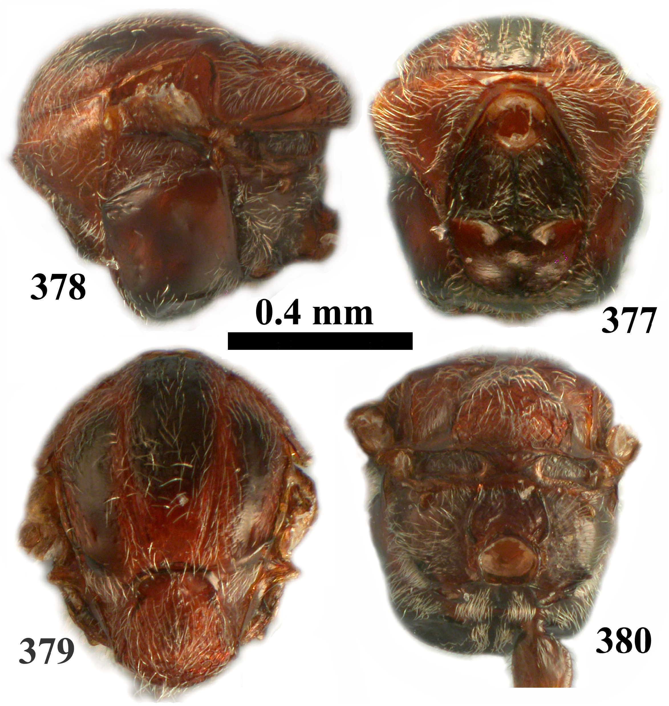
- Pronotum coriaceous, if smooth then with dense setae and piliferous points ( Figs 130 View FIGURES 130–133 , 275 View FIGURES 274–278 , 378 View FIGURES 377–380 ); mesoscutum only slightly longer than broad (subequal), pubescent ( Figs 131 View FIGURES 130–133 , 276 View FIGURES 274–278 , 379 View FIGURES 377–380 ); mesoscutum dark brown in between notauli in anterior 1/3 length of mesoscutum and along parapsidal line, with other surface sculpture; anterior parallel line and parapsidal line present ( Figs 131 View FIGURES 130–133 , 276 View FIGURES 274–278 , 379 View FIGURES 377–380 )....................................................................................... 25
25. Head ovate in frontal view ( Fig. 269 View FIGURES 269–273 ); pronotum smooth with dense setae and piliferous points ( Fig. 275 View FIGURES 274–278 ); mesoscutellar foveae fused ( Fig. 277 View FIGURES 274–278 ); 2nd metasomal tergum extending to 1/3 the length of metasoma in dorsal view ( Fig. 280 View FIGURES 279–283 )........................................................................................... pattersonae comb. nov. (asex)
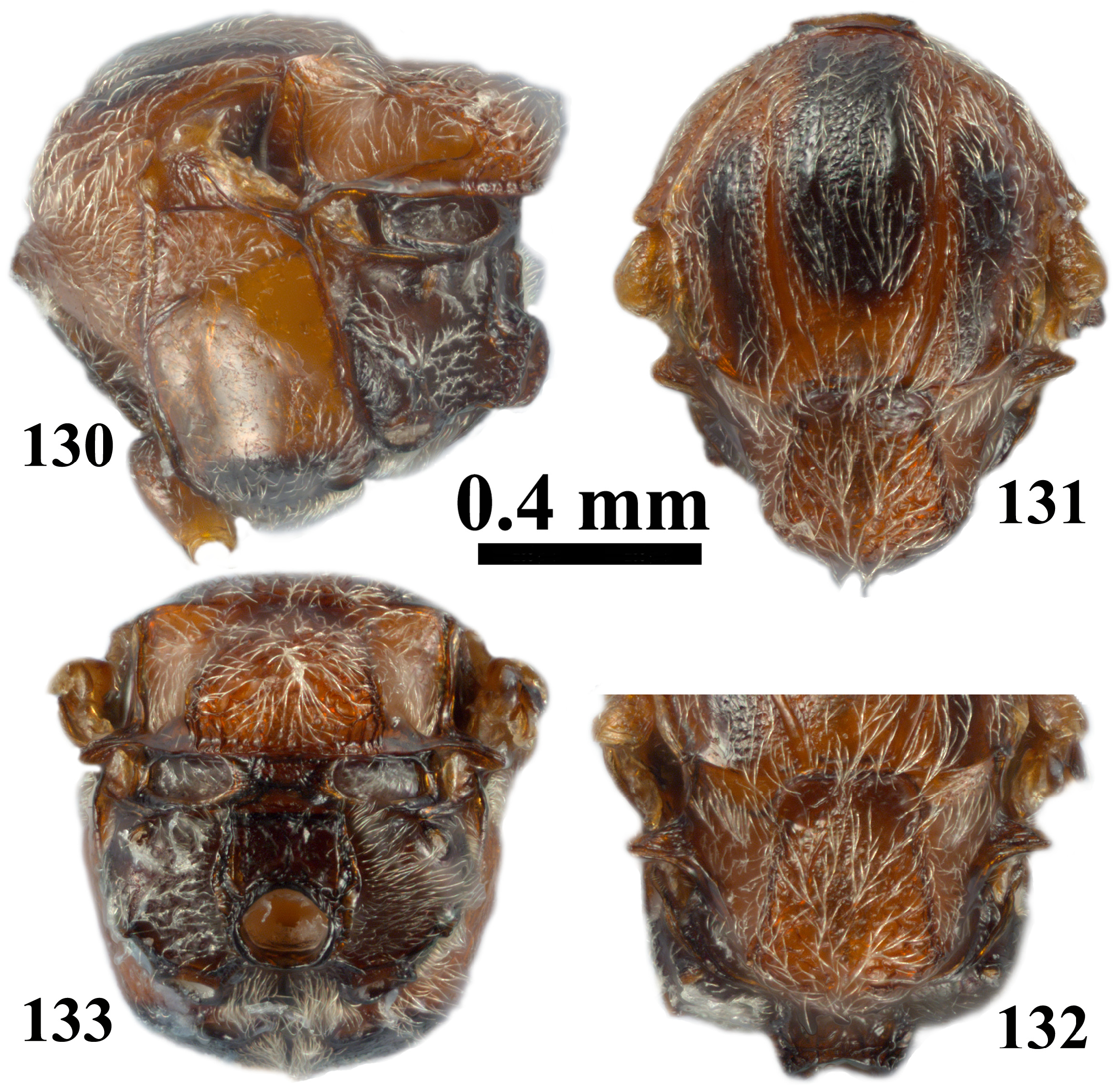
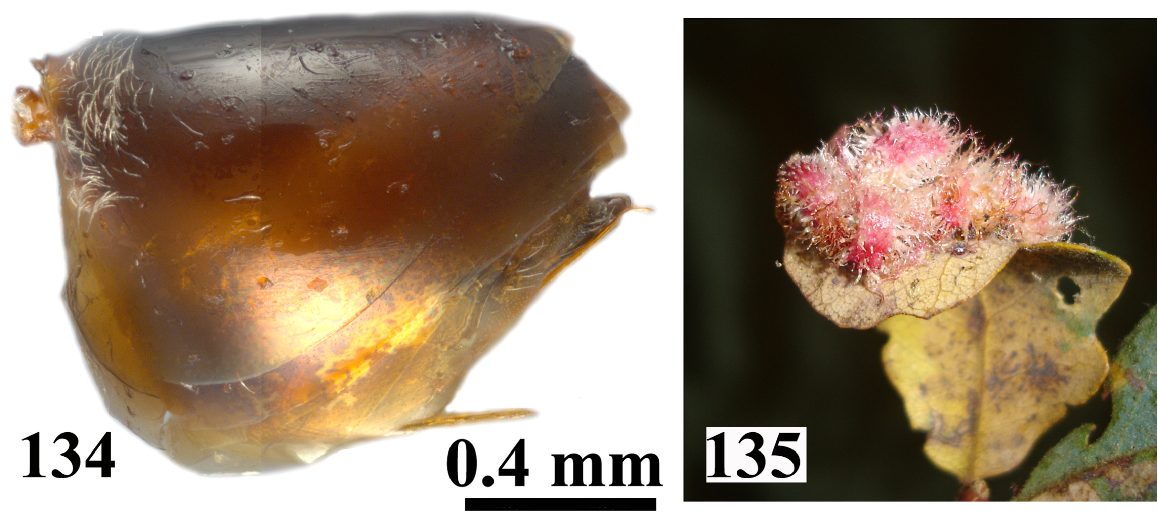
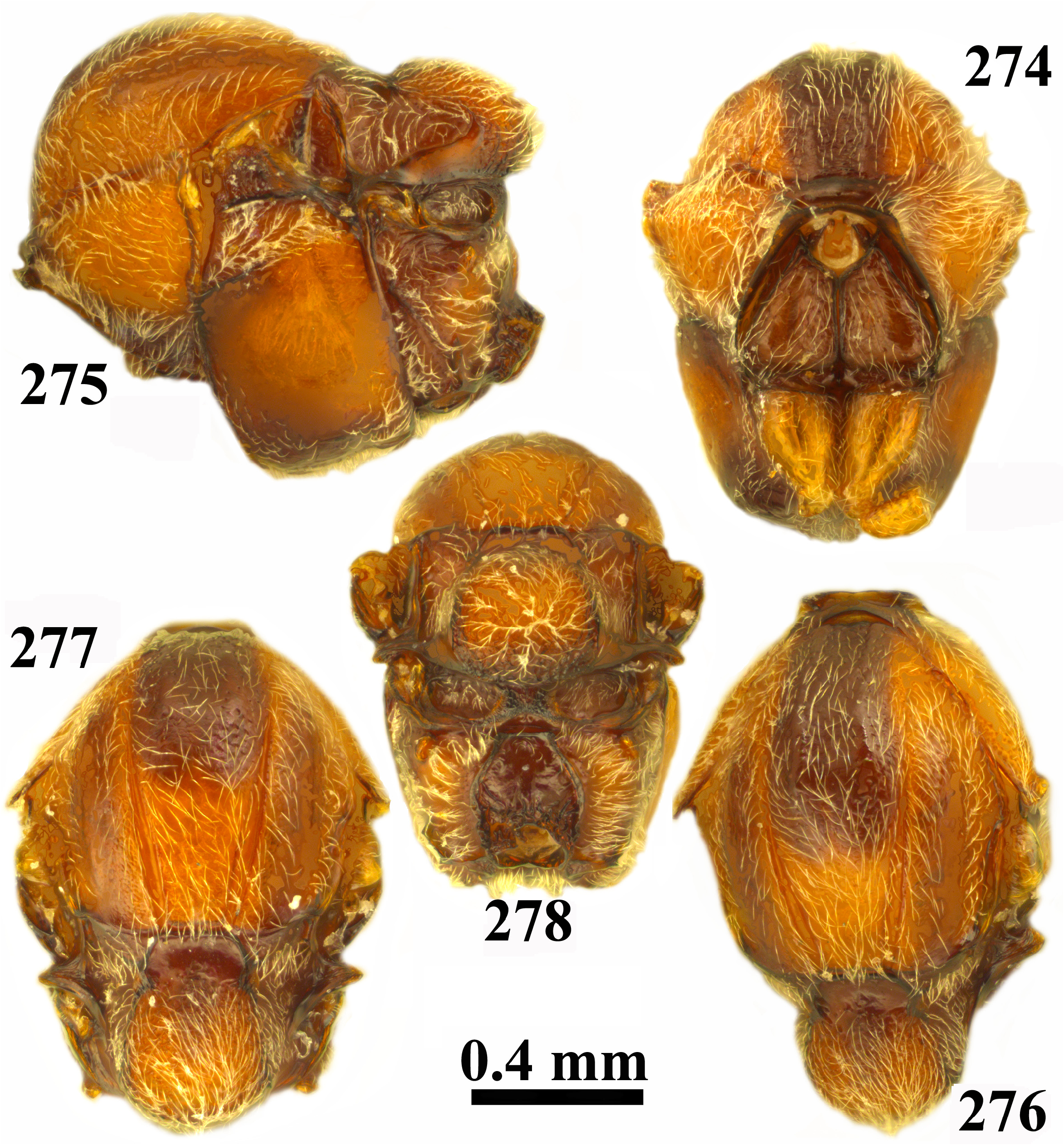
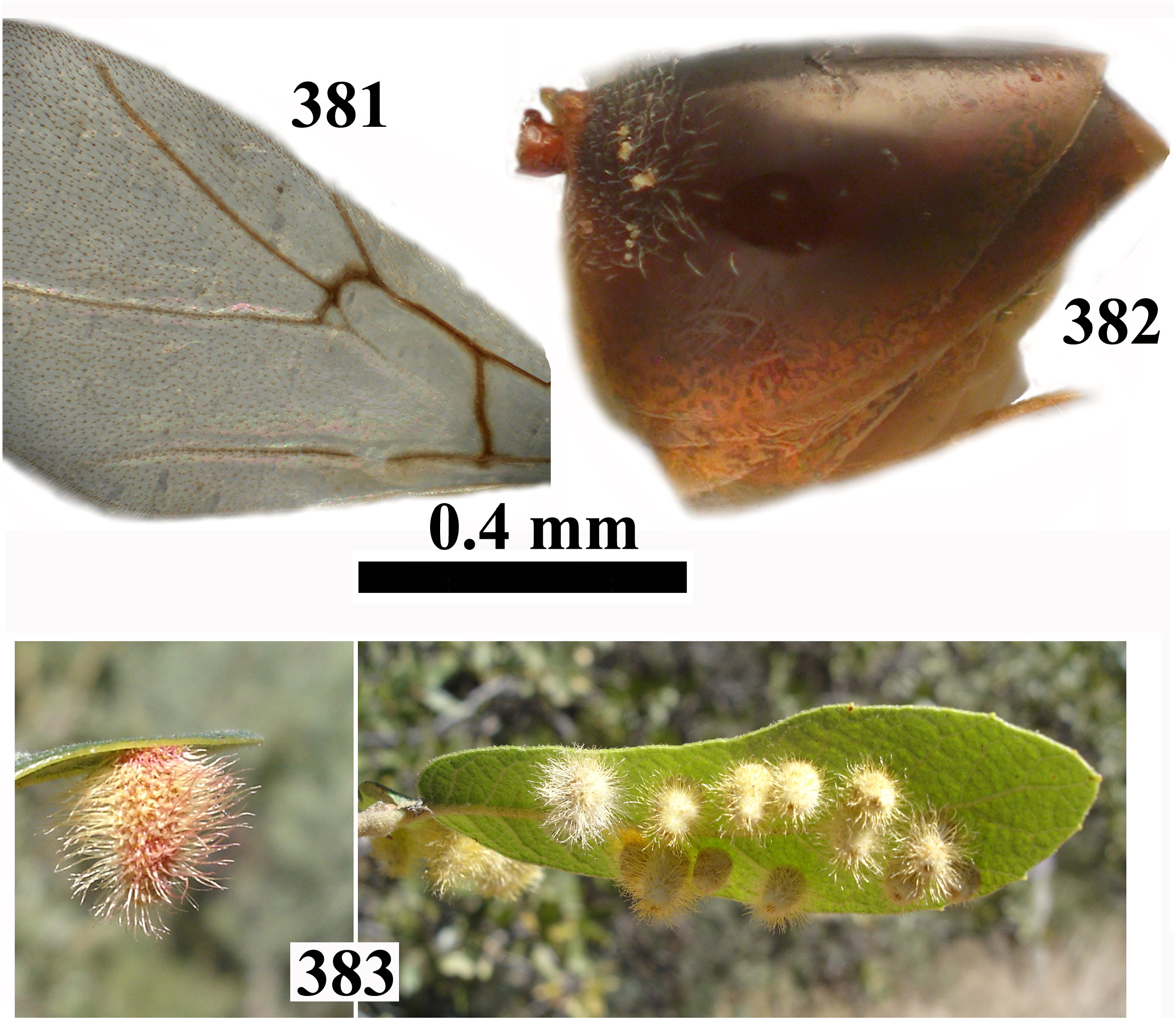
- Head trapezoid in frontal view ( Figs 124 View FIGURES 124–129 , 372 View FIGURES 372–376 ); pronotum coriaceous, if smooth then with dense setae and without piliferous points ( Figs 130 View FIGURES 130–133 , 378 View FIGURES 377–380 ); mesoscutellar foveae divided by median elevated narrow triangular area or carina ( Figs 132 View FIGURES 130–133 , 379 View FIGURES 377–380 ); 2nd metasomal tergum smooth, extending at least to 5/6 length of metasoma in dorsal view ( Figs 134 View FIGURES 134–135 , 382 View FIGURES 381–383 )................ 26
26. Pronotum coriaceous in posterolateral area, with piliferous points ( Fig. 130 View FIGURES 130–133 ); mesoscutum rugose-reticulate between notauli and laterad to notaulus in anterior half, alutaceous glabrous between notauli in posterior half ( Fig. 131 View FIGURES 130–133 ); eye 2.7× as high as length of malar space ( Fig. 124 View FIGURES 124–129 ); distal flagellomeres broader than basal ones, F1 shorter than scape+pedicel ( Fig. 128 View FIGURES 124–129 ); prominent part of ventral spine of hypopygium 6.5x as long as broad in ventral view ( Fig. 134 View FIGURES 134–135 )................................................................................................... crystallinum comb. nov. (asex)
- Pronotum smooth, without piliferous points ( Fig. 378 View FIGURES 377–380 ); mesoscutum uniformly delicately coriaceous ( Fig. 379 View FIGURES 377–380 ); eye 3.7× as high as length of mala space ( Fig. 372 View FIGURES 372–376 ); all flagellomeres uniformly broad, F1 longer than scape+pedicel ( Fig. 376 View FIGURES 372–376 ); prominent part of ventral spine of hypopygium more than 5.5× as long a broad in ventral view ( Fig. 382 View FIGURES 381–383 )...................................................................................................... sulfureum comb. nov. (asex)
27. Mesoscutum with distinct and deep piliferous points ( Figs 235–236 View FIGURES 234–237 , 471 View FIGURES 469–472 )........................................ 28
- Mesoscutum without piliferous points or shallow almost inconspicuous points ( Figs 58 View FIGURES 57–60 , 70 View FIGURES 68–71 , 79 View FIGURES 78–81 , 210 View FIGURES 209–212 , 305 View FIGURES 304–307 , 361 View FIGURES 360–363 , 390 View FIGURES 389–392 , 402–403 View FIGURES 401–404 , 420 View FIGURES 419–425 , 436 View FIGURES 435–438 )........................................................................................... 30
28. Frons bulging in frontal view, ocelli not elevated above head ( Fig. 186 View FIGURES 184–188 ); toruli at mid-height of head ( Fig. 186 View FIGURES 184–188 ); median mesoscutal line in the form of a short triangle; entire body black ( Figs 184–186 View FIGURES 184–188 )............. discularis comb. nov. (asex)
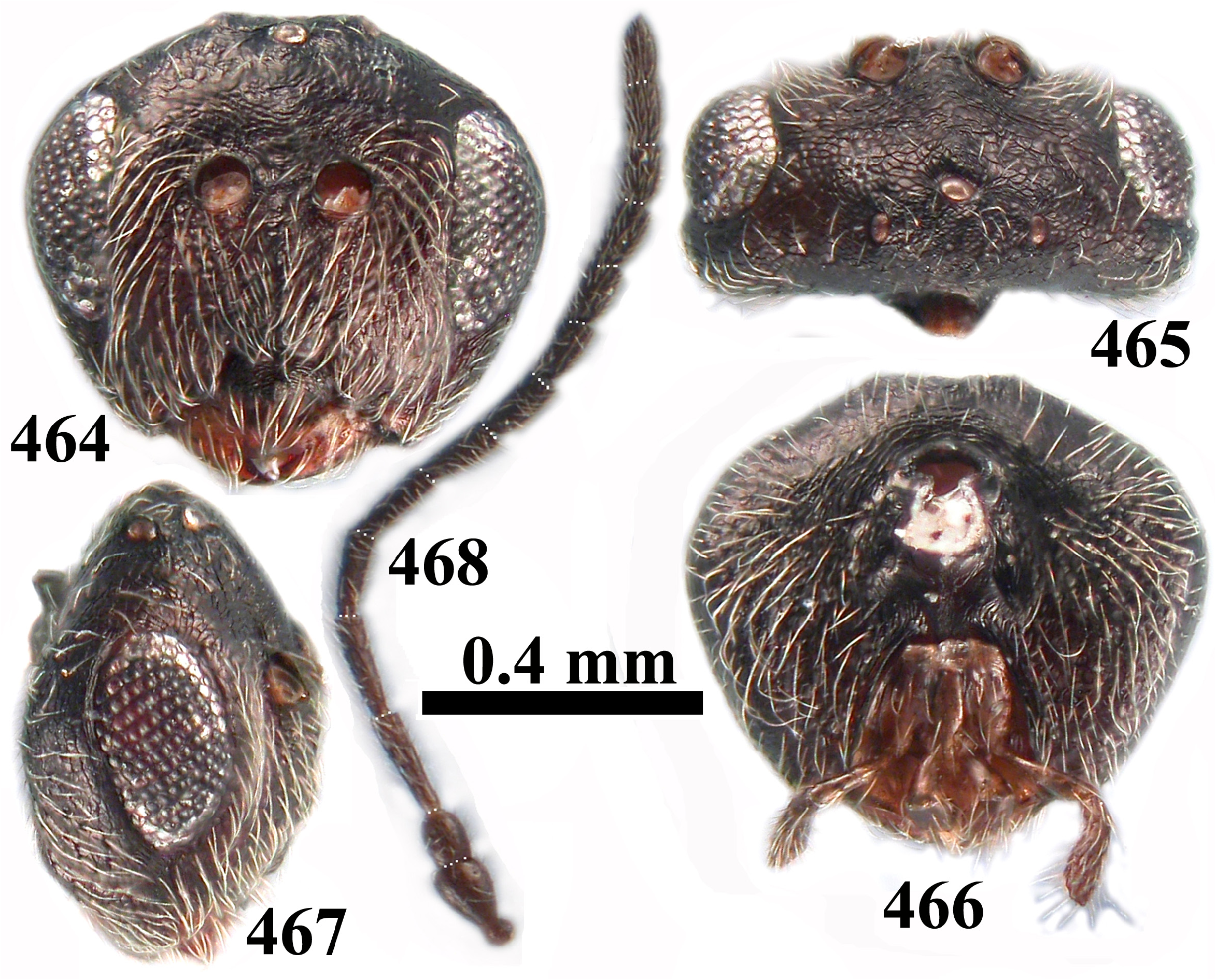
- Frons not bulging in frontal view, ocelli not elevated above head; toruli in upper half of head ( Figs 228 View FIGURES 228–233 , 464 View FIGURES 464–468 ); median mesoscutal line absent ( Figs 235 View FIGURES 234–237 , 471 View FIGURES 469–472 ); head and mesosoma never black ( Figs 228–238 View FIGURES 228–233 View FIGURES 234–237 View FIGURES 238–240 , 464–472 View FIGURES 464–468 View FIGURES 469–472 , 474 View FIGURES 473–475 )......................... 29
29. Gena broadened behind eye in frontal view ( Fig. 228 View FIGURES 228–233 ); clypeus smooth; eye 2.9× as high as length of malar space ( Fig. 228 View FIGURES 228–233 ); metapleural sulcus reaching mesopleuron at half of its height ( Fig. 234 View FIGURES 234–237 ); fore wing longer than body; metasomal terga 6 and 7 with micropunctures; prominent part of ventral spine of hypopygium around 8.0× as long as broad in ventral view ( Fig. 238 View FIGURES 238–240 ); light specimens, rusty brown and rarely uniformly dark brown ( Figs 228–238 View FIGURES 228–233 View FIGURES 234–237 View FIGURES 238–240 )................... kingi comb. nov. (asex)
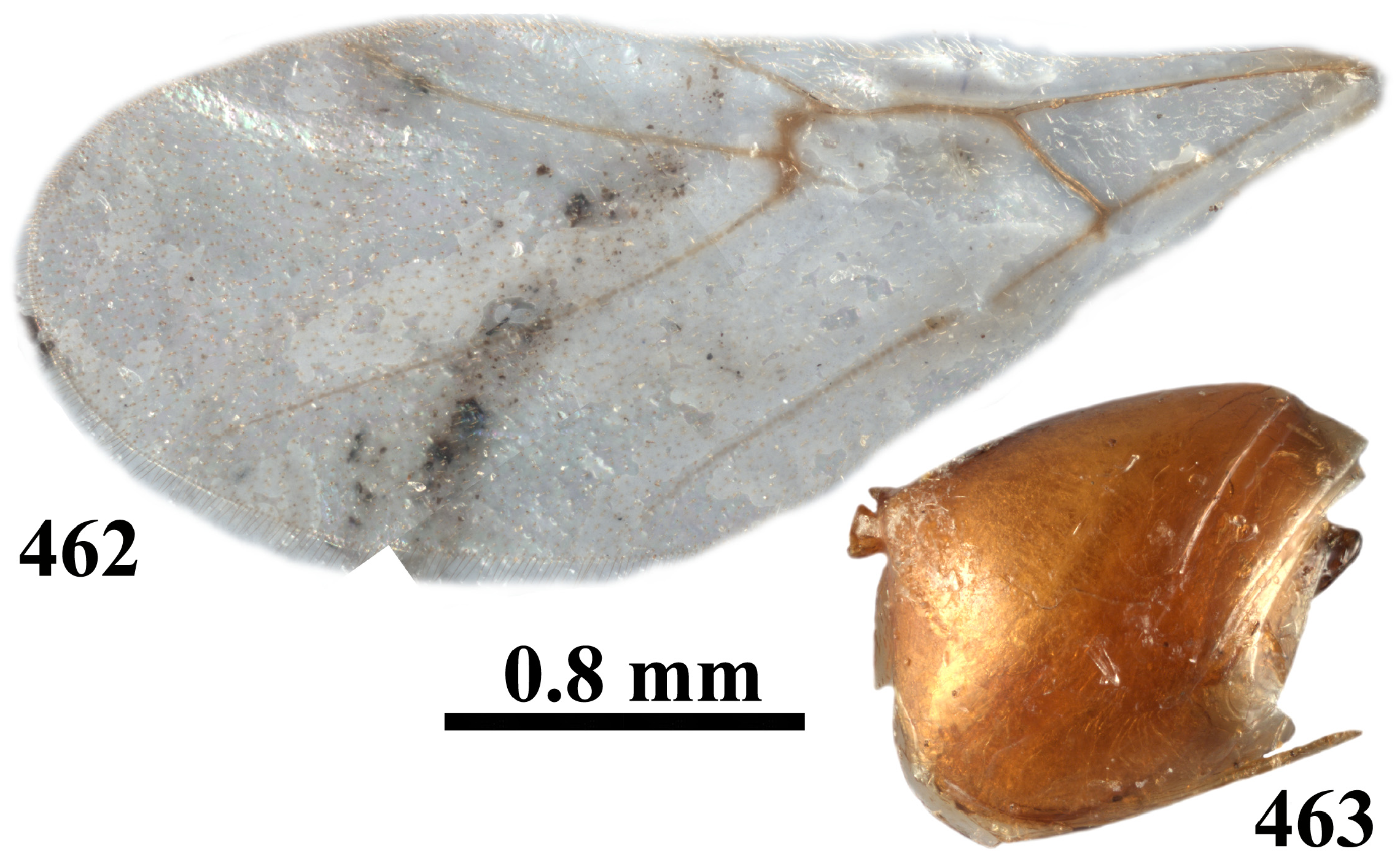
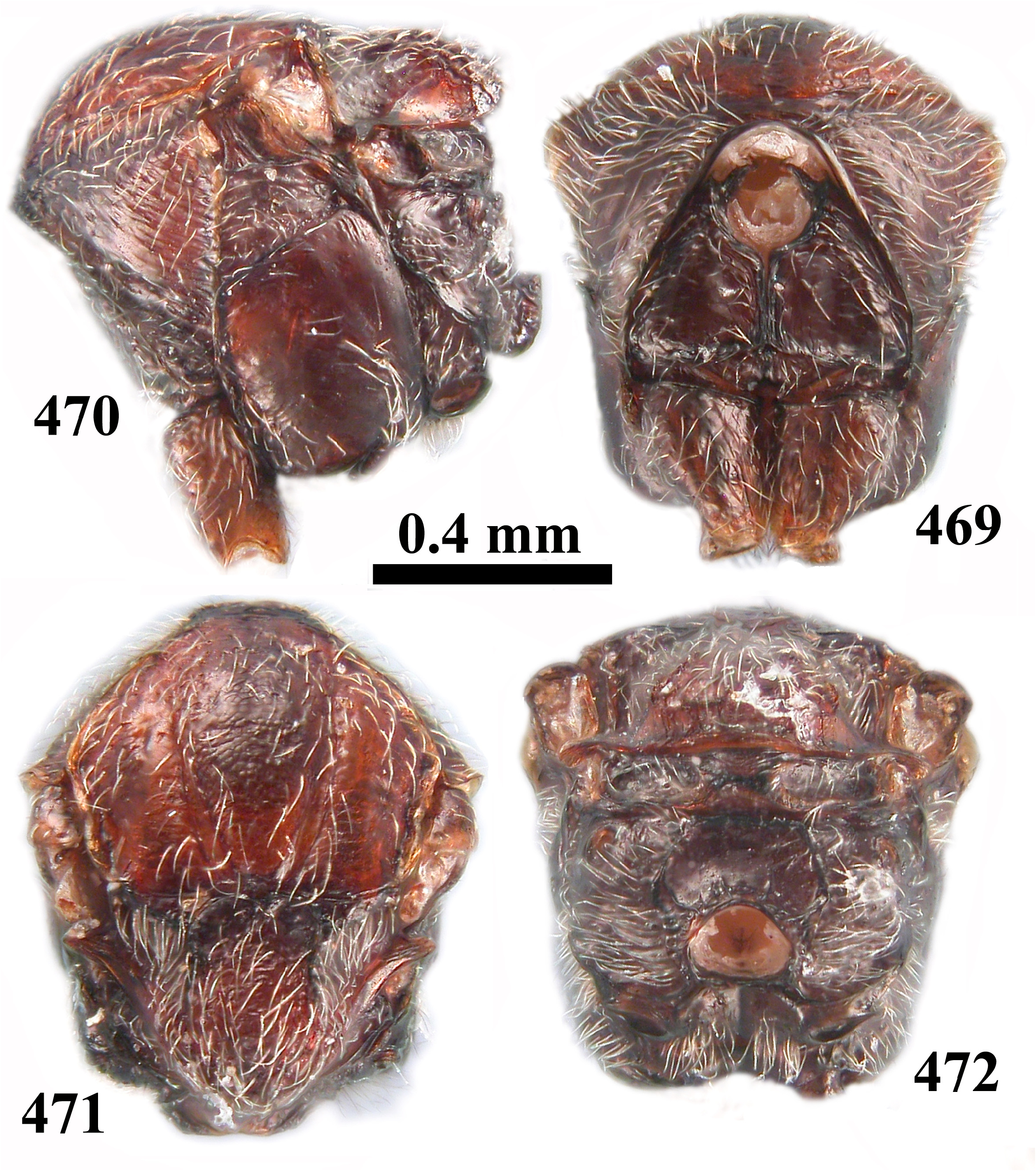
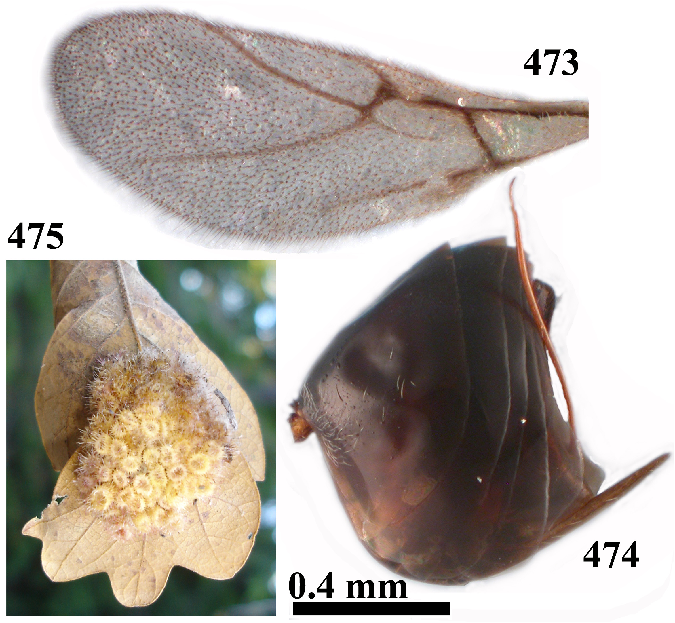
- Head not broadened behind eye in frontal view; clypeus with short interrupted delicate transverse striae; eye 1.9× as high as length of malar space ( Fig. 464 View FIGURES 464–468 ); metapleural sulcus reaching mesopleuron at upper 1/3 of its height ( Fig. 470 View FIGURES 469–472 ); fore wing only slightly longer than length of body ( Fig. 473 View FIGURES 473–475 ); all metasomal terga smooth, without micropunctures; prominent part of ventral spine of hypopygium around 5.0× as long as broad ( Fig. 474 View FIGURES 473–475 ); dark specimens, head dark brown to black, mesosoma reddish brown with some darker tints ( Figs 464–472 View FIGURES 464–468 View FIGURES 469–472 , 474 View FIGURES 473–475 ).................................... tubifaciens comb. nov. (asex)
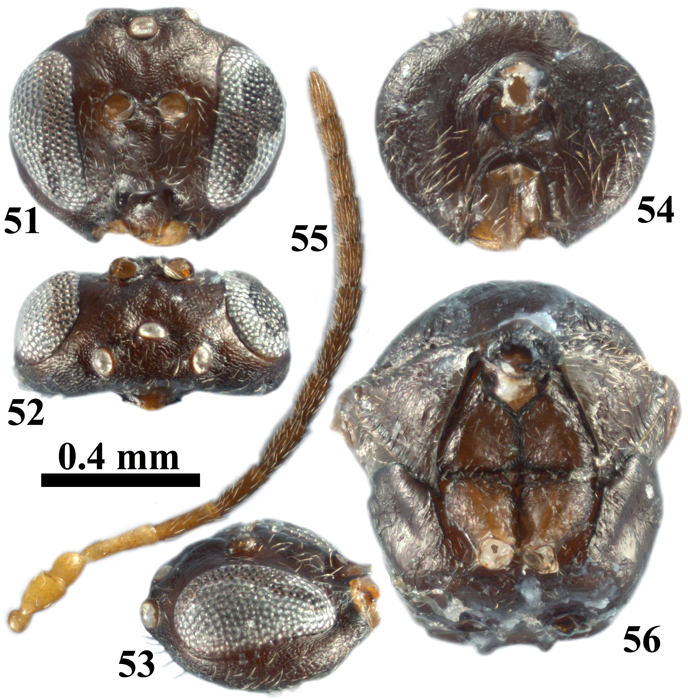
30. Eyes strongly converging ventrally; transfacial distance shorter than height of eye; antennal toruli located at half or slightly above height of eyes; eye around 6.0× as high as length of malar space ( Fig. 51 View FIGURES 51–56 ); body black ( Figs 51–61 View FIGURES 51–56 View FIGURES 57–60 View FIGURES 61–67 )............................................................................................. atrimentum comb. nov. (sex)
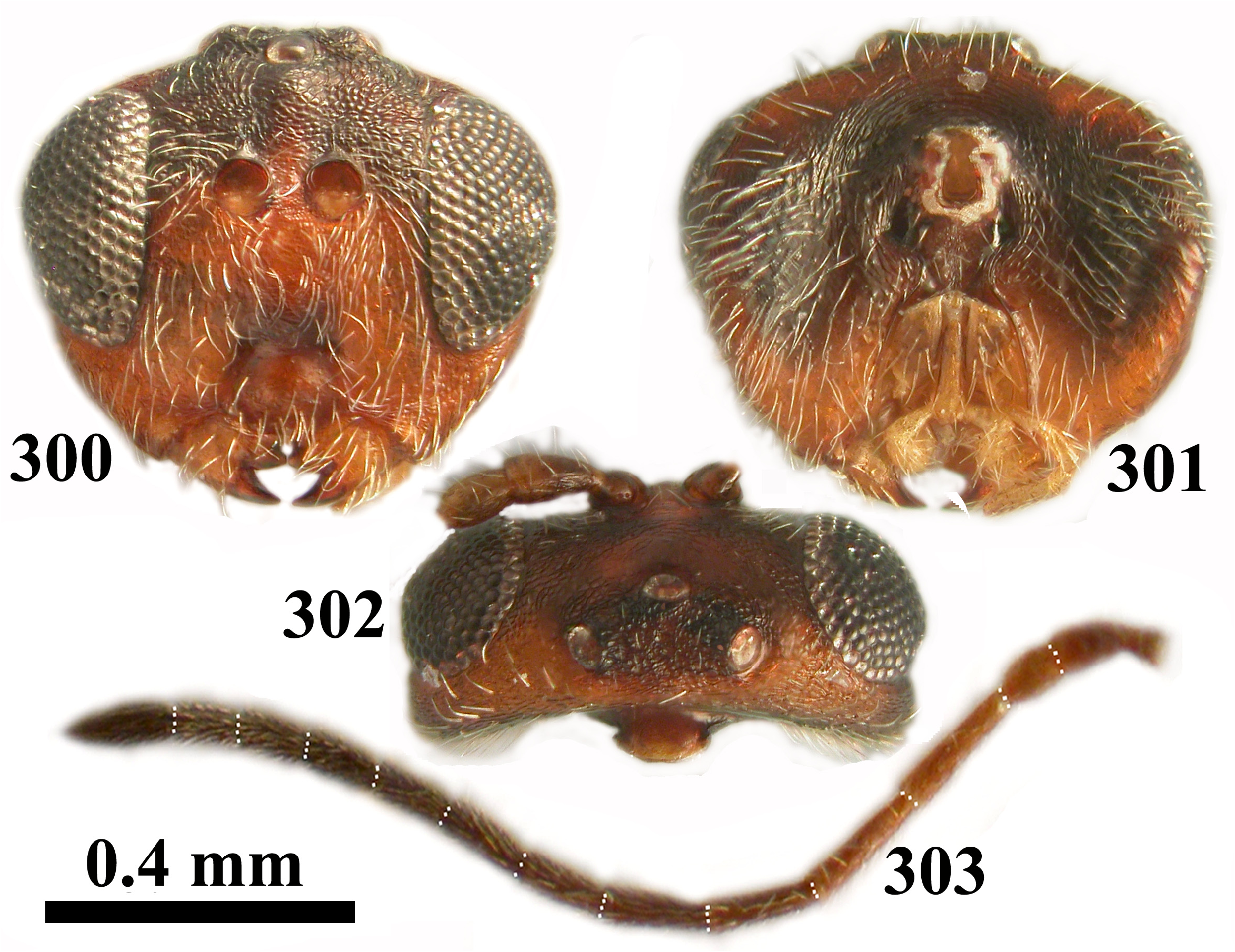
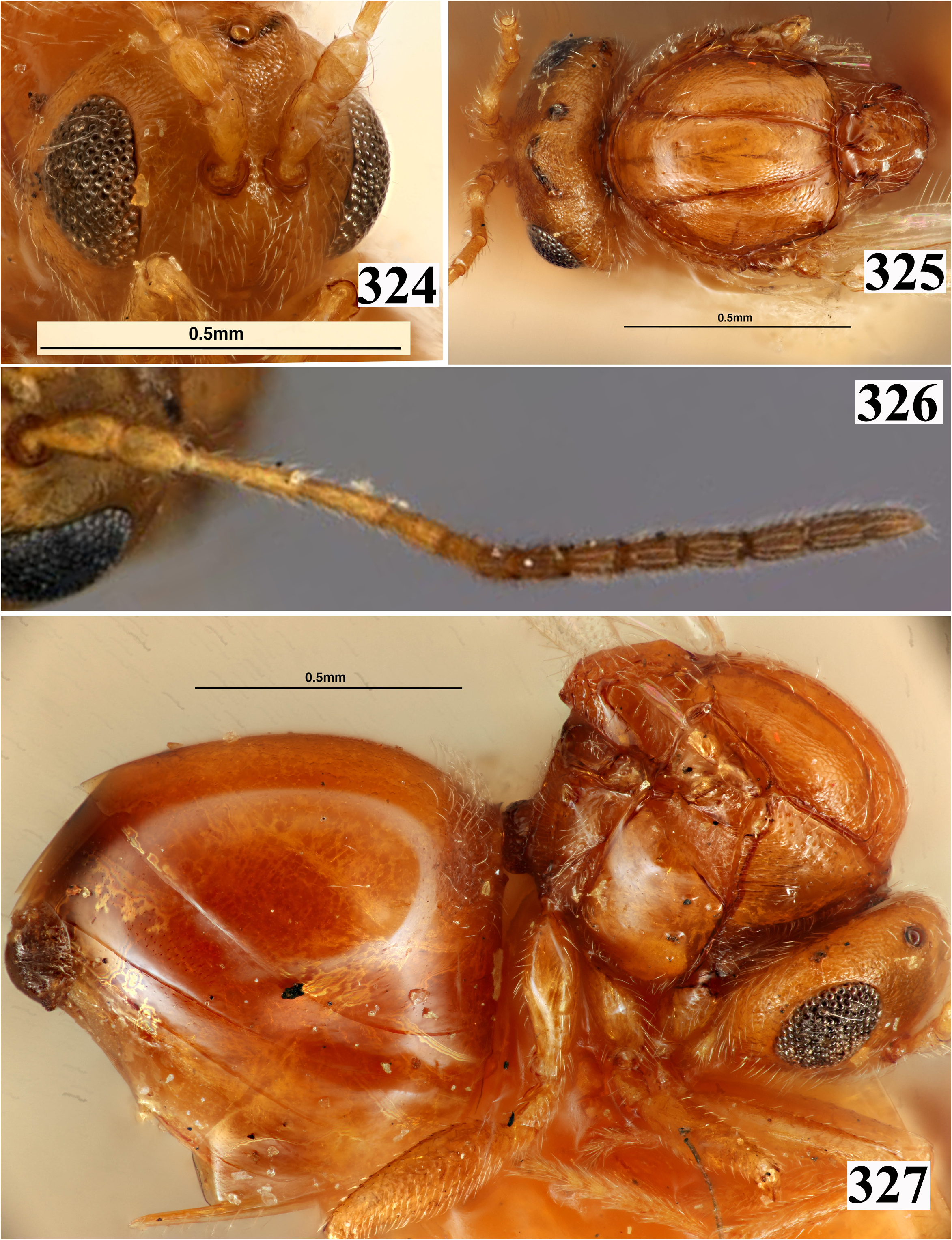
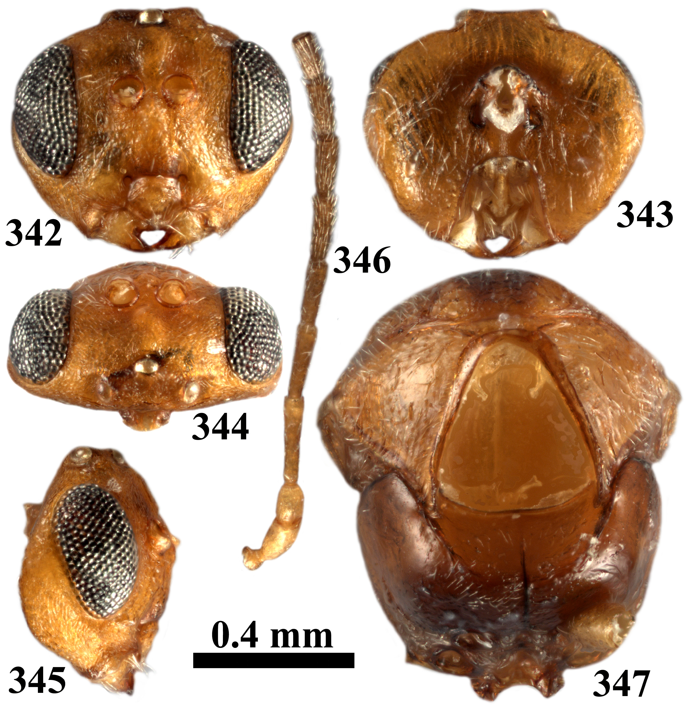
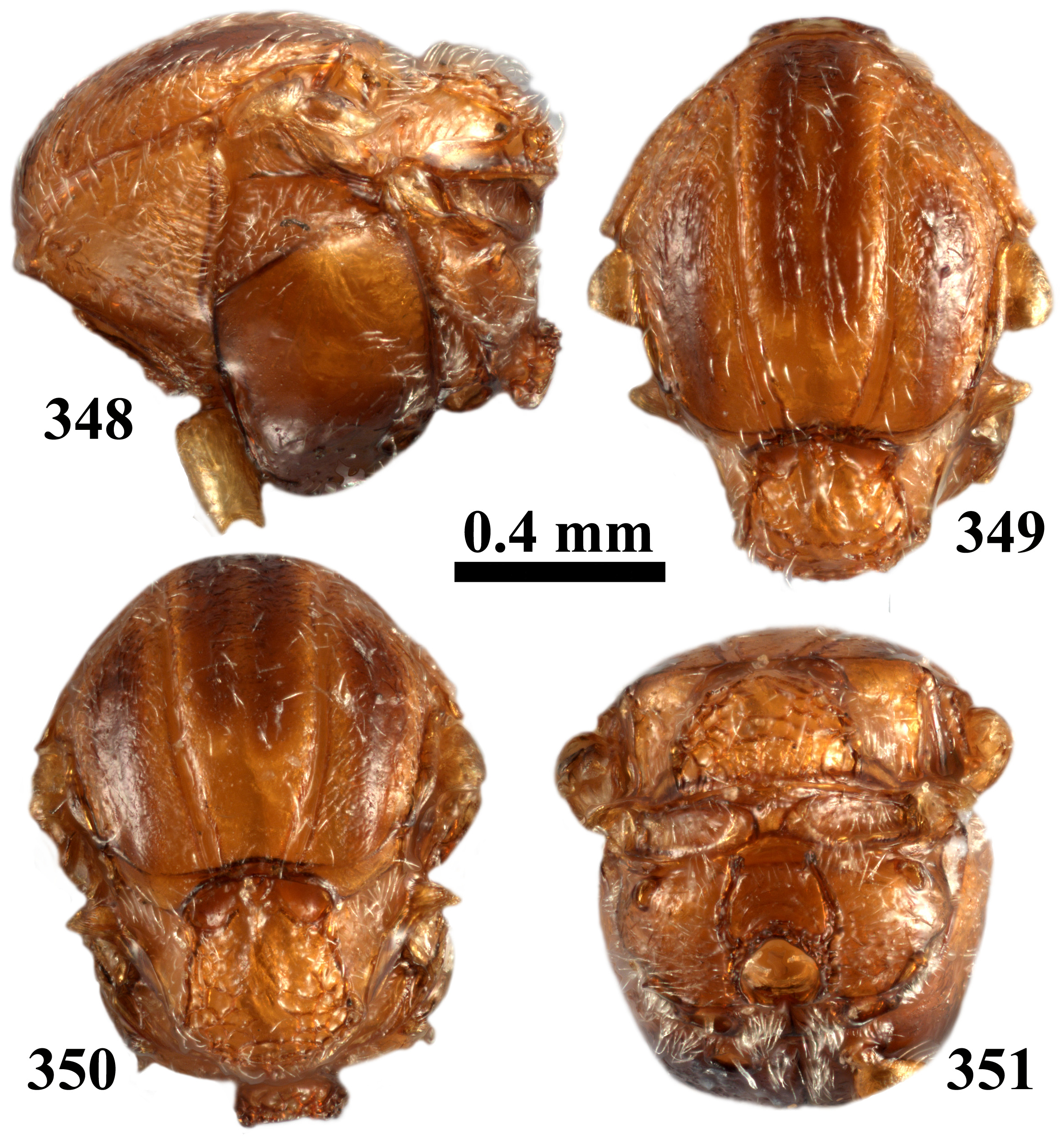
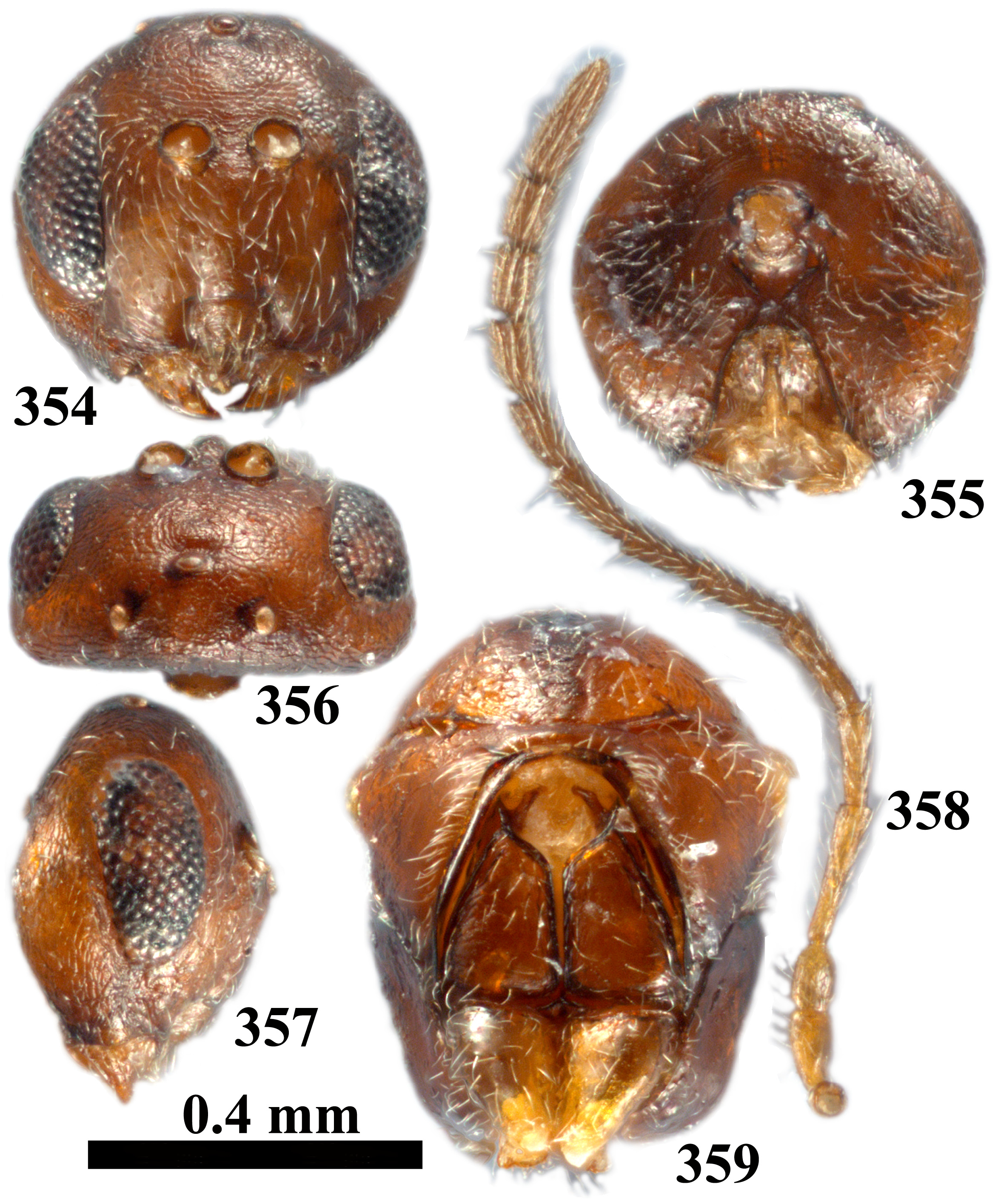
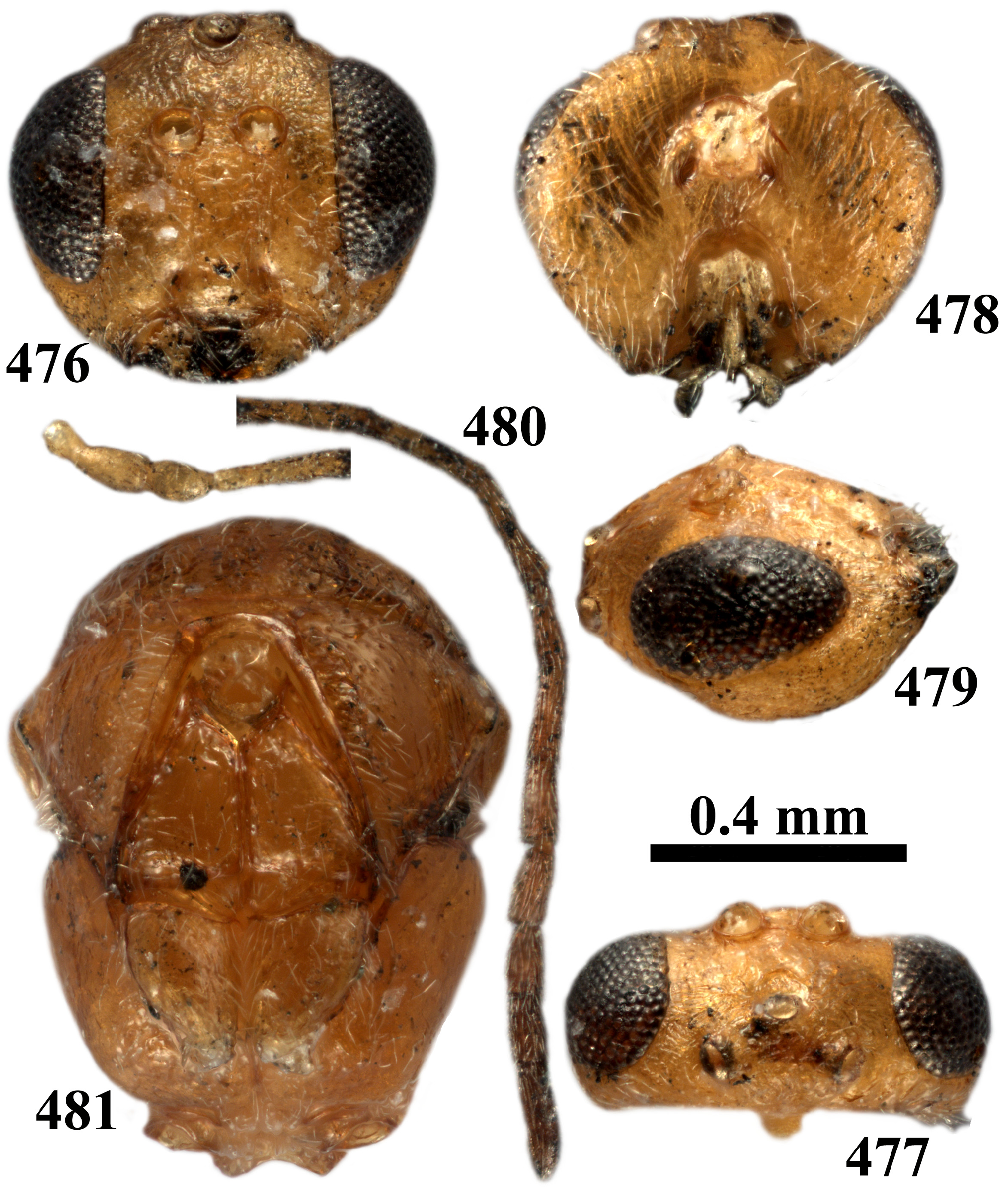
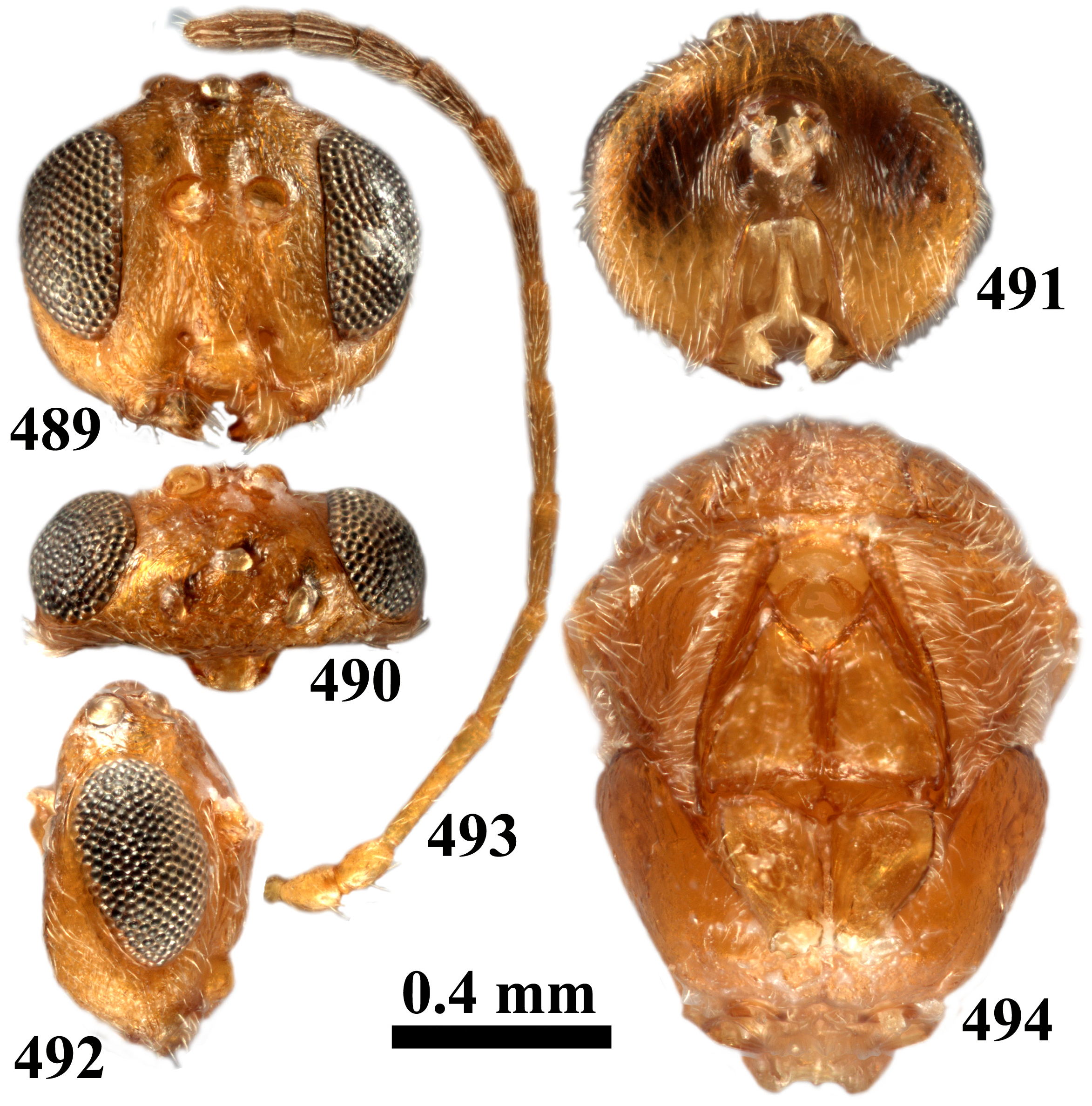
- Eyes parallel or very slightly converging ventrally; transfacial distance equal to or longer than height of eye; toruli located above mid-height of eyes; eye less than 3.0× as high as length of malar space ( Figs 69 View FIGURES 68–71 , 72 View FIGURES 72–77 , 300 View FIGURES 300–303 , 329 View FIGURES 329–334 , 342 View FIGURES 342–347 , 476 View FIGURES 476–481 , 489 View FIGURES 489–494 , 502 View FIGURES 502–507 ); body yellowish, reddish brown, brown to chestnut brown ( Figs 68–70 View FIGURES 68–71 , 72–82 View FIGURES 72–77 View FIGURES 78–81 View FIGURES 82–83 , 203–212 View FIGURES 203–208 View FIGURES 209–212 , 256–265 View FIGURES 256–261 View FIGURES 262–265 , 324–327 View FIGURES 324–327 , 329–338 View FIGURES 329–334 View FIGURES 335–338 , 354–363 View FIGURES 354–359 View FIGURES 360–363 ) 31
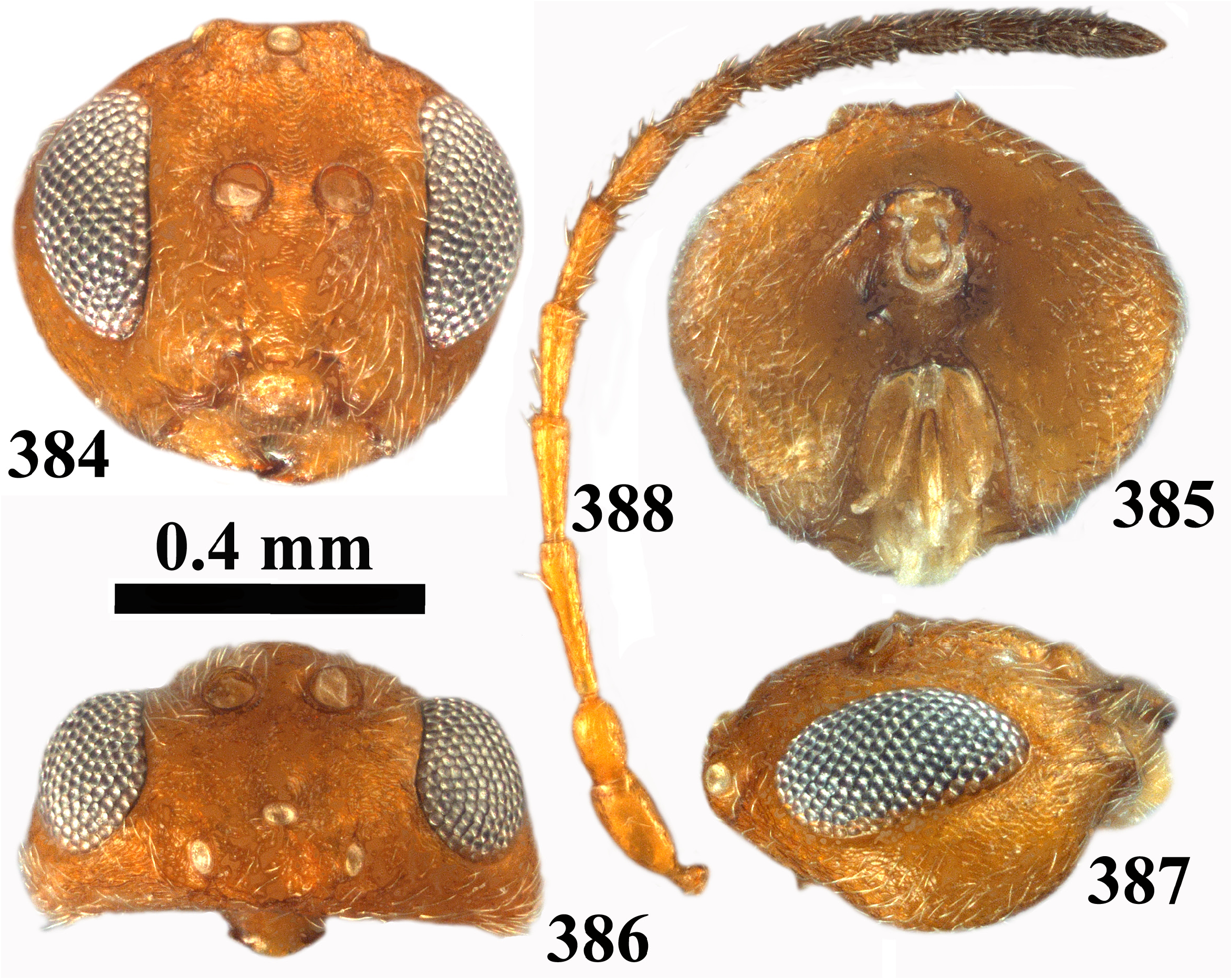
31. Head quadrangular or ovate in frontal view, with ocelli always elevated above frons ( Figs 342 View FIGURES 342–347 , 476 View FIGURES 476–481 , 489 View FIGURES 489–494 , 502 View FIGURES 502–507 ); lateral ocelli large, OOL subequal or at most 1.7× as long as diameter of ocellus ( Figs 344 View FIGURES 342–347 , 477 View FIGURES 476–481 , 490 View FIGURES 489–494 , 503 View FIGURES 502–507 )........................ 32
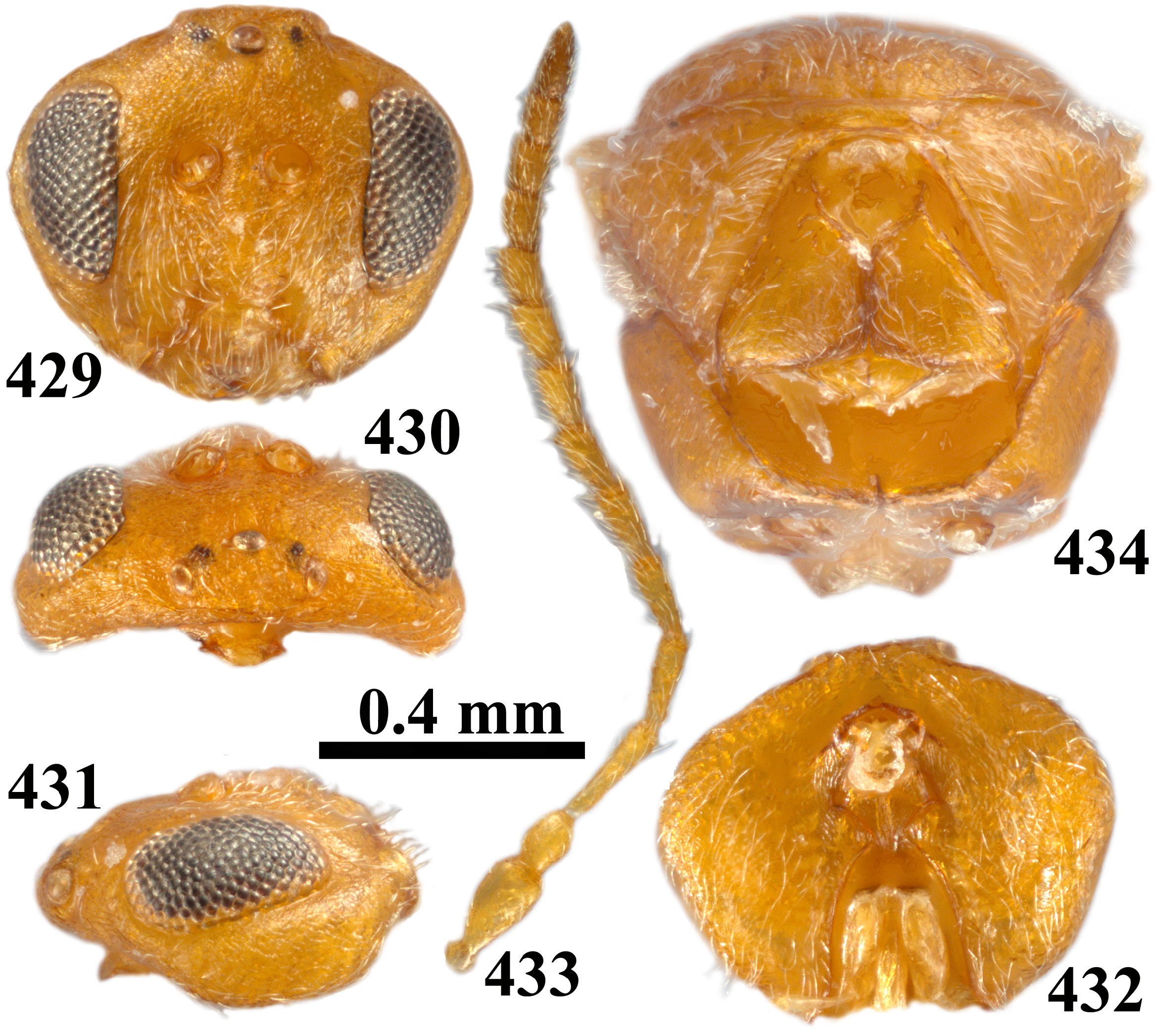
- Head rounded or trapezoid to triangular in frontal view, ocelli usually not elevated above frons ( Figs 69 View FIGURES 68–71 , 72 View FIGURES 72–77 , 203 View FIGURES 203–208 , 256 View FIGURES 256–261 , 300 View FIGURES 300–303 , 354 View FIGURES 354–359 , 384 View FIGURES 384–388 , 395 View FIGURES 395–400 , 429 View FIGURES 429–434 ); lateral ocelli smaller, OOL at least 2.5× as long as diameter of ocellus, if shorter then head and mesosoma are not yellowish or light brown ( Figs 73 View FIGURES 72–77 , 204 View FIGURES 203–208 , 257 View FIGURES 256–261 , 302 View FIGURES 300–303 , 356 View FIGURES 354–359 , 386 View FIGURES 384–388 , 397 View FIGURES 395–400 , 430 View FIGURES 429–434 )........................................ 34
32. F1 distinctly longer than scape+pedicel ( Fig. 493 View FIGURES 489–494 ); transfacial distance equal to or slightly shorter than height of eye ( Fig. 489 View FIGURES 489–494 )............................................................................ vitreum comb. rev. (asex)
- F1 subequal to or slightly longer than scape+pedicel ( Figs 346 View FIGURES 342–347 , 480 View FIGURES 476–481 , 506 View FIGURES 502–507 ); transfacial distance longer than height of eye ( Figs 342 View FIGURES 342–347 , 476 View FIGURES 476–481 , 502 View FIGURES 502–507 )....................................................................................... 33.. 33. Mesoscutum with some delicate transverse striae in anterior part between notauli; anterior parallel lines indistinct ( Figs 349–350 View FIGURES 348–351 ); mesoscutellar foveae transverse, ovate, separated by a broad carina, posteriorly delimited by a carina ( Fig. 350 View FIGURES 348–351 )............................................................................... splendens comb. nov. (asex)
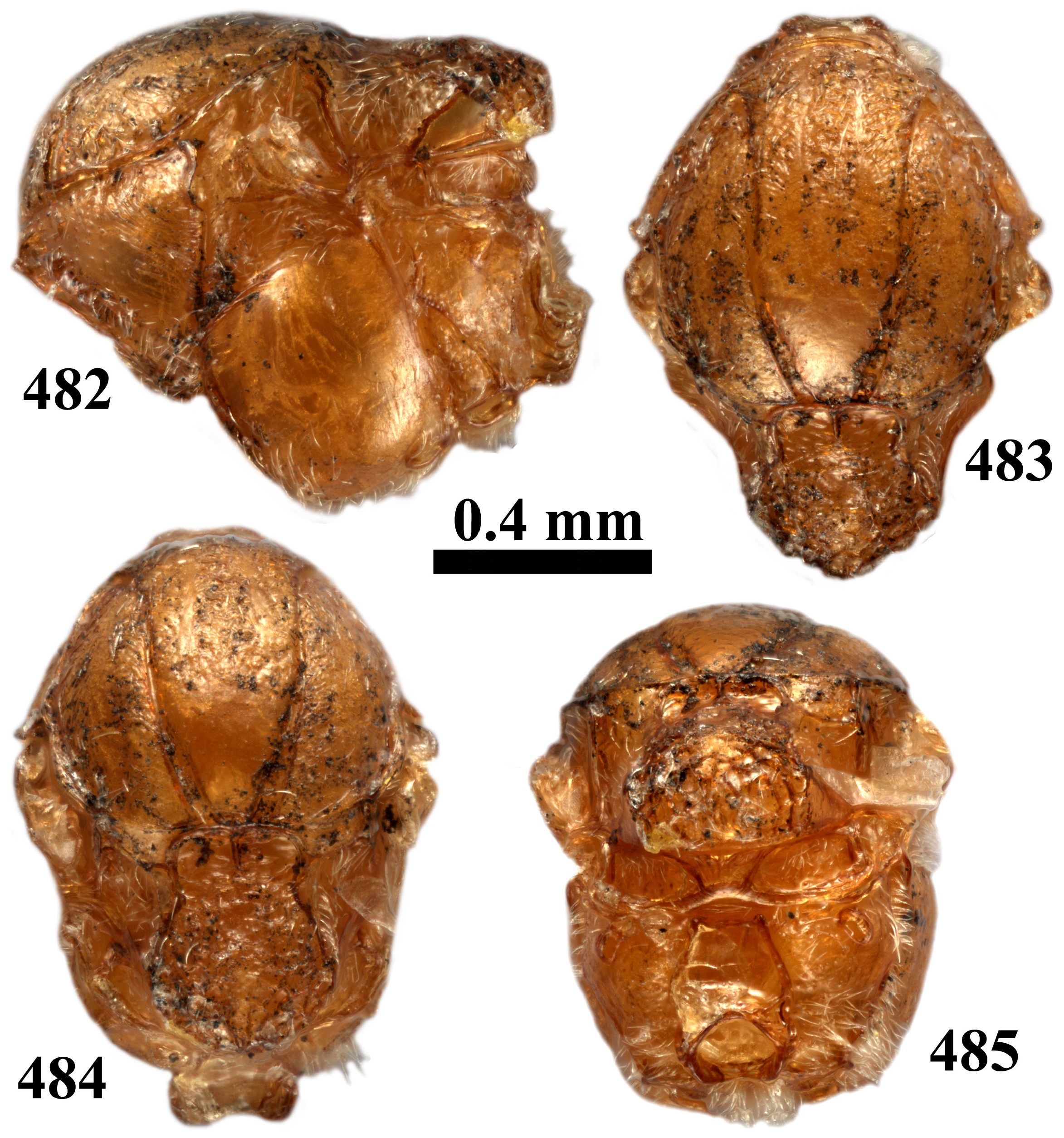
- Mesoscutum without striae, anterior parallel line in the form of a bare, smooth stripe, extending to 1/3 length of mesoscutum ( Figs 483–484 View FIGURES 482–485 ); foveae quadrangular, separated by a thin carina, posteriorly not delimited by a carina ( Fig. 484 View FIGURES 482–485 ).......................................................................................... verutum comb. rev. (asex)
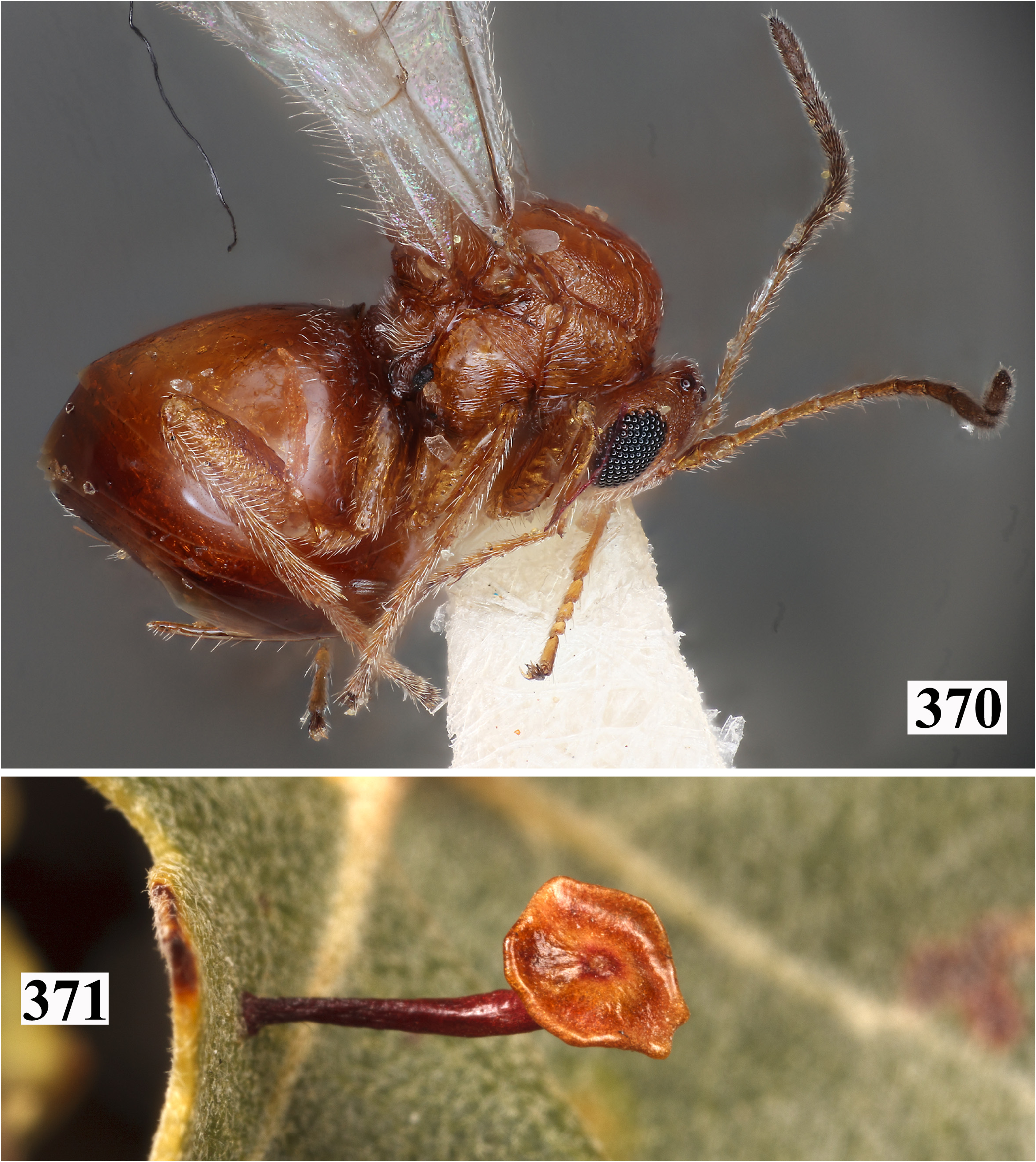
34. Mesopleuron with transverse reticulate-carinate band at mid-height ( Figs 209 View FIGURES 209–212 , 262 View FIGURES 262–265 , 370 View FIGURES 370–371 , 389 View FIGURES 389–392 , 435 View FIGURES 435–438 ).................... 35
- Mesopleuron entirely smooth or slightly sculptured on anterior margin ( Figs 68 View FIGURES 68–71 , 78 View FIGURES 78–81 , 304 View FIGURES 304–307 , 317 View FIGURES 317–320 , 327 View FIGURES 324–327 , 335 View FIGURES 335–338 , 360 View FIGURES 360–363 , 401 View FIGURES 401–404 , 419 View FIGURES 419–425 , 435 View FIGURES 435–438 ). .................................................................................................. 39
35. Gena broadened behind eye in frontal view ( Figs 384 View FIGURES 384–388 , 429 View FIGURES 429–434 ); 2nd metasomal tergum without micropunctures posteriorly and subsequent terga with or without micropunctures ( Figs 393 View FIGURES 393–394 , 439 View FIGURES 439–440 ).............................................. 36
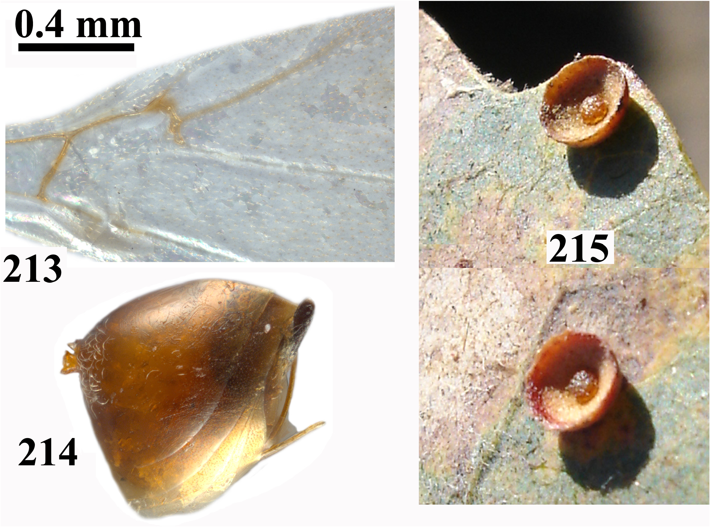
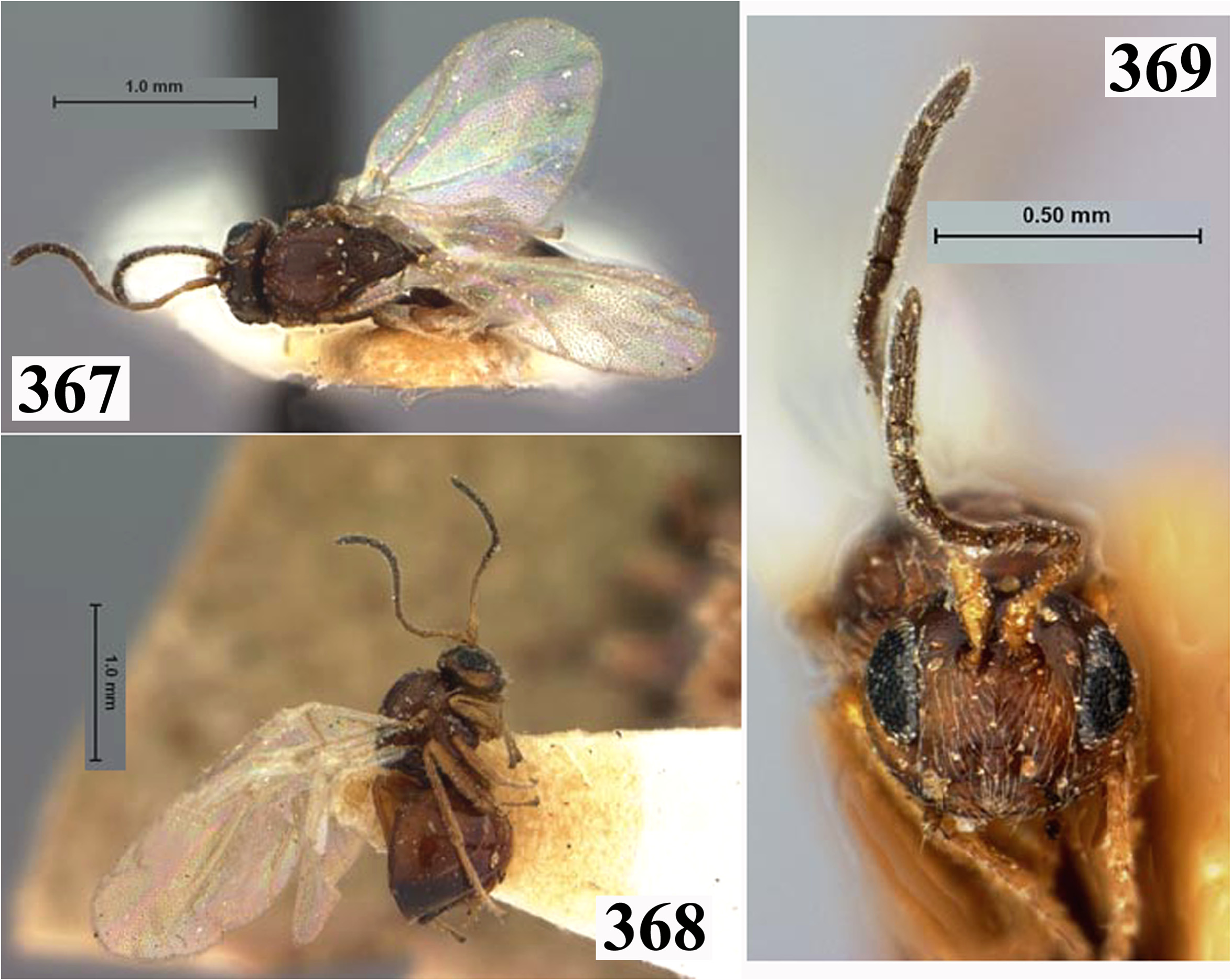
- Gena not broadened behind eye in frontal view ( Figs 203 View FIGURES 203–208 , 256 View FIGURES 256–261 , 369 View FIGURES 367–369 ); 2nd metasomal tergum with a band of micropunctures posteriorly and all subsequent terga with micropunctures ( Figs 214 View FIGURES 213–215 , 267 View FIGURES 266–268 , 368 View FIGURES 367–369 ).................................... 37
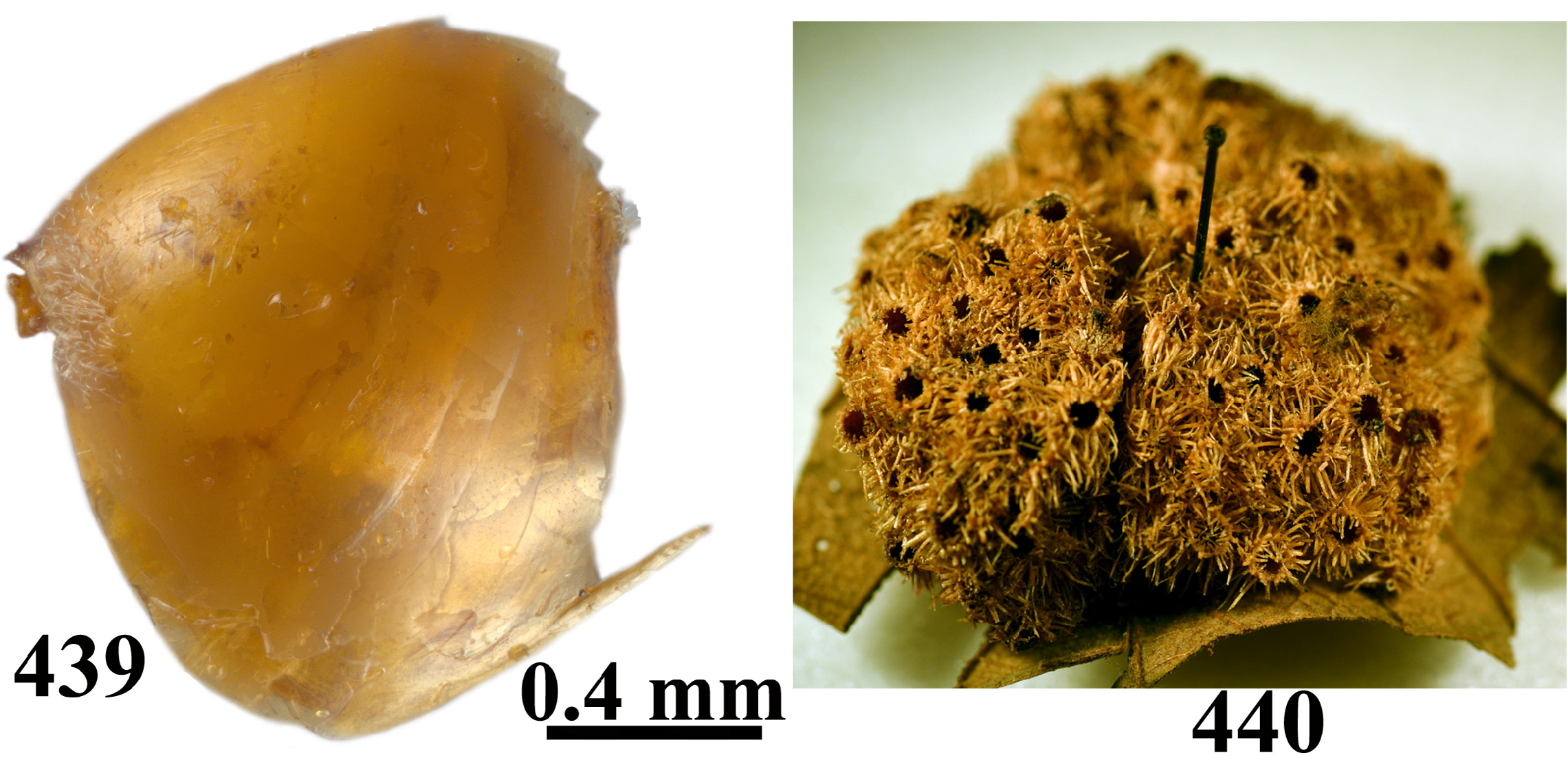
36. Mesoscutum coarsely reticulated; notaulus complete, notaular furrow smooth and deep along its length or alutaceous on anterior 1/4 ( Fig. 436 View FIGURES 435–438 ); mesoscutellar disk flat in lateral view, following curvature of mesoscutum ( Fig. 437 View FIGURES 435–438 ); subaxillular bar reaching half-length of mesoscutellum in lateral view ( Fig. 435 View FIGURES 435–438 )................................ tibiale comb. rev. (asex), part
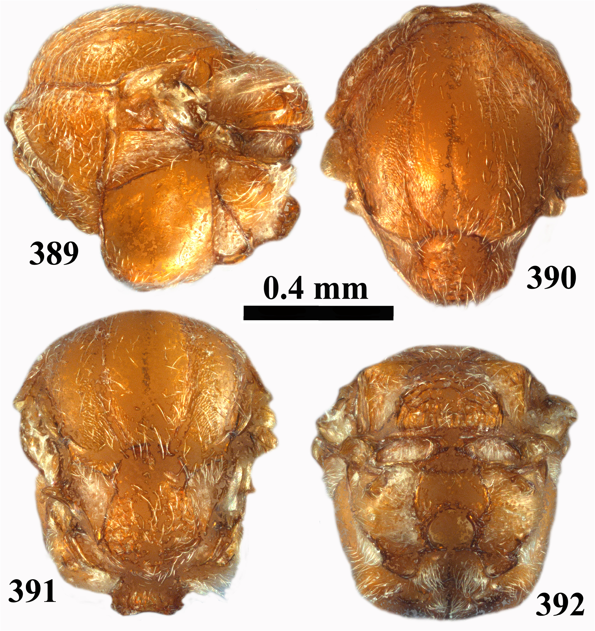
- Mesoscutum finely alutaceous; notaulus complete, but indistinct and shallow at least on anterior 3/4 of its length ( Fig. 390 View FIGURES 389–392 ); mesoscutellar disk strongly curved in lateral view; mesoscutum and mesoscutellum forming two independent lobes in lateral view ( Fig. 391 View FIGURES 389–392 ); subaxillular bar reaching 1/3 of height of mesoscutellum ( Fig. 389 View FIGURES 389–392 )........... syndicorum sp. nov. (asex)
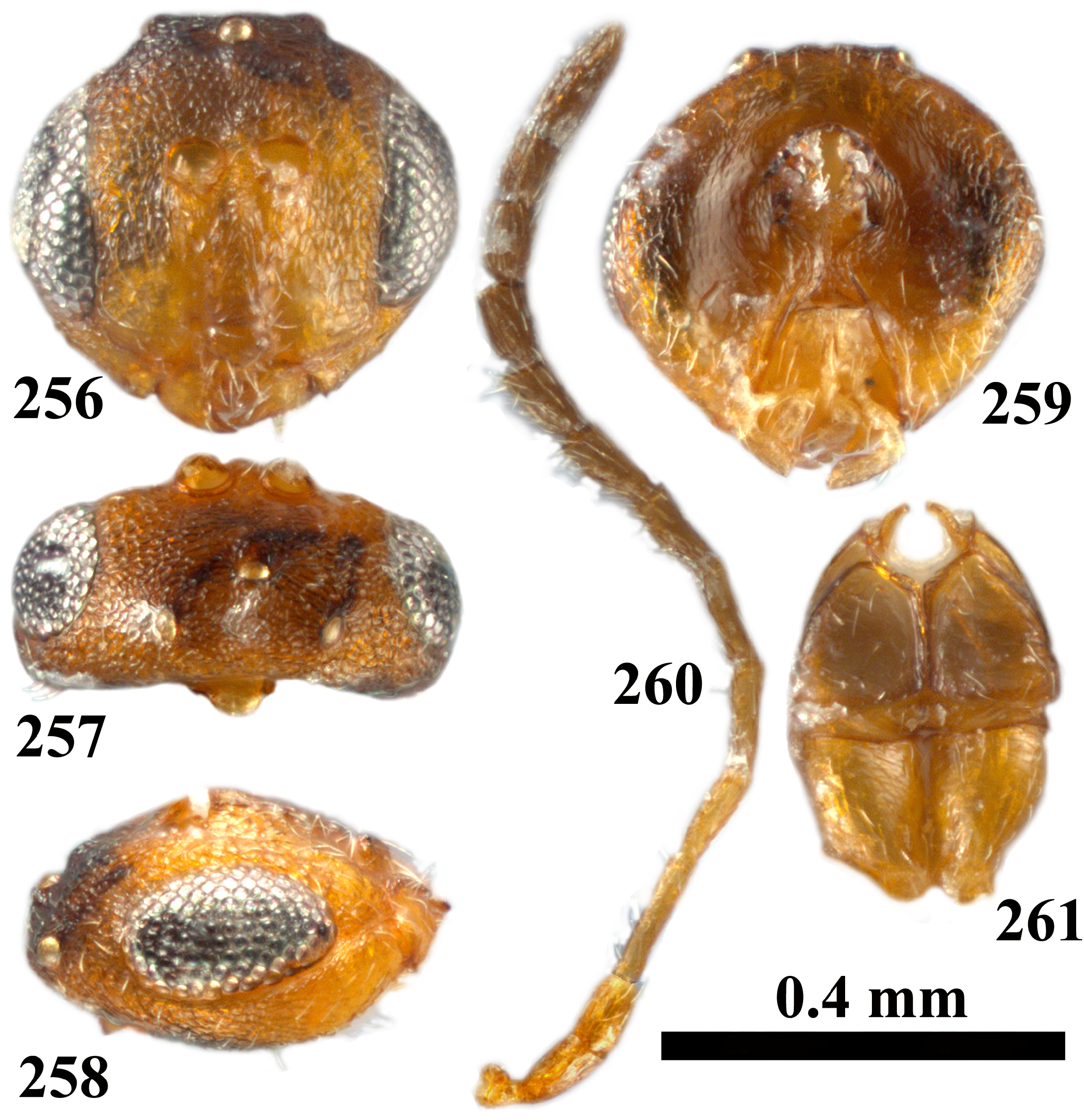
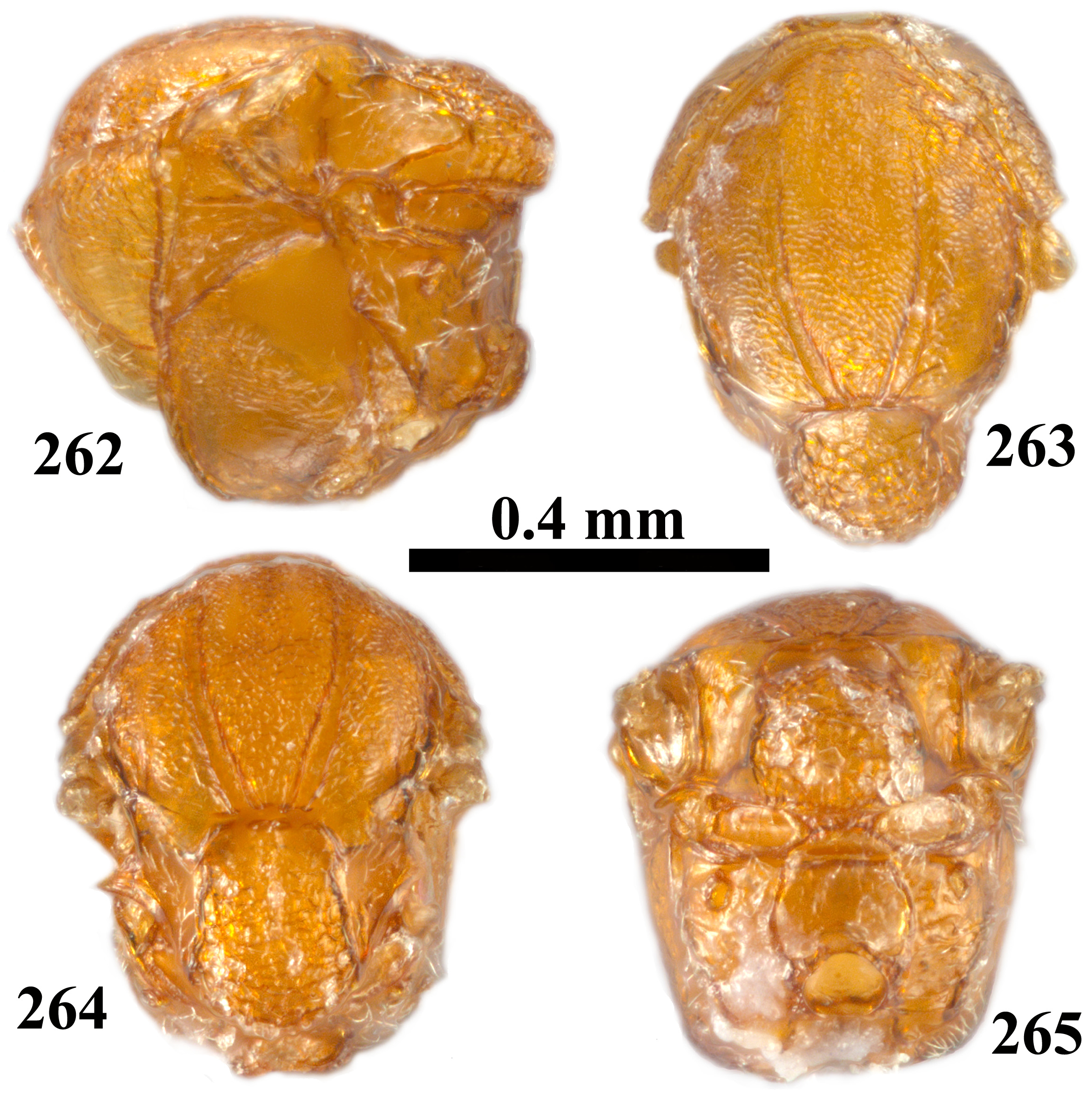
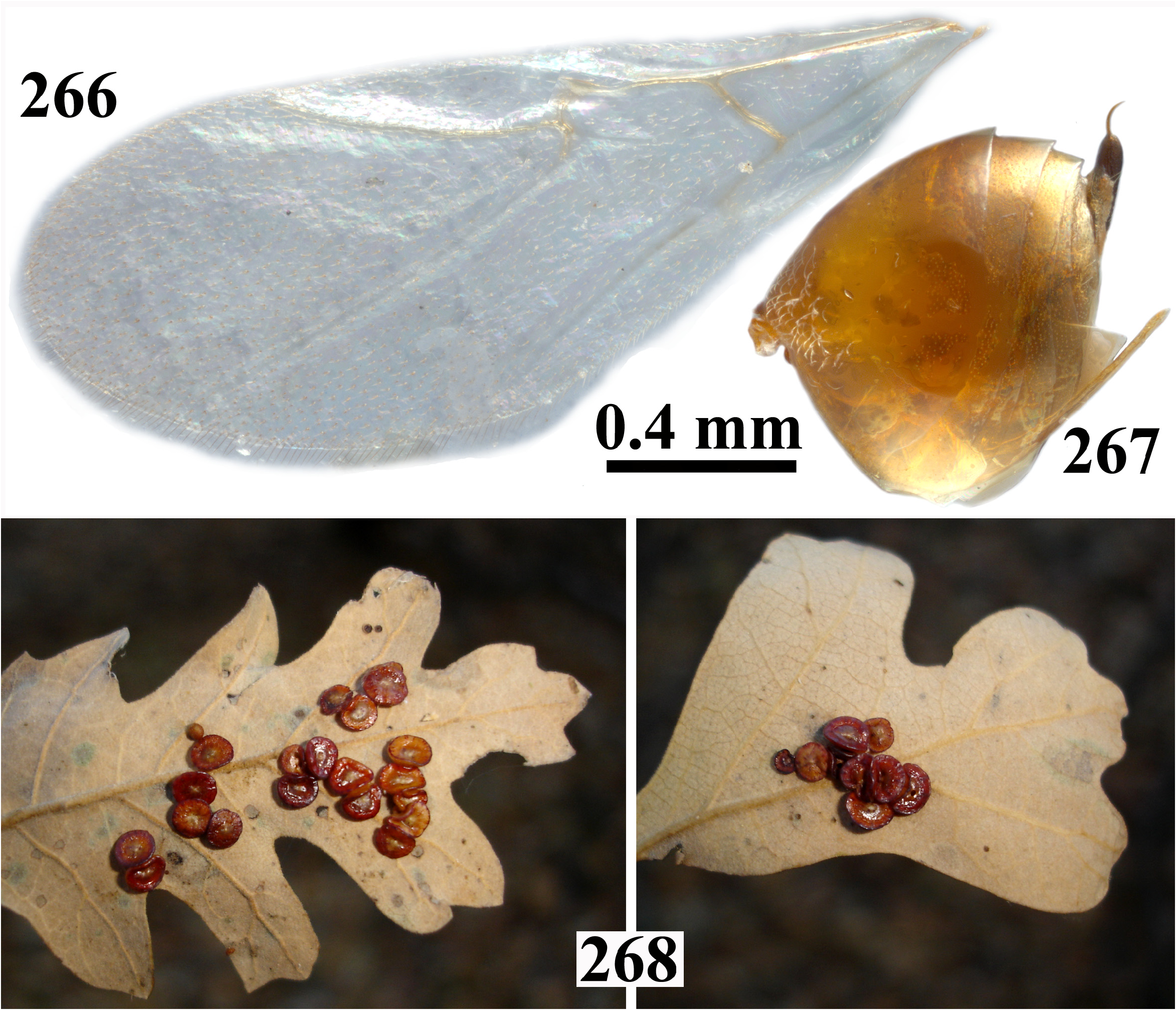
37. Malar space without striae ( Fig. 256 View FIGURES 256–261 ); notaulus complete, indistinct in anterior 1/4 of mesoscutum ( Fig. 263 View FIGURES 262–265 ); mesoscutellar foveae absent or slightly impressed ( Fig. 264 View FIGURES 262–265 ); veins of fore wing pale ( Fig. 266 View FIGURES 266–268 ); prominent part of ventral spine of hypopygium 7.0× as long as broad in ventral view ( Fig. 267 View FIGURES 266–268 )........................................ parmula comb. nov. (asex)
- Malar space with a few delicate striae radiating from clypeus ( Figs 203 View FIGURES 203–208 , 369 View FIGURES 367–369 ); notaulus complete, distinct through all its length ( Figs 210 View FIGURES 209–212 , 367 View FIGURES 367–369 ); mesoscutellar foveae defined ( Figs 211 View FIGURES 209–212 , 367 View FIGURES 367–369 ); veins of fore wing light brown to brown ( Figs 213 View FIGURES 213–215 , 367 View FIGURES 367–369 ); prominent part of ventral spine around 4.5× as long as broad in ventral view ( Figs 214 View FIGURES 213–215 , 368 View FIGURES 367–369 )......................... 38
38. Antenna with 11 flagellomeres ( Figs 368–370 View FIGURES 367–369 View FIGURES 370–371 ); eye 2.7× as high as length of malar space ( Fig. 369 View FIGURES 367–369 ); veins of fore wing brown; areolet present ( Fig. 367 View FIGURES 367–369 ); central propodeal area with rugae.............................. stellulum comb. nov. (asex)
- Antenna with 12 flagellomeres ( Fig. 207 View FIGURES 203–208 ); eye 2.2× as high as length of malar space ( Fig. 203 View FIGURES 203–208 ); veins of fore wing lighter; areolet absent ( Fig. 213 View FIGURES 213–215 ); central propodeal area without rugae ( Fig. 212 View FIGURES 209–212 )....................... gigas comb. nov. (asex)
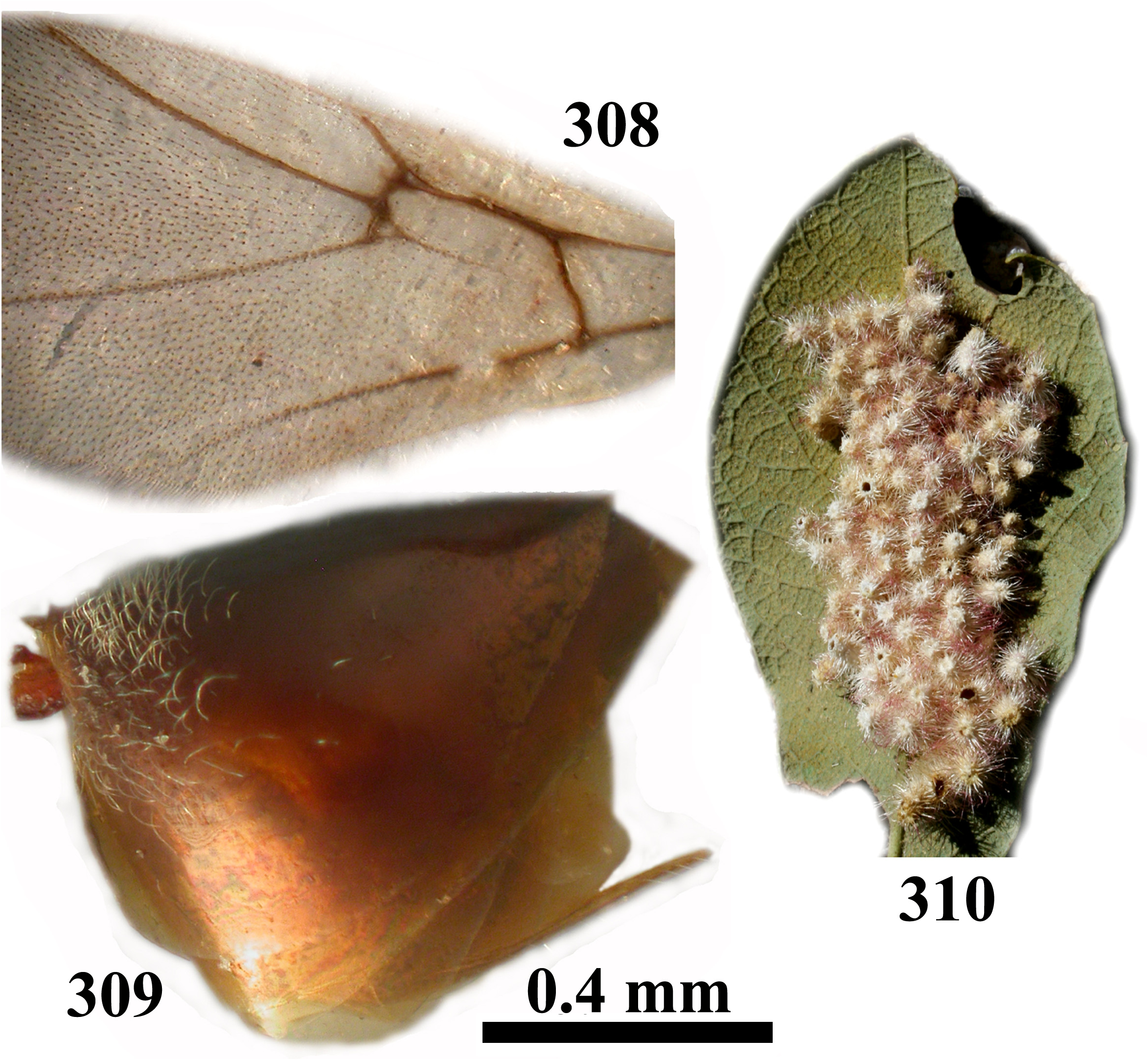
39. Head trapezoid ( Fig. 300 View FIGURES 300–303 ); gena not broadened behind eye; transfacial distance equal to height of eye ( Fig. 300 View FIGURES 300–303 ); antenna with 11 flagellomeres; F1=F2; mesoscutellar disc reticulate ( Fig. 303 View FIGURES 300–303 ); metapleural sulcus reaching mesopleuron in lower 1/3 of its height ( Fig. 304 View FIGURES 304–307 ); 2nd metasomal tergum extending to 3/4 of metasoma length in dorsal view ( Fig. 309 View FIGURES 308–310 )................................................................................................... roberti sp. nov. (asex)
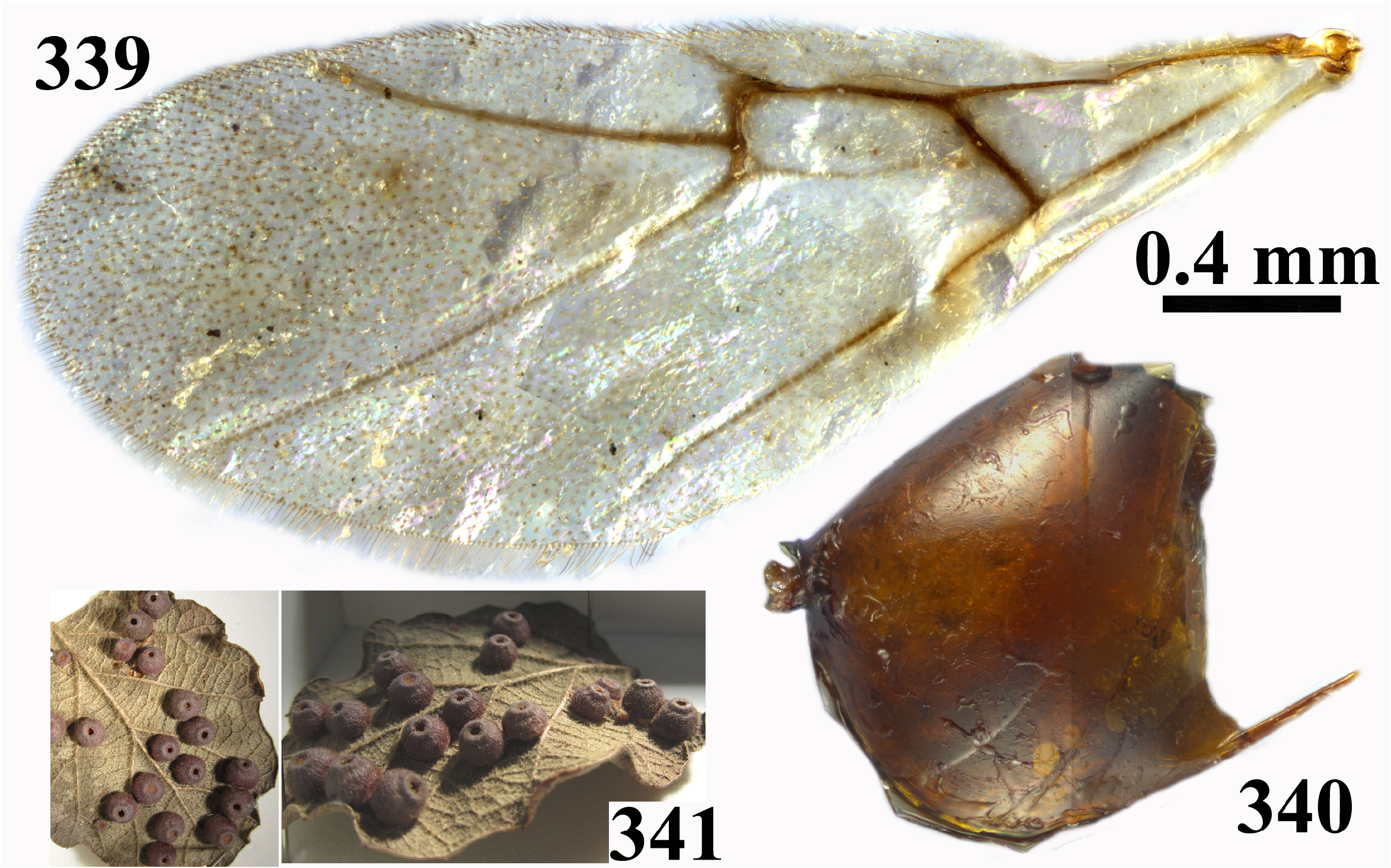
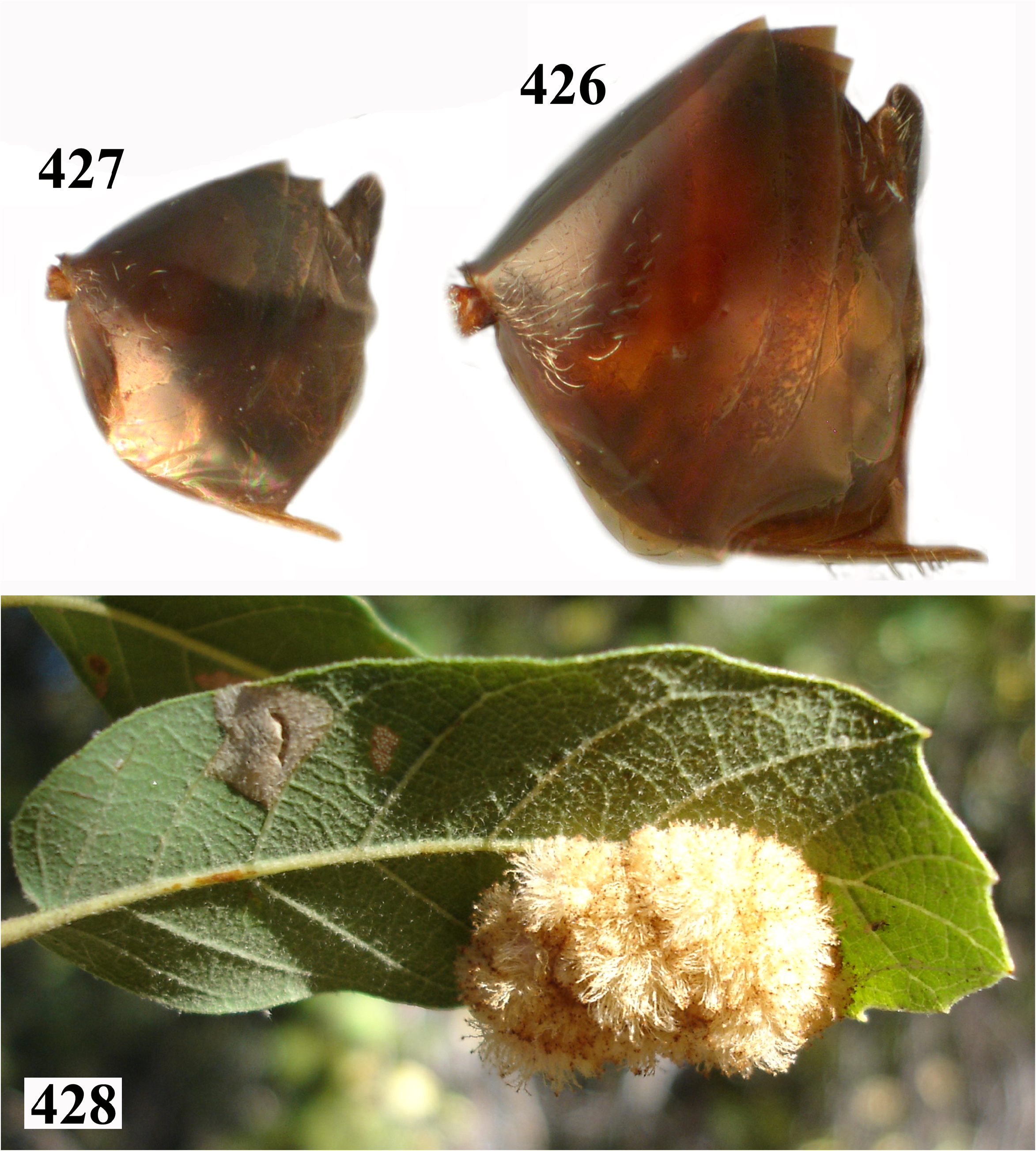
- Head rounded or ovate ( Figs 69 View FIGURES 68–71 , 72 View FIGURES 72–77 , 311 View FIGURES 311–316 , 324 View FIGURES 324–327 , 329 View FIGURES 329–334 , 354 View FIGURES 354–359 , 395 View FIGURES 395–400 , 409 View FIGURES 409–418 , 429 View FIGURES 429–434 ); gena at least slightly broadened behind eyes; transfacial distance longer than height of eye ( Figs 69 View FIGURES 68–71 , 72 View FIGURES 72–77 , 311 View FIGURES 311–316 , 324 View FIGURES 324–327 , 329 View FIGURES 329–334 , 354 View FIGURES 354–359 , 395 View FIGURES 395–400 , 409 View FIGURES 409–418 , 429 View FIGURES 429–434 ); antenna with 12 flagellomeres, sometimes suture between F11 and F12 incomplete; F1 longer than F2; distal flagellomeres broader than basal ones ( Figs 76 View FIGURES 72–77 , 315 View FIGURES 311–316 , 326 View FIGURES 324–327 , 333 View FIGURES 329–334 , 358 View FIGURES 354–359 , 399 View FIGURES 395–400 , 413, 418 View FIGURES 409–418 , 433 View FIGURES 429–434 ); mesoscutellar disc smooth or with a different sculpture; metapleural sulcus reaching mesopleuron at least at 1/2 of its height ( Figs 70 View FIGURES 68–71 , 80 View FIGURES 78–81 , 319 View FIGURES 317–320 , 325 View FIGURES 324–327 , 337 View FIGURES 335–338 , 362 View FIGURES 360–363 , 403 View FIGURES 401–404 , 420, 424 View FIGURES 419–425 , 437 View FIGURES 435–438 ); 2nd metasomal tergum extending to 1/2–2/3 length of metasoma in dorsal view ( Figs 68 View FIGURES 68–71 , 82 View FIGURES 82–83 , 321 View FIGURES 321–323 , 327 View FIGURES 324–327 , 340 View FIGURES 339–341 , 365 View FIGURES 364–366 , 406 View FIGURES 405–408 , 426–427 View FIGURES 426–428 , 439 View FIGURES 439–440 )............................ 40
40. Head transversely ovate in frontal view ( Figs 329 View FIGURES 329–334 , 395 View FIGURES 395–400 , 409 View FIGURES 409–418 , 429 View FIGURES 429–434 ); slightly elevated median area of lower face alutaceous to delicately coriaceous, matte ( Figs 329 View FIGURES 329–334 , 395 View FIGURES 395–400 , 409 View FIGURES 409–418 , 429 View FIGURES 429–434 ); lateral sides of pronotum longitudinally striated at least on posterior half, rest of pronotum alutaceous, matte ( Figs 335 View FIGURES 335–338 , 401 View FIGURES 401–404 , 419 View FIGURES 419–425 , 435 View FIGURES 435–438 ); mesoscutum coarsely reticulated, each individual cell formed by the reticula bulging ( Figs 336 View FIGURES 335–338 , 402 View FIGURES 401–404 , 420 View FIGURES 419–425 , 436 View FIGURES 435–438 ); mesopleuron finely striated to alutaceous on anterior margin ( Figs 335 View FIGURES 335–338 , 401 View FIGURES 401–404 , 419 View FIGURES 419–425 , 435 View FIGURES 435–438 )............................................................................................... 41
- Head rounded ( Figs 69 View FIGURES 68–71 , 72 View FIGURES 72–77 , 311 View FIGURES 311–316 , 324 View FIGURES 324–327 , 354 View FIGURES 354–359 ); slightly elevated median area of lower face smooth, shining ( Figs 69 View FIGURES 68–71 , 72 View FIGURES 72–77 , 311 View FIGURES 311–316 , 324 View FIGURES 324–327 , 354 View FIGURES 354–359 ); lateral sides of pronotum with fine longitudinal striations on posterior margin, never reaching half the length of pronotum, rest of the pronotum shining, smooth with piliferous points on dorsal margin ( Figs 68 View FIGURES 68–71 , 78 View FIGURES 78–81 , 317 View FIGURES 317–320 , 327 View FIGURES 324–327 , 360 View FIGURES 360–363 ); mesoscutum finely reticulated, the cells formed by the reticula flat ( Figs 70 View FIGURES 68–71 , 79 View FIGURES 78–81 , 318 View FIGURES 317–320 , 325 View FIGURES 324–327 , 361 View FIGURES 360–363 ), mesopleuron completely smooth ( Figs 68 View FIGURES 68–71 , 78 View FIGURES 78–81 , 317 View FIGURES 317–320 , 327 View FIGURES 324–327 , 360 View FIGURES 360–363 )........................................................................................... 44
41. Mesoscutellum 1.3× longer than broad and margined posteriorly by a strong circumscutellar carina ( Fig. 337 View FIGURES 335–338 )............................................................................................. serranoae sp. nov. (asex)
- Mesoscutellum slightly longer than broad and circumscutellar carina absent ( Figs 403 View FIGURES 401–404 , 420, 424 View FIGURES 419–425 , 437 View FIGURES 435–438 )................. 42
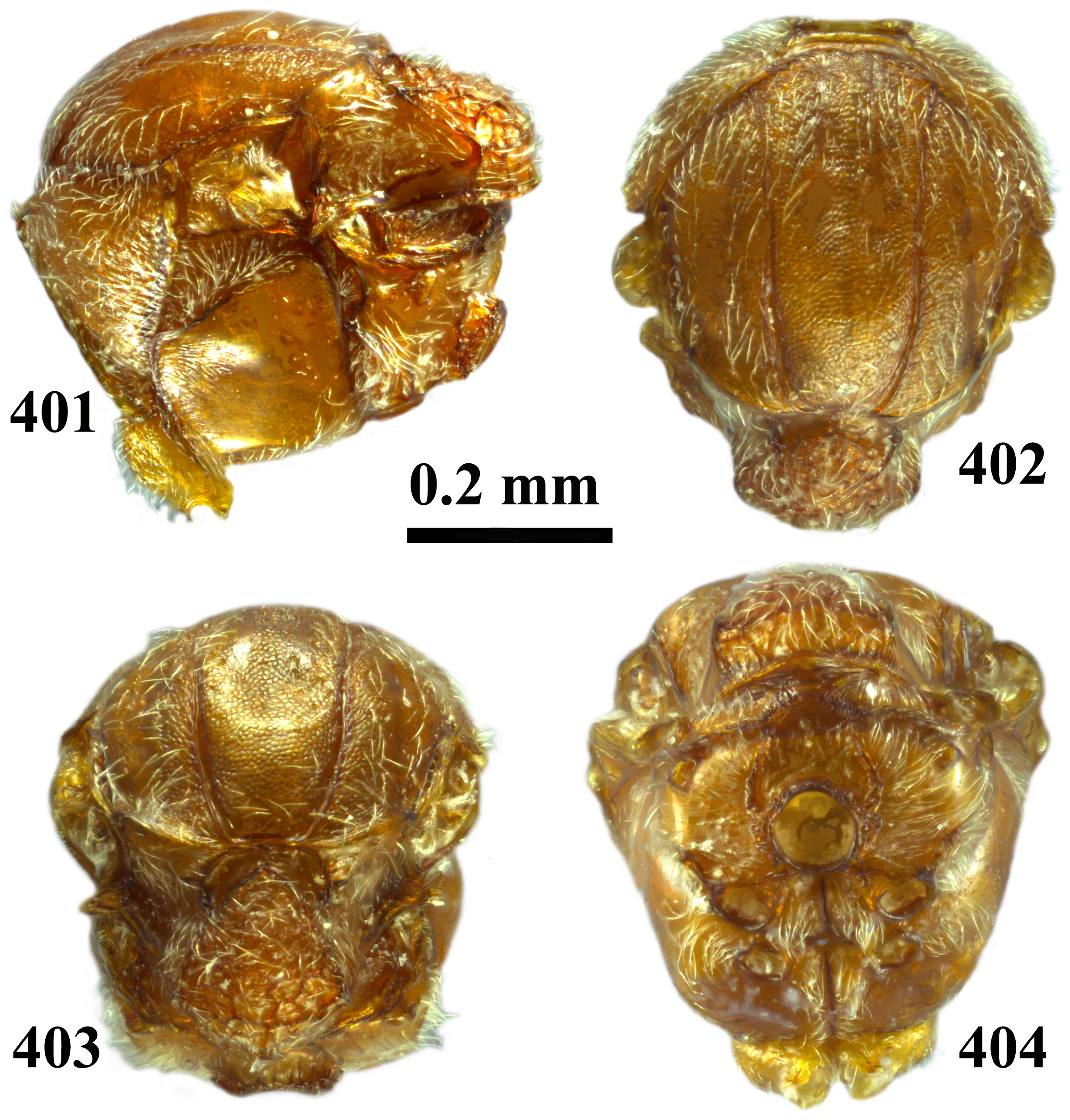
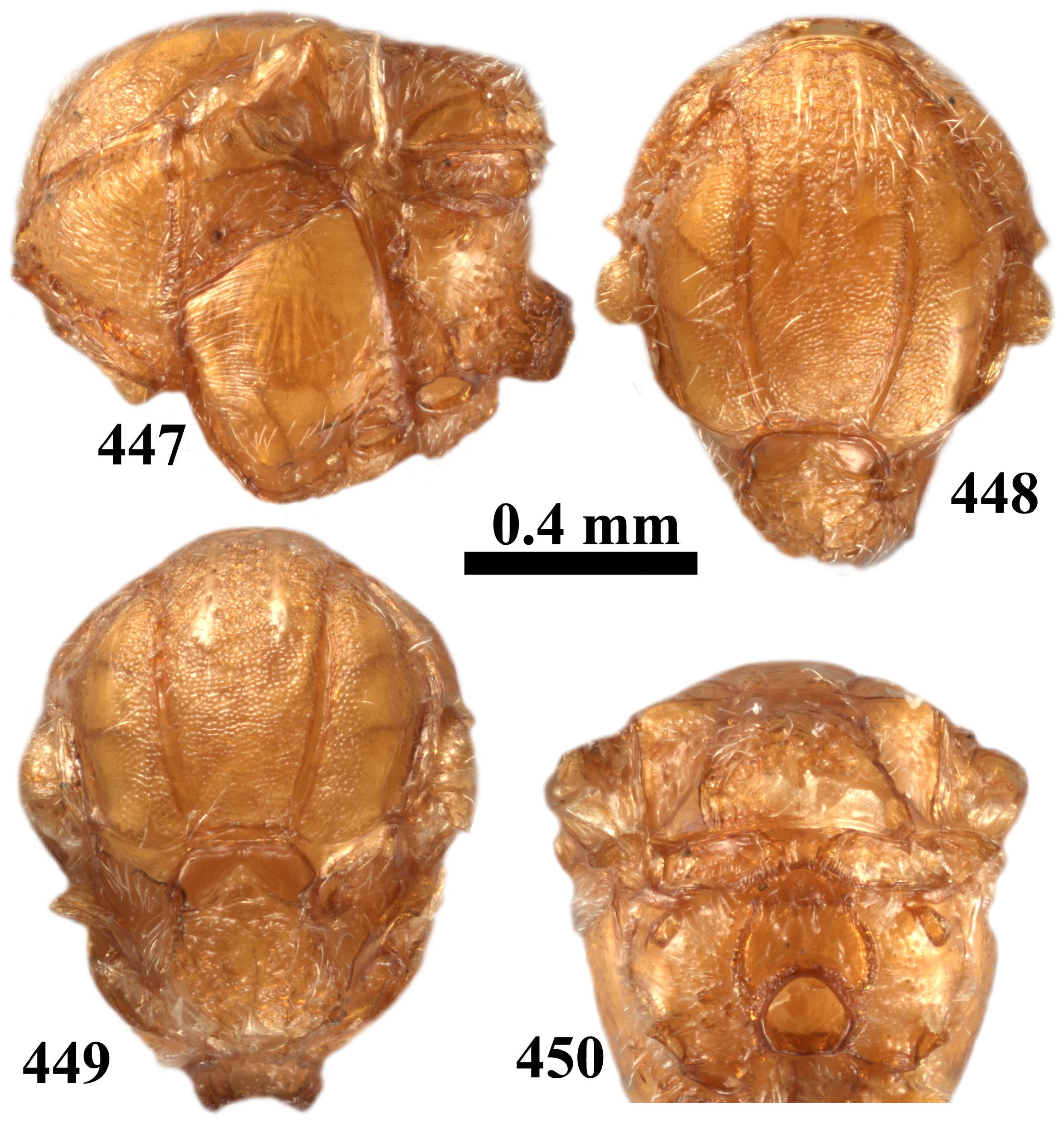
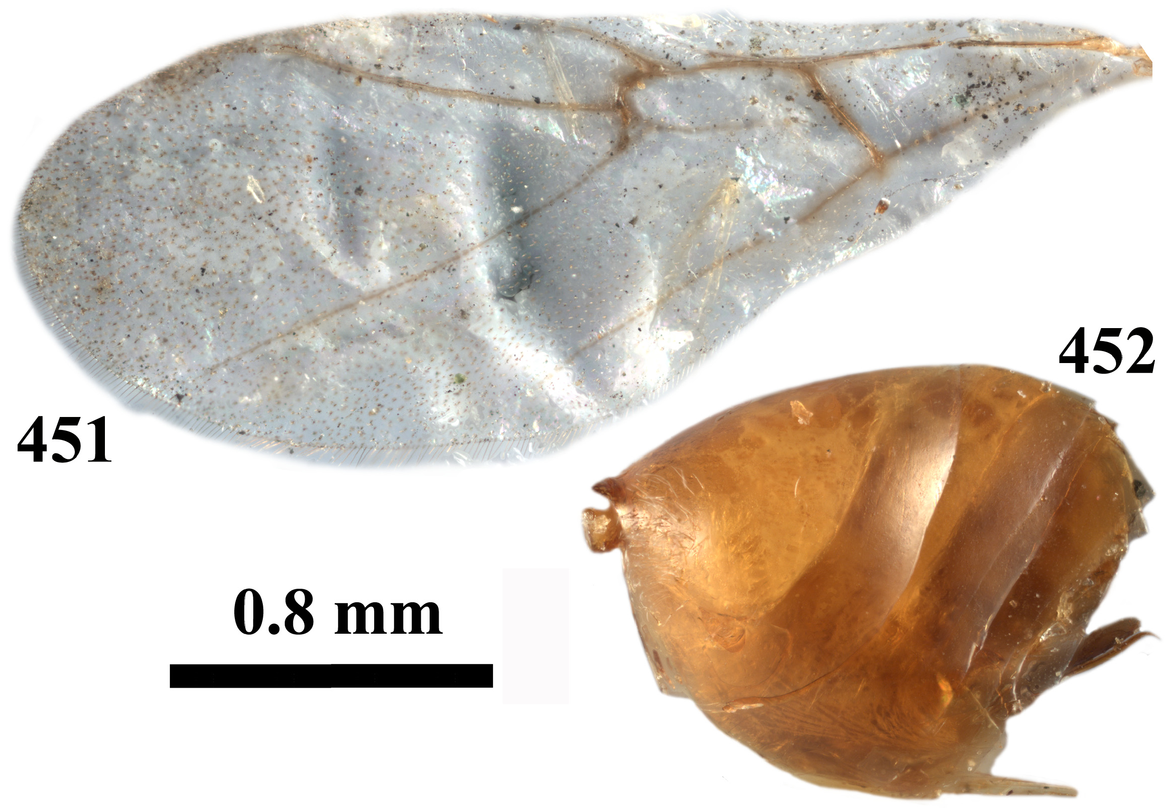
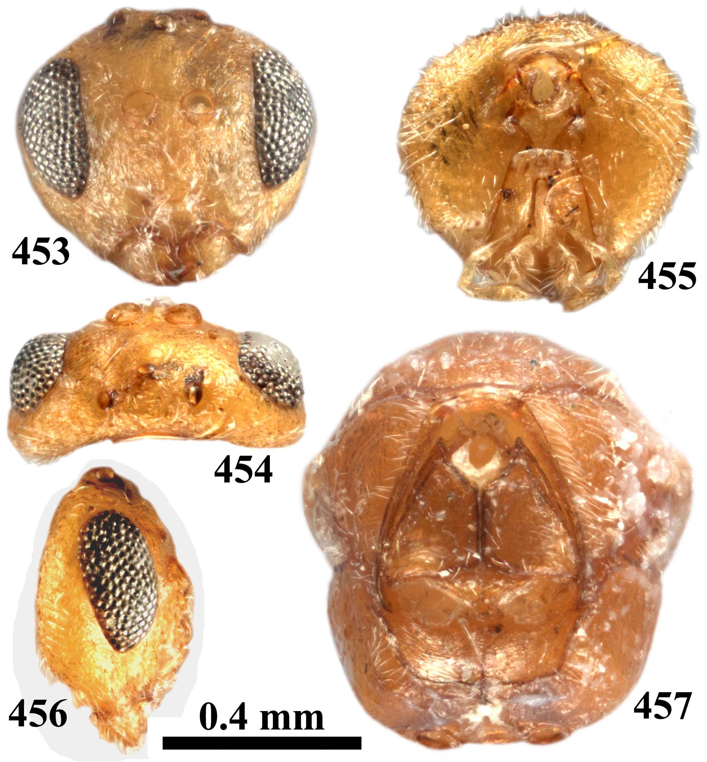
42. Frons bulging in frontal view ( Fig. 429 View FIGURES 429–434 ); POL subequal to OOL ( Fig. 430 View FIGURES 429–434 ); gena broadened behind eye in frontal view ( Fig. 429 View FIGURES 429–434 ); mesoscutum uniformly reticulate-coriaceous ( Fig. 436 View FIGURES 435–438 ); central mesoscutellar disk coriaceous ( Fig. 437 View FIGURES 435–438 ); radial cell around 4.8× as long as broad; third and subsequent metasomal terga with sparse micropunctures ( Fig. 439 View FIGURES 439–440 ); body yellowish to light brown ( Figs 429–439 View FIGURES 429–434 View FIGURES 435–438 View FIGURES 439–440 )...................................................... tibiale comb. rev. (asex), part
- Frons not bulging in frontal view ( Figs 395 View FIGURES 395–400 , 409, 414 View FIGURES 409–418 ), POL at least 1.4× longer than OOL ( Figs 397 View FIGURES 395–400 , 410, 415 View FIGURES 409–418 ); gena not or slightly broadened behind eye in frontal view ( Figs 395 View FIGURES 395–400 , 409, 414 View FIGURES 409–418 ); mesoscutum alutaceous to reticulate ( Figs 402 View FIGURES 401–404 , 420, 424 View FIGURES 419–425 ); center of mesoscutellar disk smooth or finely rugose ( Figs 403 View FIGURES 401–404 , 420, 424 View FIGURES 419–425 ); radial cell around 3.7× as long as broad ( Figs 405 View FIGURES 405–408 , 422 View FIGURES 419–425 ); all metasomal terga without micropunctures ( Figs 406 View FIGURES 405–408 , 426–427 View FIGURES 426–428 ); body chestnut brown or rusty brown ( Figs 395–406 View FIGURES 395–400 View FIGURES 401–404 View FIGURES 405–408 , 409–427 View FIGURES 409–418 View FIGURES 419–425 View FIGURES 426–428 , 429–439 View FIGURES 429–434 View FIGURES 435–438 View FIGURES 439–440 )................................................................................... 43
43. Mesoscutum sometimes with dark brown areas around anterior parallel lines and parapsidal lines ( Figs 420, 424 View FIGURES 419–425 ); frons coarsely coriaceous ( Figs 409–410 View FIGURES 409–418 ); mesoscutellar foveae divided by a fine carina ( Fig. 420 View FIGURES 419–425 ); chestnut brown, head always darker than mesosoma ( Figs 409–427 View FIGURES 409–418 View FIGURES 419–425 View FIGURES 426–428 )............................................................ tetyanae sp. nov. (asex)
- Frons finely alutaceous ( Fig. 395 View FIGURES 395–400 ); mesoscutellar foveae divided by a triangular elevated coriaceous central carina ( Fig. 403 View FIGURES 401–404 ); rusty brown to light brown, head and mesosoma of the same colour and without dark areas ( Figs 395–404 View FIGURES 395–400 View FIGURES 401–404 , 406 View FIGURES 405–408 )...................................................................................... tecturnarum comb. nov. (asex)
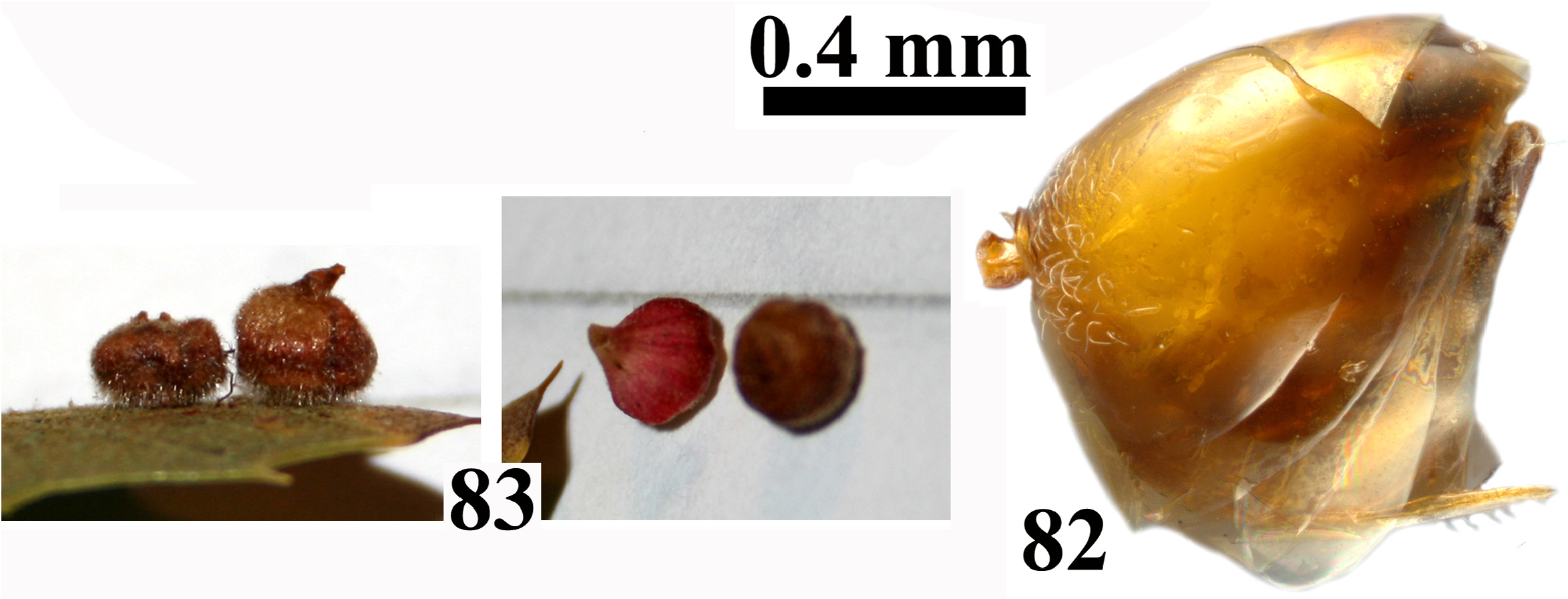
44. Frons bulging in frontal view; eyes strongly converging ventrally; eye 3.0× as high as length of malar space ( Fig. 72 View FIGURES 72–77 ); central part of mesoscutellum smooth ( Fig. 80 View FIGURES 78–81 ); prominent part of ventral spine of hypopygium around 3.8× as long as broad in ventral view ( Fig. 82 View FIGURES 82–83 ); yellowish to light brown ( Figs 72–82 View FIGURES 72–77 View FIGURES 78–81 View FIGURES 82–83 ).................................... caepula comb. nov. (asex)
- Head rounded; eyes parallel or very slightly converging ventrally; height of eye less than 2.6× as high as length of malar space ( Figs 69 View FIGURES 68–71 , 311 View FIGURES 311–316 , 324 View FIGURES 324–327 , 354 View FIGURES 354–359 ); central part of mesoscutellum coriaceous to rugose ( Figs 70 View FIGURES 68–71 , 319 View FIGURES 317–320 , 325 View FIGURES 324–327 , 362 View FIGURES 360–363 ); prominent part of ventral spine of hypopygium at least 5.5× as long as broad in ventral view ( Figs 68 View FIGURES 68–71 , 321 View FIGURES 321–323 , 327 View FIGURES 324–327 , 365 View FIGURES 364–366 ); brown, sometimes with darker marks ( Figs 68–70 View FIGURES 68–71 , 311–315 View FIGURES 311–316 , 317–321 View FIGURES 317–320 View FIGURES 321–323 , 324–327 View FIGURES 324–327 , 354–363 View FIGURES 354–359 View FIGURES 360–363 , 365 View FIGURES 364–366 )............................................... 45
45. Mesoscutellar foveae absent, only slightly impressed on anterior part, with the bottom finely rugose like the rest of the disk, without central carina ( Fig. 70 View FIGURES 68–71 ); metasoma longer than high in lateral view ( Fig. 68 View FIGURES 68–71 )............ bakkeri , comb. nov. (asex)
- Mesoscutellar foveae conspicuous and bottom smooth, fused or divided by a central carina or a triangular elevated area ( Figs 319 View FIGURES 317–320 , 325 View FIGURES 324–327 , 362 View FIGURES 360–363 ); metasoma as high as long or higher in lateral view ( Figs 321 View FIGURES 321–323 , 327 View FIGURES 324–327 , 365 View FIGURES 364–366 )............................. 46
46. Mesoscutellum disk faintly reticulate, with an elevated central area; mesoscutellar foveae not delimited posteriorly ( Fig. 325 View FIGURES 324–327 )............................................................................. scutellum comb. nov. (asex)
- Mesoscutellar disk coarsely rugose, without elevated area; mesoscutellar fovea completely delimited by rugose sculpture or by a carina ( Figs 319 View FIGURES 317–320 , 362 View FIGURES 360–363 )............................................................................... 47
47. Notaulus incomplete ( Fig. 361 View FIGURES 360–363 ); mesoscutellar foveae delimited all around by strong black carina; mesoscutellum rugose-coriaceous ( Fig. 362 View FIGURES 360–363 ); all metasomal terga without micropunctures ( Fig. 365 View FIGURES 364–366 ); first flagellomeres lighter than subsequent ( Fig. 358 View FIGURES 354–359 )............................................................................. stellare comb nov. (asex)
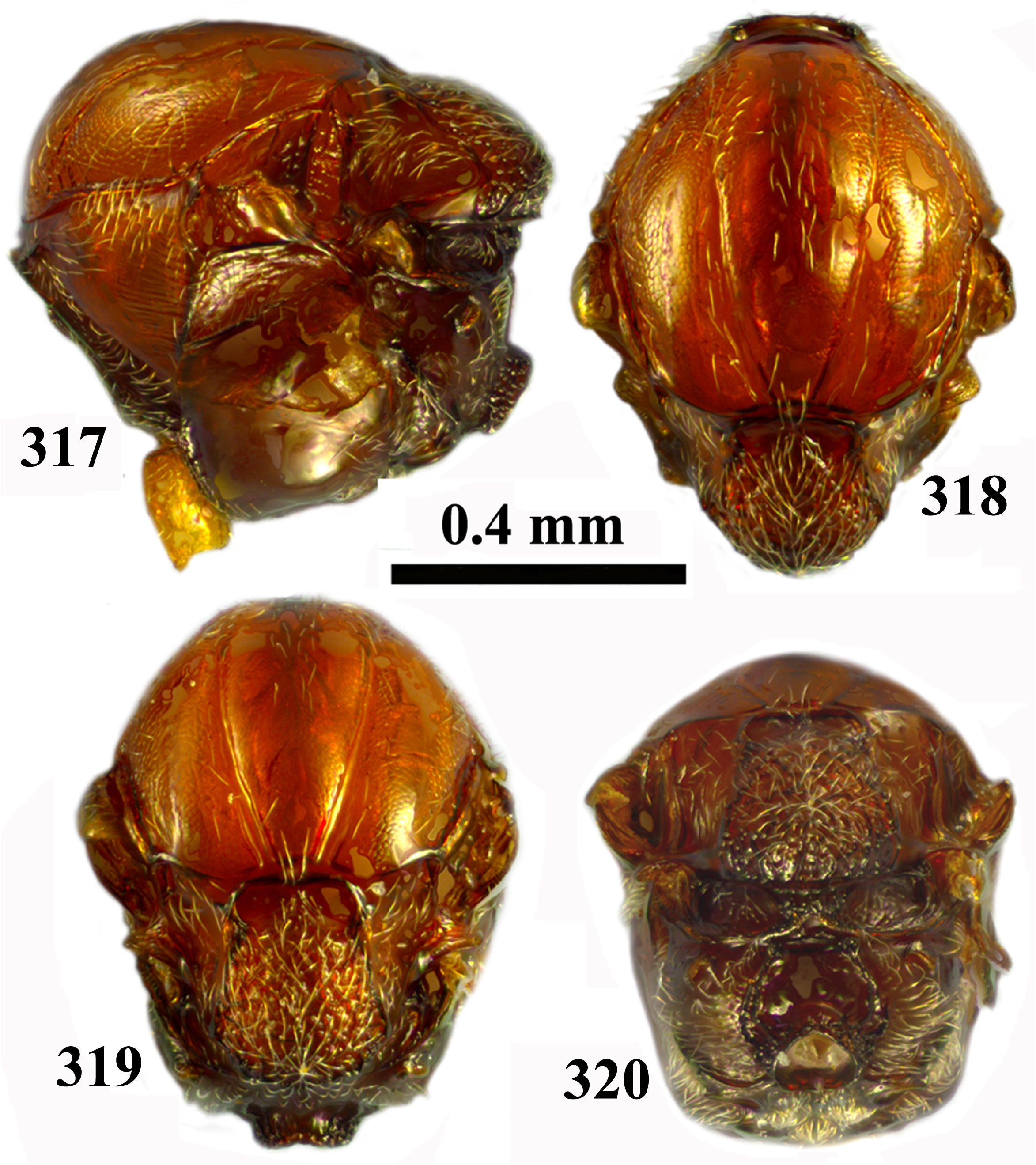
- Notaulus complete ( Fig. 318 View FIGURES 317–320 ); mesoscutellar foveae separated by triangular elevated coriaceous central carina; mesoscutellum rugose ( Fig. 319 View FIGURES 317–320 ); third and subsequent terga with sparse micropunctures ( Fig. 321 View FIGURES 321–323 ); all flagellomeres dark ( Fig. 315 View FIGURES 311–316 )........................................................................................ rucklei sp. nov. (asex)


No known copyright restrictions apply. See Agosti, D., Egloff, W., 2009. Taxonomic information exchange and copyright: the Plazi approach. BMC Research Notes 2009, 2:53 for further explanation.
|
Kingdom |
|
|
Phylum |
|
|
Class |
|
|
Order |
|
|
Family |