
Leviathanochelys aenigmatica, Castillo-Visa & Luján & Galobart & Sellés, 2022
|
publication ID |
https://doi.org/10.1038/s41598-022-22619-w |
|
DOI |
https://doi.org/10.5281/zenodo.7342030 |
|
persistent identifier |
https://treatment.plazi.org/id/10183C77-406C-B02E-FE18-FC0F93CE7300 |
|
treatment provided by |
Tatiana |
|
scientific name |
Leviathanochelys aenigmatica |
| status |
gen. et sp. nov. |
Leviathanochelys aenigmatica gen. etsp. nov.
urn:lsid:zoobank.org:act:28CDDCDB-AC31-45B6-98B1-448E3282A041.
urn:lsid:zoobank.org:act:2B8F389C-6437-450F-812C-E7CD8EFF07A2.
Etymology. The generic name is composed of the following words: Leviathan , in reference to the Biblical marine beast, in allusion to the body size of the new species; and chelys, Latinized name from the ancient Greek Χ έλυς (“khélūs” meaning turtle in feminine gender). The specific nomination aenigmatica, Latinized adjective from the Greek noun αἴνιγμα (“aínigma” meaning enigma, conundrum or riddle) is in reference to the peculiar anatomical characteristics of its pelvis and carapace.
Holotype. MCD9884 . Posterior portion of the carapace including the neural plates 5–8, both left and right fragmentary costals 5–8 and a putative vertebral centra, nearly unidentifiable (MCD9884a); and a partial pelvic girdle, including: the left pubis (MCD9884b); right pubis (MCD9884c); left ischium (MCD9884d); right ischium (MCD9884e); left ilium (MCD9884f); and right ilium (MCD9884g).
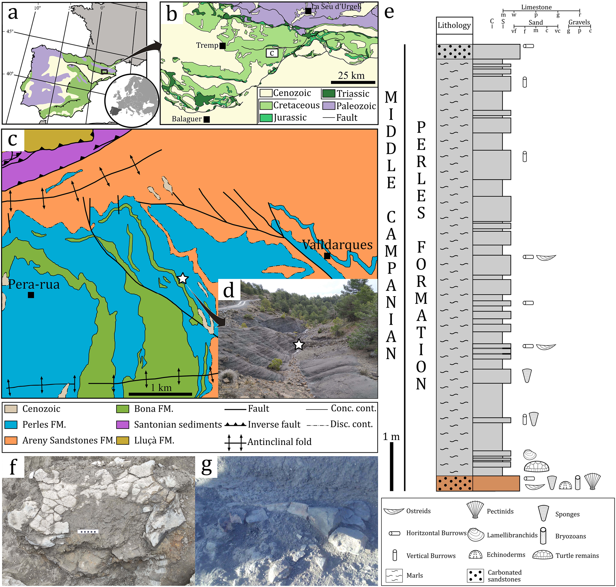
Type locality and age. Cal Torrades, Coll de Nargó (Lleida Province, Catalonia, North-eastern Spain). Lower part of the Perles Formation, Middle Campanian, Upper Cretaceous 35.
Diagnosis. Large-sized basal chelonioid defined by the following and unique combination of characters: reduction of the costal plates ossification without a sutural contact between costals and peripherals; carapacial plate margins (costals 5–8 and neurals 5–8) finely sutured; hexagonal/octagonal neural plates, longer than wide, that prevent the costals 6–7 from contacting one another; posterior costal plates that are rectangular-shaped, much wider than long; oval articular area of the ilium, located near the lateral margin of the right costal 8; H-shaped pelvis; enlarged and flat lateral pubic process; conspicuously ornamented, textured surface surrounding the acetabular region; extremely elongated iliac neck; and the absence of carapacial scute sulci, keels, or ornamentation on the external part of the carapace, and absence of the ilium’s posterior notch. Leviathanochelys aenigmatica is further diagnosed by having two autapomorphic characters as follows: accessory process on the anteromedial margin of the pubis; and acetabulum strongly ventrolaterally directed.
Nomenclatural acts. The electronic version of this article in Portable Document Format (PDF) will represent a published work according to the International Commission on Zoological Nomenclature (ICZN), and hence the new names contained in the electronic version are effectively published under that Code from the electronic edition alone. This published work and the nomenclatural acts it contains have been registered in ZooBank, the online registration system for the ICZN. The ZooBank LSIDs (Life Science Identifiers) can be resolved and the associated information viewed through any standard web browser by appending the LSID to the prefix http://zoobank.org/. The LSID for this publication is: urn:lsid:zoobank.org:pub:8288E740-AE81- 4F71-8931-86A730182034. The online version of this work is archived and available from the following digital repositories: PubMed Central and CLOCKSS.
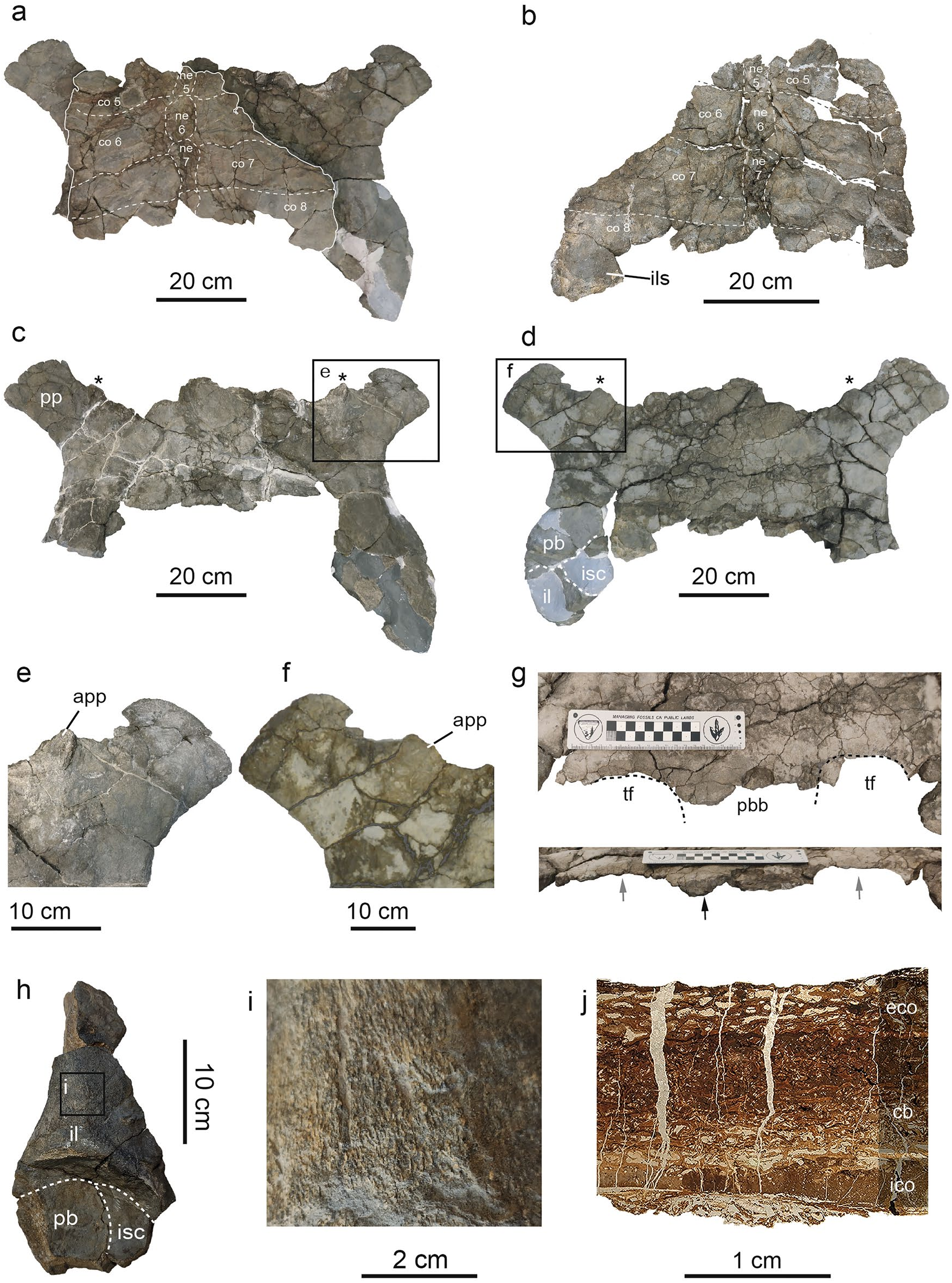
Description. Only a posterior fragment of the carapace is preserved ( Fig. 2a,b View Figure 2 ), consisting of the remains of the neurals 5–8, and both right and left costals 5–8. The smooth dorsal surface of the carapace is slightly convex, whereas the ventral one is almost flat. No epidermal scute marks are present ( Fig. 2a View Figure 2 ), and there is no evidence of keel or medial shallow depression along the medial axis of the preserved neural or costal plates ( Fig. 2a View Figure 2 ). The left portion of the carapace is the most complete, being their lateral edge slightly sinuous and smooth. The preserved costal plates are subrectangular, much wider mediolaterally than long anteroposteriorly, and finely sutured ( Fig. 2a View Figure 2 ). Their distal edges are slightly sinuous and smooth, which indicates that these plates would have had the lateral extensions rod-shaped to join with the peripheral plates. Viscerally, the right portion of the costal 8 preserves an oval concavity to anchor the ilium to the carapace by ligaments ( Fig. 2b View Figure 2 , Fig. S1a View Figure 1 ). The neural series only preserves four elements ( Fig. 2a,b View Figure 2 ): Neural 5 only preserves its posteriormost part, neural 6 is the largest plate and is octagonal-shaped, while neural 7 is hexagonal with short sides in front, and neural 8 is represented by its anteriormost part. It is noteworthy that the neural 7 is highly reduced posteriorly, which would indicate that the total number of elements of the neural series would be equal to or less than nine. In either case, both neurals 6–7 prevent the costals 6–7 from contacting one another along the midline ( Fig. 2a,b View Figure 2 ). Remains of the thoracic vertebrae attachments can be discerned viscerally in both neural plates 6–7 ( Fig. 2a,b View Figure 2 ), highlighting extremely crushed vertebral centra preserved over the neural 7.
As for the preserved pelvic bones ( Fig. 2c,d,h View Figure 2 , Fig. S1b–f View Figure 1 ), they form a nearly flat and H-shaped pelvic girdle ( Fig. 2c,d View Figure 2 ). Both pubes are almost complete but lacking most of the anteromedial and posteromedial processes. These bones are flat, smooth, and completely fused to each other. Because of the fragmentary nature of the pelvis, it is not possible to accurately evaluate the expansion of the anteromedial pubic process. The lateral pubic process is flat, square-shaped, and prominent ( Fig. 2c–f View Figure 2 , Fig. S1b View Figure 1 ): it extends anterolaterally being deflected about 50º from the sagittal plane of the pubic symphysis ( Fig. 2c View Figure 2 ). Apubic accessory process is located between the lateral and medial pubic processes, which is slightly protruding anteriorly ( Fig. 2e,f View Figure 2 ): it shows a striated pattern on its surface and is slightly convex dorsally and ventrally.
Due to the absence of most of the posterior margins of the pubes, it is not possible to evaluate with confidence if the thyroid fenestra was completely separated ( Fig. 2g View Figure 2 ). However, it is certain that an expanded pubioischiadic bridge would have divided the thyroid fenestrae, at least partially along its medial plane, given that the area for accommodating such process is thicker than the surrounding lateral areas ( Fig. 1g View Figure 1 ). The acetabular contour is oval-shaped and slightly constricted anteroposteriorly ( Fig. 2d View Figure 2 , Fig. S1b View Figure 1 ). The acetabulum concavity is completely directed ventrally, and slightly tilted laterally ( Fig. 2d View Figure 2 ). Both the lateral and medial external surfaces surrounding the acetabular region are strongly ornamented with irregular anastomosed ridges. There is no posterior notch in the acetabulum.
Both left and right ilia are partially preserved, but given the fragmentary nature of the right ilium, the following description is mainly based on the best-preserved left ilium ( Fig. 2h View Figure 2 , Fig. S1c–e View Figure 1 ). It preserves most of the acetabular region and the iliac neck, which is elongated, and when complete, would have reached an anteroposterior length greater than two times the anteroposterior acetabulum’s length. Ventrally, the medial margin of the iliac neck is nearly straight, while the lateral one is convex, proximally straight, and distally deflected posteriorly. The iliac neck slightly bends dorsomedially ( Fig. 2h View Figure 2 , Fig. S1c–f View Figure 1 ). Moreover, its medial, lateral and ventral external surfaces are strongly sculptured with anastomosing anteroposteriorly-oriented ridges ( Fig. 2i View Figure 2 ).
Both ischia are poorly preserved. In fact, only fragments of both left and right ischia, which contribute to the posteromedial region of the acetabulum, are available. According to the preserved graphic documents, it can be stated that the ischium contributed significantly to the acetabulum, and that its main body was likely projected medially ( Fig. S1f View Figure 1 ).
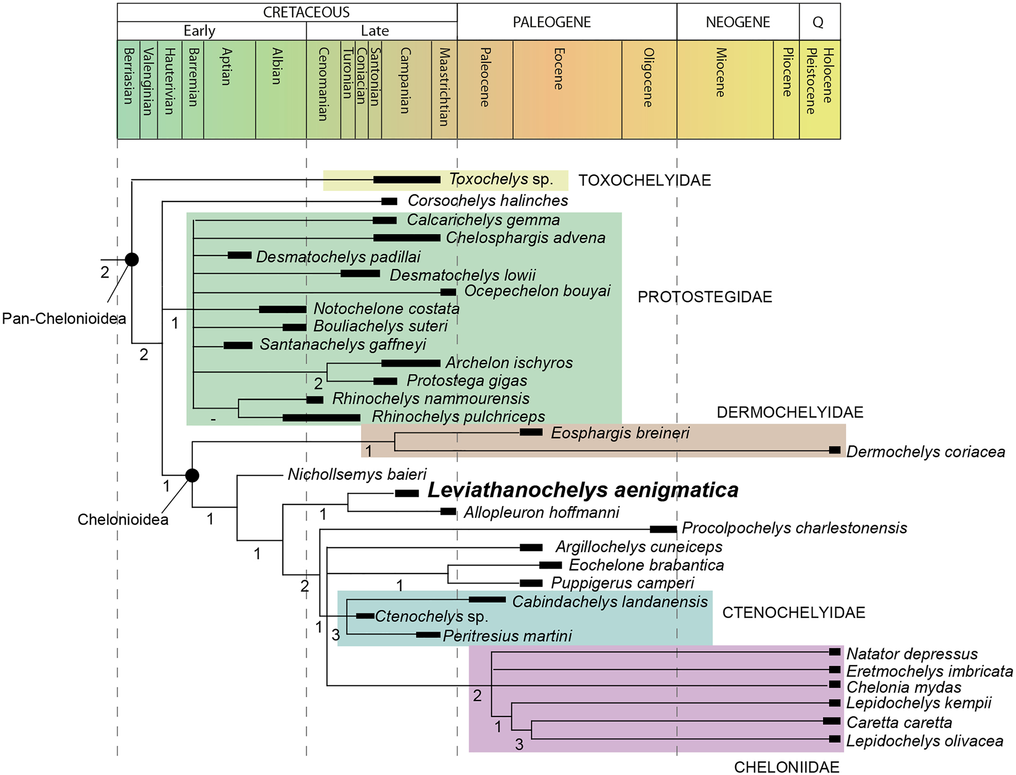
Phylogeneticrelationships. The parsimony analyses resulted in 20 most parsimonious trees of 1647 steps in length, with a Consistency Index of 0.250 and a Retention Index of 0.686 ( Fig. 3 View Figure 3 ). The Strict Consensus topology recovered Leviathanochelys aenigmatica as the sister taxon of the basal chelonioid Allopleuron hoffmanni ( Fig. 3 View Figure 3 ). Nonetheless, it is noteworthy that the only common synapomorphy grouping Leviathanochelys and Allopleuron is “the attachmentof thepelvis to shellby ligaments, insteadof astrong sutural contact” (ch. 318:0) 37. However, this feature is highly plesiomorphic since it represents the typical condition for all Testudinata, to the exception of Pleurodira, Proterochersidae and, maybe also Australochelyidae 38, 39.
The identification of L. aenigmatica as a pan-chelonioid is supported by the absence of contact between costal and peripheral plates (ch. 212:1), while the recovery of Leviathanochelys and Allopleuron as basal members of the Chelonioidea superfamily is established by: the absence of discernible carapacial scutes (ch. 188) 37; and the presence of apartially or completely separated thyroid fenestra (ch. 319) 37, a featurethat is also shared with Peritresius martini and Erquelinnesia gosseleti taxa.
Despite being out of thescope of the present study to deeply analyse all the phylogeneticrelationships of the recovered topology, it is worth noting that our phylogenetic results located Protostegidae as stem Chelonioidea . As previously mentioned, the phylogenetic position of this group of Cretaceous marine turtles is a matter of an intense debate 5, nonetheless, our results concur with some of the most recent studies 37. On the other hand, Ctenochelyidae is recovered as members of Chelonioidea , being its location more inclusive than in other recent phylogeneticstudies 5, 11.
No known copyright restrictions apply. See Agosti, D., Egloff, W., 2009. Taxonomic information exchange and copyright: the Plazi approach. BMC Research Notes 2009, 2:53 for further explanation.
|
Kingdom |
|
|
Phylum |
|
|
Class |
|
|
Order |
|
|
SuperFamily |
Chelonioidea |
|
Family |
|
|
Genus |