
Polana (Varpulana) McKamey, 2006
|
publication ID |
https://doi.org/ 10.11646/zootaxa.4767.4.1 |
|
publication LSID |
urn:lsid:zoobank.org:pub:C3172464-EE05-43BF-90D2-EBE0A7E45826 |
|
DOI |
https://doi.org/10.5281/zenodo.3796715 |
|
persistent identifier |
https://treatment.plazi.org/id/100D87DD-F250-FF89-BEFA-C5B3E7919019 |
|
treatment provided by |
Plazi |
|
scientific name |
Polana (Varpulana) McKamey, 2006 |
| status |
|
Polana (Varpulana) McKamey, 2006 View in CoL
Type-species Polana alata DeLong & Freytag, 1972 View in CoL
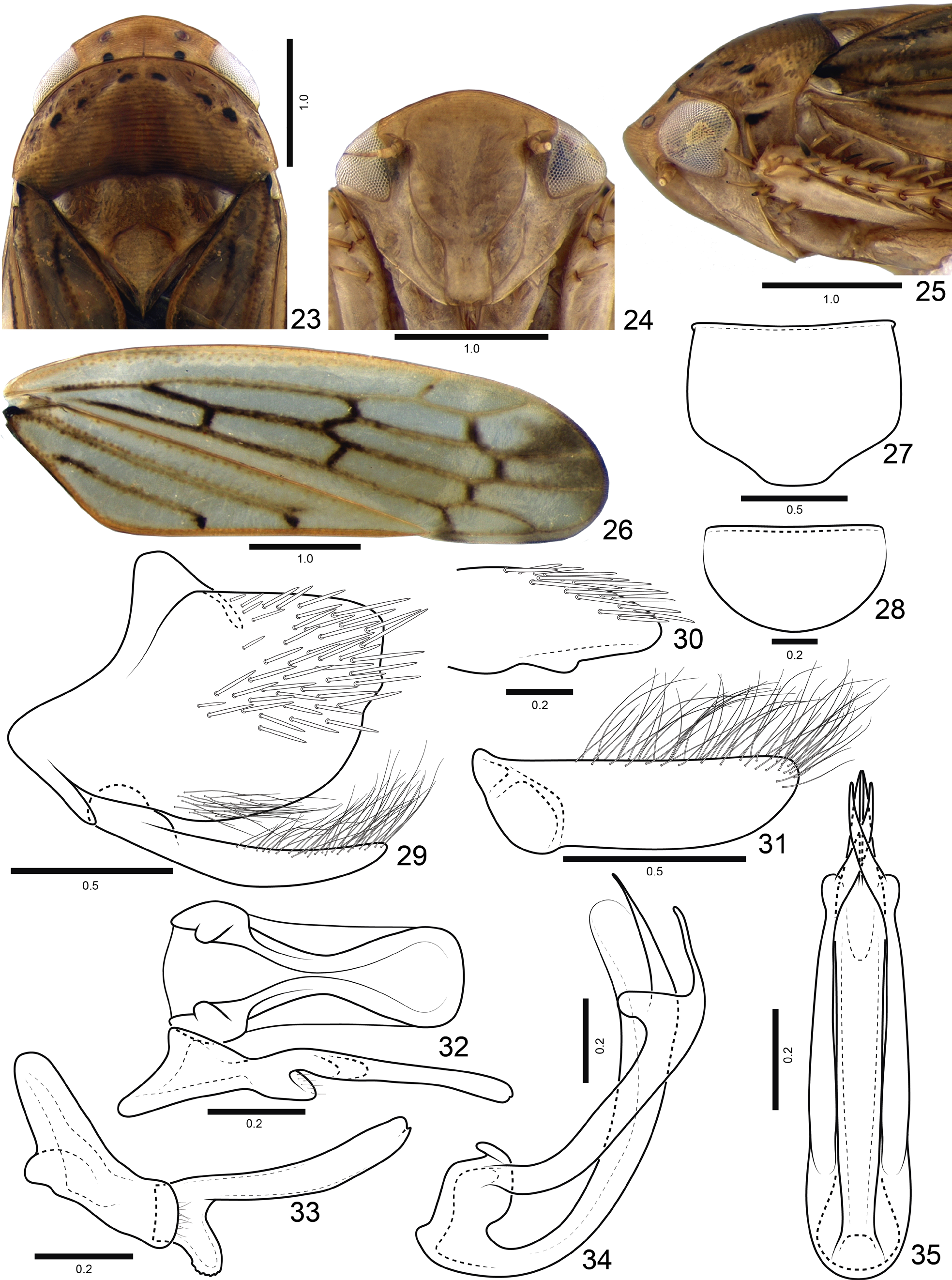
Diagnosis. Male sternite VIII partially hiding subgenital plates. Male pygofer ( Figs 51, 52 View FIGURES 45–57 ) commonly with short, thin process on dorsal margin at base; ventral margin with tuft of hair-like setae; posterior margin with or without rounded protrusion near apex of pygofer. Subgenital plate ( Fig. 9 View FIGURES 1–13 ) with hair-like setae on external margin. Style ( Fig. 33 View FIGURES 23–35 ) with conspicuous protrusion on ventral margin near base of blade. Aedeagus ( Figs 12 View FIGURES 1–13 , 34 View FIGURES 23–35 ) with apodemal processes; shaft elongate, with or without processes at mid-length or subapically; apex membranous.
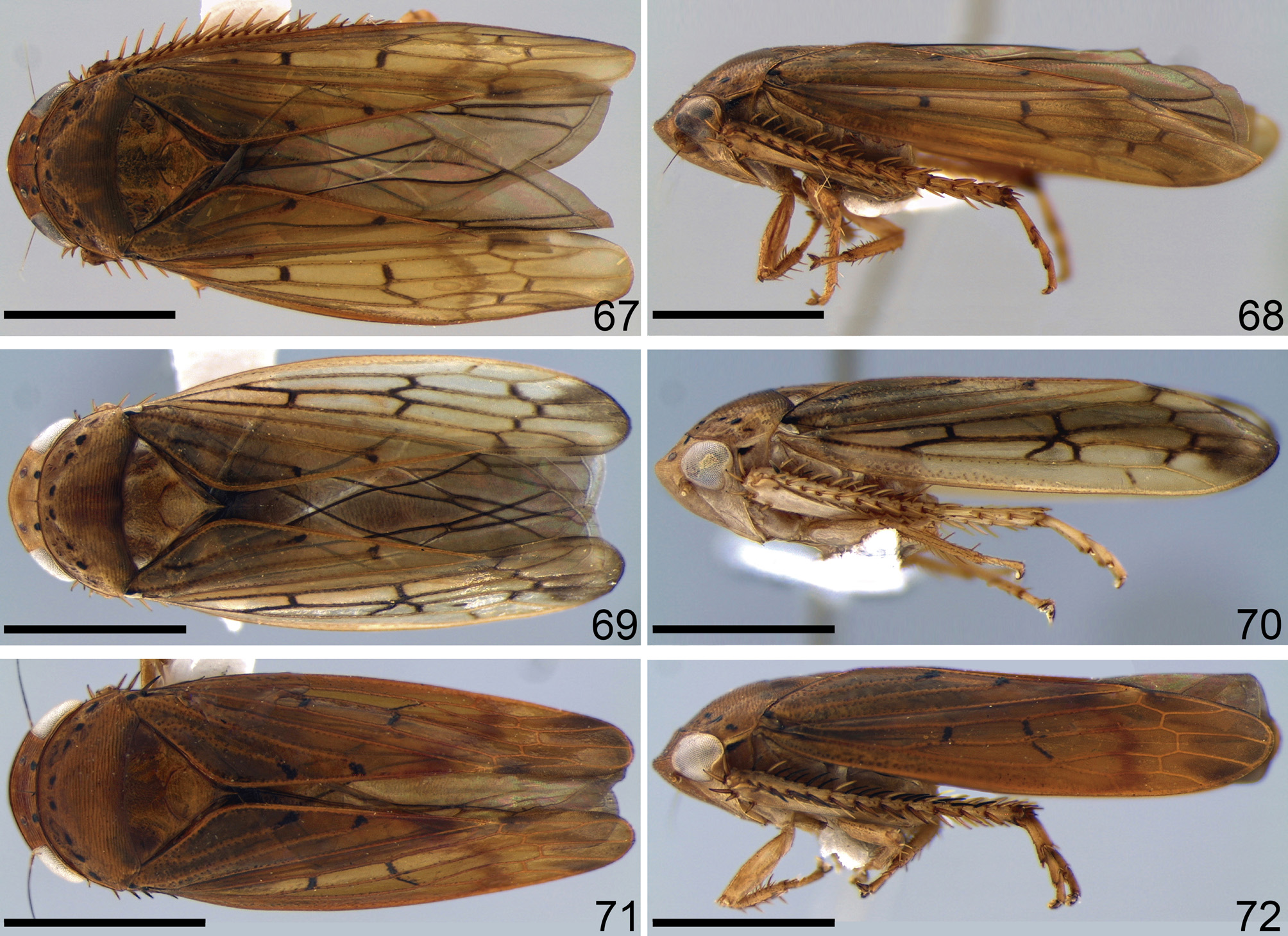
Description. Body ( Figs 67–72 View FIGURES 67–72 ) approximately oval, not flattened dorsoventrally, small, between 6.1 to 9.2mm. Head ( Figs 1 View FIGURES 1–13 , 23 View FIGURES 23–35 , 45 View FIGURES 45–57 ), in dorsal view, not produced; median length of crown approximately half as long as interocular width; crown with transverse parallel striae, anterior margin of crown approximately parallel to posterior margin; transocular width of head slightly narrower than maximum pronotum width; ocelli equidistant between eyes and median line and near to anterior than to posterior margin of crown. Head ( Figs 2 View FIGURES 1–13 , 24 View FIGURES 23–35 , 46 View FIGURES 45–57 ), in ventral view, with face wider than long; frontogenal suture distant from eye margins by half width of clypeus and surpassing antennal ledge, extending to anterior margin of crown; antennal ledge carinate, oriented obliquely downwards in relation to frons and extending over frons by short distance; frons approximately as long as wide medially, surface with texture shagreen, not excavated below anterior margin of crown; epistomal suture indistinct medially; clypeus not inflated, approximately 1.3 times longer than wide, lateral margins parallel, apex carinate and slightly emarginated; maxillary plate produced ventrally as far as clypeus apex; gena with ventrolateral margins slightly convex at midlength. Head ( Figs 3 View FIGURES 1–13 , 45, 47 View FIGURES 45–57 ), in lateral view, with crown-face transition rounded and with several parallel striae. Pronotum, in lateral view, slightly declivous, with transverse striae on disc and posterior third. Forewing ( Figs 4 View FIGURES 1–13 , 26 View FIGURES 23–35 , 48 View FIGURES 45–57 ) without extra crossveins; venation distinct; appendix well developed and bordering first and second apical cells, narrower than maximum width of first apical cell.
Profemur, with AV and PV rows formed by 4–5 and 1–2 setae respectively. Protibia, in cross-section, more or less cylindrical, with longitudinal carina adjacent to PD row; AV row formed by long setae, gradually increasing in thickness and length towards apex; AD formed by many small undifferentiated setae; PD row with 4 long setae and intercalary small undifferentiated setae; PV row developed, with very small setae near base and 4–5 long setae on apical two thirds. Hind leg with femoral setal formula 2:2:1; tibial rows PD, AD, and AV with 22–25, 11–12, and 12–15 macrosetae, respectively; AD row with intercalary small setae between macrosetae; PV row with setae of apical half formed by sequence of 1 thicker and 3–4 thinner setae; first tarsomere with two rows of 6–7 setae on plantar surface; apex with 4–5 patellae flanked by tapered lateral setae; second tarsomere pecten with 2 platellae flanked by 2 tapered lateral setae on inner and 1 on external corner.
Male terminalia. Sternite VIII ( Figs 5 View FIGURES 1–13 , 49 View FIGURES 45–57 ) commonly wider than long, partially hiding subgenital plates. Valve ( Figs 6 View FIGURES 1–13 , 28 View FIGURES 23–35 ) wider than long; integument thickening present only on dorsal margin. Pygofer with ( Figs 7 View FIGURES 1–13 , 29 View FIGURES 23–35 ) or without ( DeLong & Freytag, 1972: 294, fig. 298) short processes, on dorsal margin, at base; with tuft of hair-like setae near ventral margin ( Fig. 7 View FIGURES 1–13 ); macrosetae dispersed on posterodorsal quadrant; posterior margin with ( Figs 30 View FIGURES 23–35 , 52 View FIGURES 45–57 ) or without ( Domahovski & Cavichioli, 2017a: 540, fig. 22) short and rounded protrusion near apex of pygofer. Subgenital plate ( Figs 9 View FIGURES 1–13 , 31 View FIGURES 23–35 ) with hair-like setae on external margin. Style with conspicuous protrusion on ventral margin near base of blade ( Figs 11 View FIGURES 1–13 , 33 View FIGURES 23–35 ). Aedeagus ( Figs 12 View FIGURES 1–13 , 34 View FIGURES 23–35 , 56 View FIGURES 45–57 ) preatrium not developed; dorsal apodeme rounded, not developed laterally; atrium with processes; shaft elongate, cylindrical, with or without processes at mid-length or subapically; apex membranous.
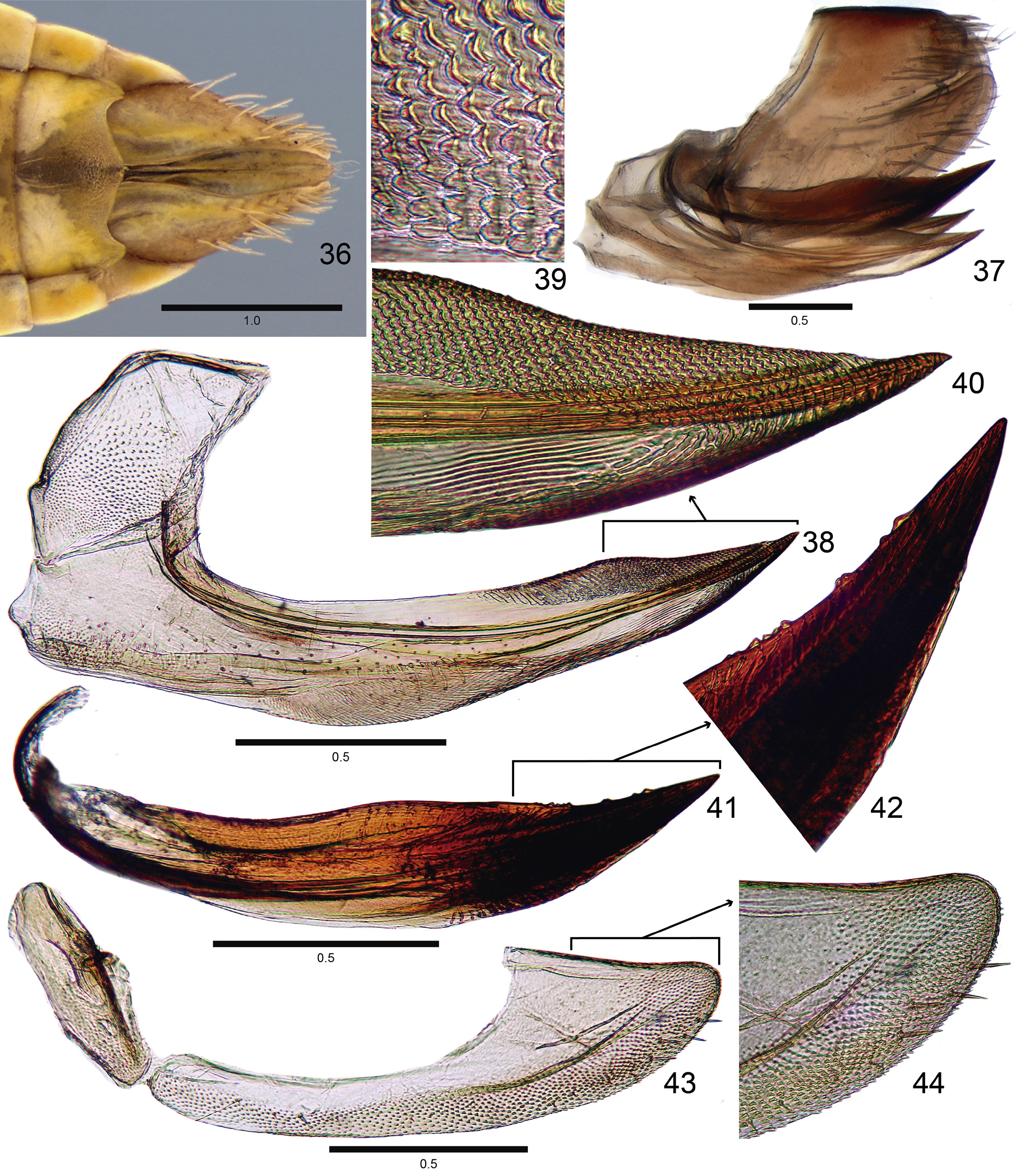
Female terminalia. Pygofer ( Figs 15 View FIGURES 14–22 , 37 View FIGURES 36–44 , 59 View FIGURES 58–66 ) about 1.5 times longer than maximum height; with apex rounded; macrosetae dispersed on dorsoapical fourth and ventroapical half. Internal sternite VIII membranous. First valvifer ( Figs 16 View FIGURES 14–22 , 38 View FIGURES 36–44 , 60 View FIGURES 58–66 ) higher than long. First valvula apical portion with lateral carina bearing small teeth; in lateral view ( Figs 16 View FIGURES 14–22 , 38 View FIGURES 36–44 , 60 View FIGURES 58–66 ) slightly curved dorsally; basal portion produced anterad and rounded; ventral margin with oblique striae medially; apical third with dorsal sculptured area formed by scale-like processes arranged in oblique lines ( Figs 17 View FIGURES 14–22 , 39 View FIGURES 36–44 , 61 View FIGURES 58–66 ); apex abruptly tapered and acute. Second valvula, in cross-section, with apical portion triangular, expanded laterally forming lateral carina, in lateral view ( Figs 19 View FIGURES 14–22 , 41 View FIGURES 36–44 , 63 View FIGURES 58–66 ), higher near mid-length; dorsal margin with few very small teeth on apical fourth; apical portion gradually narrowed to acute apex. Second valvifer ( Figs 21 View FIGURES 14–22 , 43 View FIGURES 36–44 , 65 View FIGURES 58–66 ) about 2.5 times longer than high. Gonoplac ( Figs 21 View FIGURES 14–22 , 43 View FIGURES 36–44 , 65 View FIGURES 58–66 ), about 3.5 times longer than high; dorsoapical and ventroapical margins convergent; dorsoapical margin straight, short, with one third length of gonoplac; ventral margin and apical potion ( Figs 22 View FIGURES 14–22 , 44 View FIGURES 36–44 , 66 View FIGURES 58–66 ) with dentiform cuticular projections and few short setae; apex subacute.
Coloration. Head and thorax ( Figs 69, 71 View FIGURES 67–72 ) yellow or brown. Head ( Fig. 1 View FIGURES 1–13 ) frequently with pair of black spots behind ocelli. Pronotum ( Fig. 23 View FIGURES 23–35 ) frequently with small maculae near anterior margin. Forewings ( Fig. 4 View FIGURES 1–13 ) commonly with small black maculae on corium and apex of anal veins, with or without larger black maculae on costal margin at mid-length of wing.
Distribution. Brazil (Distrito Federal (new record) and states of Bahia, Espírito Santo, Minas Gerais, Paraná, Piauí (new record), Rio de Janeiro, Rio Grande do Sul, Santa Catarina and São Paulo), Mexico (Department of Tabasco) and ( Colombia, departments of Amazonas and Putumayo).


No known copyright restrictions apply. See Agosti, D., Egloff, W., 2009. Taxonomic information exchange and copyright: the Plazi approach. BMC Research Notes 2009, 2:53 for further explanation.
|
Kingdom |
|
|
Phylum |
|
|
Class |
|
|
Order |
|
|
Family |
|
|
Genus |
Polana (Varpulana) McKamey, 2006
| Domahovski, Alexandre Cruz & Cavichioli, Rodney Ramiro 2020 |
Polana alata
| DeLong & Freytag 1972 |